5 - Acute Kidney Injury (AKI)
 Call Kai
 Learn
 Practice Test
 Spaced Repetition
 Match
 Flashcards
 Knowt Play
1/121

Earn XP
Description and Tags
8 Questions
122 Terms
Acute Kidney Injury (AKI)?
Is an abrupt decrease in kidney filtration/excretory function identified by:
Increase in serum creatinine and/or
Decrease in urine output
What might an AKI reflect?
Impaired kidney function without structural injury
Structural kidney injury
or both
Consequences of AKI?
Accumulation of metabolic waste
Fluid, electrolyte and acid-base disturbances
Can mild AKI be asymptomatic?
Yes
It can be asymptomatic
Serum creatinine criteria for AKI?
Increase in serum creatinine of ≥ 0.3 mg/dL within 48 hours
or
Increase in serum creatinine ≥ 1.5-fold from baseline, which is known or presumed to have occurred within the prior 7 days
Urine output criteria for AKI?
Urine output of <0.5 ml/kg/hr for ≥ 6 hours
Patient susceptibility risk factors for AKI?
Chronic kidney disease
Older age
Diabetes
Hypertension / vascular disease
Heart failure or chronic liver disease
Acute illness or exposures risk factors for AKI?
Critical illness, sepsis, or shock
Hypovolemia / hemorrhage
Major surgery, trauma, or burns
Nephrotoxic or renal-hemodynamic medications

What are the 3 traditional categories of AKI?
Prerenal
Intrinsic
Postrenal
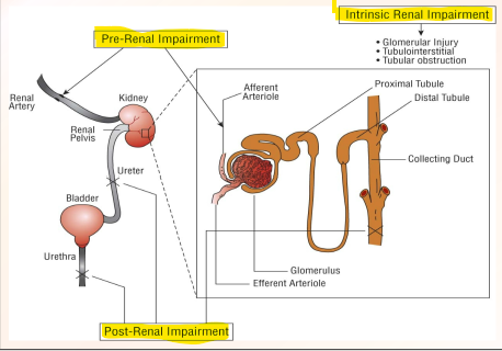
What is the other category of AKI?
Pseudo AKI
Prerenal AKI?
Reduced kidney perfusion without established parenchymal injury
Common causes of prerenal AKI?
Volume depletion → renal/GI losses, hemorrhage
Hypotension / Shock
Reduced effective renal perfusion → ex; HF
What has compensatory responses to reduced perfusion?
Sympathetic nervous system
RAAS
ADH + Thirst
Intrarenal autoregulation
Compensatory response of the sympathetic nervous system to reduced perfusion?
Supports blood pressure
Stimulates renin release
Compensatory response of the RAAS to reduced perfusion?
Helps maintain glomerular pressure
Increase Na+ and water retention
Compensatory response of the ADH + Thirst to reduced perfusion?
Conserve water / increase water intake
Compensatory response of the intrarenal autoregulation to reduced perfusion?
Adjusts arteriolar tone to help preserve GFR
What factors limit autoregulatory compensation?
Older age, atherosclerosis, long-standing hypertension
Chronic kidney disease
Medications → can interfere with compensatory arteriolar responses
slide 27
MOA of ACEI / ARBs?
Block angiotensin II mediated efferent constriction
Decrease intraglomerular pressure and GFR
What is expected after starting / increasing doses of ACEI/ARBs?
A small stable rise in SCr may occur
What is a concerning rise in SCr with ACEI/ARBs for Prerenal AKI?
>30% rise within 4 weeks
Evaluate for volume depletion, hypotension, NSAID use, or renal artery stenosis
Management of ACEI/ARBs if concerning rises in SCr for Prerenal AKI?
Correct reversible factors first
If kidney function does not improve → reduce/hold the ACEI/ARB
MOA of NSAIDs?
Inhibit vasodilatory prostaglandins → causing afferent arteriolar vasoconstriction
Decrease in intraglomerular pressure and GFR
Who is at higher risk of Prerenal AKI when using NSAIDs?
Reduced renal perfusion/volume depletion
CKD
Older age
Concurrent ACEI/ARB
Diuretic use
Prevention of AKI with NSAIDs?
Avoid or minimize NSAID use in high risk patients
Correct volume depletion when present
Management of NSAIDs with prerenal AKI?
Discontinue NSAID when possible
Correct the contributing volume depletion or hypotension
Clinical pearl with NSAIDs?
Can cause Acute interstitial nephritis (AIN) through a separate mechanism
MOA of calcineurin inhibitors?
Renal arteriolar vasoconstriction, particularly of the afferent arteriole
Decrease in renal blood flow and GFR
Ex → Tacrolimus, cyclosporine
Who is at higher risk of Prerenal AKI when using calcineurin inhibitors?
Higher CNI exposure
Volume depletion
Concurrent nephrotoxins
Prevention of Prerenal AKI with calcineurin inhibitors?
Monitor CNI exposure, volume depletion and concurrent nephrotoxins
Management of calcineurin inhibitors with Prerenal AKI?
Assess CNI concentration, volume status and interacting/nephrotoxic medications
Reduce CNI exposure when toxicity is suspected
When can compensatory responses fail to maintain GFR?
Severe or prolonged hypoperfusion → or medications that interfere with arteriolar responses
Early GFR changes may occur without tubular injury
Severe/prolonged ischemia → tubular injury → acute tubular necrosis (ATN)
What are the major causes of intrinsic AKI?
Tubular injury → Acute tubular necrosis (ATN), and Crystal induced nephropathy
Glomerular disease
Vascular injury/disease
Interstitial injury (AIN) → Drugs and infections
Acute tubular necrosis (ATN)?
Most common pattern of intrinsic AKI
Involves injury to renal tubular epithelial cells
What are common settings associated with tubular injury?
Sepsis
Ischemia
Nephrotoxins
How are prerenal AKI and ischemic ATN related?
They exist on a continuum of hypoperfusion
Severe or prolonged hypoperfusion can progress from prerenal AKI to ischemic ATN
How does myoglobin cause intrinsic AKI?
Endogenous nephrotoxin
Rhabdomyolysis → skeletal muscle breakdown → myoglobin release
Filtered myoglobin causes direct tubular injury and pigment-cast obstruction
Volume depletion and renal vasoconstriction further increase AKI ris
Aminoglycosides?
Exogenous nephrotoxin
Can cause ATN → intrinsic AKI
Presents as → Nonoliguric → urine output remains within a normal rang despite the kidneys being damaged/failing
Usually develops after several days of therapy
Mg2+ wasting and other tubular abnormalities may occur
Higher risk of ATN with aminoglycosides in intrinsic AKI?
Prolonged aminoglycoside therapy and concurrent nephrotoxins
Prevention of ATN and intrinsic AKI with aminoglycosides?
Use extended interval dosing when appropriate
Adjust dosing and monitor levels based on kidney function
Management of aminoglycosides for ATN and intrinsic AKI?
Discontinue when possible
Supportive care
Vancomycin?
Exogenous nephrotoxin
Can cause ATN → intrinsic AKI
MOA → Tubular cell injury; vancomycin-containing casts may contribute
Presents as → Rising SCr, usually after several days
High vancomycin concentrations may be a cause or consequence of AKI
Higher risk of ATN with vancomycin in intrinsic AKI?
Greater vancomycin exposure
Critical illness
Concurrent nephrotoxins
Prevention of ATN and intrinsic AKI with vancomycin?
Appropriate dosing and therapeutic drug monitoring
Minimize unnecessary nephrotoxins
Management of vancomycin for ATN and intrinsic AKI?
Reduce or discontinue when possible
Supportive care
Amphotericin B?
Exogenous nephrotoxin
Can cause ATN → intrinsic AKI
MOA → Direct tubular injury; renal vasoconstriction may also contribute
Presents as → Rising Scr with electrolyte wasting → K, Mg
Lipid formulations have less nephrotoxicity
Higher risk of ATN with Amphotericin B in intrinsic AKI?
Higher cumulative exposure
Dehydration
CKD
Prevention of ATN and intrinsic AKI with Amphotericin B?
Prefer lipid / liposomal formulations when clinically appropriate → IV isotonic crystalloid hydration
Management of vancomycin for ATN and intrinsic AKI?
Discontinue or change therapy when possible
Maintain hydration
Avoid other nephrotoxins
Tenofovir?
Exogenous nephrotoxin
Can cause ATN → intrinsic AKI
Presents as → AKI with phosphate wasting, glycosuria, proteinuria, or bicarbonate loss
Tubular dysfunction may occur before overt AKI
Higher risk of ATN with Tenofovir in intrinsic AKI?
Greater drug exposure
CKD
Concurrent nephrotoxins
Prevention of ATN and intrinsic AKI with Tenofovir?
Adjust TDR dosing for kidney function → monitor for tubular toxicity
Tenofovir alafenamide (TAF) → is less nephrotoxic than tenofovir disoproxil fumarate (TDF)
When is iodinated contrast used with?
IV contrast enhanced CT
Intra-arterial angiography
Contrast-associated AKI (CA-AKI)?
AKI occurring after contrast exposure
Regardless of cause
Contrast-induced AKI (CI-AKI)?
AKI actually caused by contrast
When contrast causes AKI → tubular injury and altered renal hemodynamics may contribute
What increases the risk of contrast-associated AKI?
AKI or advanced CKD
Volume depletion
Greater contrast exposure
Risk differs between routine IV CT and intra-arterial angiography.
How is contrast-associated AKI prevented and treated?
Prevention → Depends on patient risk and procedure
Treatment → Primarily supportive care once AKI develops
Crystal induced nephropathy?
Drugs or metabolites may precipitate when urinary concentrations exceed solubility
Tubular injury → Intrinsic AKI
What increases risk for crystal induced nephropathy?
Low urine flow / volume depletion
Greater drug exposure
CKD
How can drug crystals cause intrinsic AKI?
Urine pH affects drug crystallization risk differently depending on the drug
Crystals can cause → tubular injury, inflammation, and intratubular obstruction
How can acyclovir cause crystal-induced AKI, and how is it prevented?
Risk → Crystals form with high urinary concentrations
Prevention → Hydration, avoid rapid IV administration, and adjust dose for kidney function
How can high-dose methotrexate cause crystal-induced AKI, and how is it prevented?
Risk → Methotrexate is less soluble in acidic urine
Prevention → IV hydration and urine alkalinization
Clinical features of crystal-induced nephropathy?
May be asymptomatic except for increase in SCr
Possible symptoms → Hematuria, flank pain, or decreased urine output
Urinalysis may show → Drug-specific crystals or crystal-containing casts
Acute interstitial Nephritis (AIN)?
Immune-mediated inflammation of the tubulointerstitium
Medications are the most common cause
Intrinsic AKI
What drugs are common causes of AIN?
Antibiotics → especially Beta-lactams → penicillins, cephalosporins, sulfonamides, rifampin, and fluoroquinolones
NSAIDs
Proton pump inhibitors
Immune checkpoint inhibitors
How is acute interstitial nephritis (AIN) clinically recognized?
Classic triad → Rash, fever, eosinophilia → often absent
Urinalysis → Pyuria common; WBC casts uncommon
Urine eosinophils → Not reliable for diagnosis
If uncertain → Kidney biopsy may be needed
How is acute interstitial nephritis (AIN) prevented and treated?
Remove the cause → Stop the offending drug when possible
Glucocorticoids → May be considered in selected patients, evidence and optimal regimen are limited
Key point → Earlier recognition and treatment/removal of the cause may improve kidney recovery
Postrenal AKI?
Obstruction of urine flow
Lower tract → BPH, bladder dysfunction/urinary retention, urethral obstruction
Upper tract → Ureteral stones, tumors, retroperitoneal fibrosis
AKI usually requires bilateral obstruction or obstruction of a solitary functioning kidney

Hydronephrosis?
Dilation of the renal collection system
Often results from urinary tract obstruction

Is hydronephrosis synonymous with obstruction?
No
May occur without significant obstruction
Obstruction may occasionally occur without hydronephrosis
Pseudo-AKI?
Increased SCr without a true decrease in GFR
Causes of Pseudo-AKI?
Decreased creatinine secretion → trimethoprim, cimetidine, cobicistat
Increased creatinine production
Creatinine-assay interference
Increased SCr does not always mean decreased kidney filtration
What are the first questions to ask when evaluating AKI?
1. Is this AKI?
Does the patient meet diagnostic criteria?
2. What was baseline kidney function?
Prior SCr/eGFR, CKD history, urinalysis, and imaging when relevant
3. What is the likely cause?
Hemodynamics/volume status, infection, obstruction, medications, or other exposures
Pharmacist role → Identify medication causes and adjust drugs requiring kidney-function-based dosing
What do SCr, eCrCl, and eGFR indicate during AKI?
SCr is an → indirect marker of filtration
eCrCl and eGFR are estimates
During rapidly changing kidney function, SCr lags behind GFR changes, making steady-state equations unreliable
How do kidney function estimates behave during AKI?
Worsening AKI → Estimates may overestimate current kidney function
Recovery → Estimates may underestimate current kidney function
What is Blood Urea Nitrogen (BUN), and why does it rise in AKI?
Urea is a product of protein metabolism
BUN rises as kidney clearance decreases
What does a BUN ratio greater than 20:1 suggest?
Low perfusion increases urea reabsorption
A ratio greater than 20:1 supports → prerenal AKI but is not diagnostic
What else may cause BUN to rise?
GI bleeding
Increased protein intake/nutrition
Catabolism
Glucocorticoids
What is oliguric AKI?
Markedly reduced urine output
Traditionally less than 400–500 mL/day in adults
What is anuria, and how is it related to oliguria?
Anuria is extreme oliguria
Approximately less than 100 mL/day
What do urine output patterns tell you?
Urine output patterns describe urine volume but do not establish the cause of AKI
Nonoliguric AKI?
AKI with relatively preserved urine output
Which urine microscopy findings support acute tubular necrosis (ATN)?
Renal tubular epithelial cells
Granular or muddy brown casts
What do WBC casts and RBC casts suggest?
WBC casts → Acute interstitial nephritis (AIN) or pyelonephritis
RBC casts → Glomerular disease
What do crystal-containing casts suggest in AKI?
Crystalline nephropathy
How is FENa interpreted in AKI?
FENa less than 1% → May support prerenal physiology (kidneys retain sodium)
FENa greater than 1% → May support tubular injury in the appropriate setting
When can FENa be misleading in AKI?
Diuretics
CKD
Sepsis
Glomerular disease
Obstruction
Is FEUrea a reliable substitute for FENa after diuretic use?
No
FEUrea is not reliably superior after diuretic exposure
Interpret both indices in the clinical context
What are FENa and FEUrea?
FENa → Fractional excretion of sodium — percentage of filtered sodium excreted in urine
FEUrea → Fractional excretion of urea — percentage of filtered urea excreted in urine
What biomarkers help assess kidney function or injury?
Cystatin C → Filtration marker that can complement SCr when estimating changes in filtration
NGAL and others → Kidney stress/injury markers that may detect tubular injury earlier in selected settings
What imaging is used when postrenal AKI or obstruction is suspected?
Bladder scan/postvoid residual
Renal ultrasound
CT → Used when clinically indicated.
Other urologic procedures → Depend on the suspected obstruction
When is kidney biopsy considered in AKI?
When intrinsic AKI remains unexplained
Most useful when results could change management
Major procedural risk → Bleeding
What are the general principles for preventing AKI?
Identify patients at increased risk
Correct volume depletion or hypotension
Avoid unnecessary nephrotoxic drugs and combinations
If nephrotoxic drugs are necessary → limit exposure and duration and monitor appropriately
Use drug-specific preventive measures when available
What is used for contrast-associated AKI prophylaxis?
IV isotonic saline to support hydration
Avoid volume expansion in patients who are hypervolemic
Who should receive prophylaxis before IV contrast for CT?
Ongoing AKI or eGFR less than <30 mL/min/1.73 m²
Consider prophylaxis at → eGFR 30–44 in selected high-risk patients
Who is high risk for AKI prophylaxis before intra-arterial angiography?
eGFR less than 30 mL/min/1.73 m²
eGFR less than 45 plus significant proteinuria and diabetes or another major comorbidity
What is the general treatment approach for AKI?
Identify and treat the cause
Stop or adjust contributing medications when possible
Maintain adequate blood pressure and kidney perfusion
Correct volume depletion; avoid excess fluid accumulation
Monitor recovery compared with baseline kidney function
How is prerenal AKI treated when volume depleted or hypotensive?
Volume depletion → IV isotonic crystalloid
Hypotension/shock → Treat the cause and support kidney perfusion
How is prerenal AKI treated with heart failure or medication-related causes?
Heart failure/congestion → Improve cardiac function and relieve venous congestion
Review medications contributing to hypotension or reduced GFR