Pathology Exam #3 Objectives
 Call Kai
 Learn
 Practice Test
 Spaced Repetition
 Match
 Flashcards
 Knowt Play
1/172

There's no tags or description
Looks like no tags are added yet.
173 Terms
Chronic hypertension.
What condition is hyaline arteriolosclerosis commonly caused by?
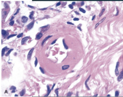
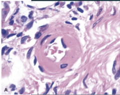
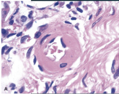
Homogeneous, eosinophilic ("pink") hyaline thickening of arteriolar walls.
What is the microscopic hallmark of hyaline arteriolosclerosis?
Luminal narrowing and downstream ischemia.
What does hyaline arteriolosclerosis lead to?

Kidneys → benign nephrosclerosis.
Which organ is commonly affected by hyaline arteriolosclerosis, and what condition does it cause?
Malignant hypertension.
In what condition is hyperplastic arteriolosclerosis typically seen?

"Onion-skin" concentric laminated thickening.
May include fibrinoid necrosis
What is the characteristic microscopic appearance of hyperplastic arteriolosclerosis?

Severe luminal compromise and ischemic injury.
What does hyperplastic arteriolosclerosis result in?

Fatty streaks
What is the earliest lesion of atherosclerosis?

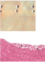
Flat yellow macules composed of lipid-loaded foamy macrophages
Do not disrupt blood flow
What are fatty streaks in Atherosclerosis composed of?
1. Cells: SMCs, macrophages, T cells
2. ECM: collagen, elastic fibers, proteoglycans
3. Lipids: intracellular (foam cells) and extracellular
What are the three main components of an atheromatous plaque microscopically?

Empty needle-shaped spaces.
What do cholesterol clefts look like microscopically in an atheromatous plaque?

A fibrous cap
What structure overlies the necrotic lipid core in an atheromatous plaque?
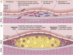
70% narrowing → stable angina.
What degree of arterial narrowing defines critical stenosis, and what condition can it cause?
Rupture, ulceration, and hemorrhage into the plaque.
What are examples of acute plaque changes in atherosclerosis?
1. MI
2. stroke
3. aneurysm
4. gangrene
What major clinical consequences can atherosclerosis lead to? (4)
Predictable, exertional chest pain relieved by rest or nitroglycerin.
What characterizes stable angina?
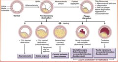
Angina at rest caused by coronary vasospasm.
What characterizes Prinzmetal (variant) angina, and what causes it?

Increasing severity of angina due to plaque disruption and thrombosis.
What characterizes unstable angina, and what is its underlying cause?

Red-blue discoloration.
What is the gross appearance of an MI at 12-24 hours?
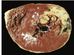
Hyperemic border with yellow-tan softening.
What is the gross appearance of an MI at 3-7 days?
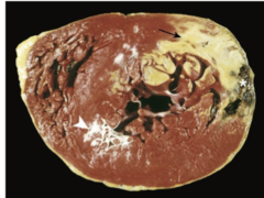
Granulation tissue.
What is the gross appearance of an MI at 10-14 days?
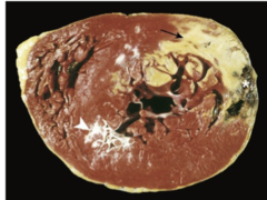
Gray-white scar formation.
What gross changes occur weeks after an MI?

Coagulative necrosis begins.
What microscopic change occurs 4-12 hours after an MI?

Neutrophil infiltration
What microscopic findings appear at 12-24 hours after an MI?

Dense neutrophil infiltration.
What occurs microscopically at 1-3 days after an MI?

Macrophage infiltration and phagocytosis.
What occurs microscopically at 3-7 days after an MI?

Granulation tissue formation.
What microscopic changes occur at 7-10 days after an MI?

Dense collagenous scar
What microscopic features are seen more than 2 months after an MI?

Ischemic heart disease (IHD) and hypertension (HTN).
What are common causes of left-sided heart failure? (2)
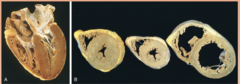
Pulmonary congestion and edema, and heart failure cells (hemosiderin-laden macrophages).
What pulmonary findings are associated with left-sided heart failure?

1. Dyspnea
2. Orthopnea
3. Paroxysmal nocturnal dyspnea (PND).
What symptoms are typical of left-sided heart failure? (3)
Left-sided heart failure or pulmonary disease.
What are the main causes of right-sided heart failure? (2)
Nutmeg liver (congestive hepatopathy).
What liver change is associated with right-sided heart failure?
1. Splenomegaly
2. Ascites
3. Peripheral edema
What systemic findings can occur in right-sided heart failure? (3)
The degree of pulmonary outflow obstruction.
What determines the severity of Tetralogy of Fallot?

1. VSD
2. Overriding aorta
3. RV hypertrophy
4. pulmonic stenosis
What are the four features of Tetralogy of Fallot?

Early cyanosis
Does Tetralogy of Fallot cause early or late cyanosis?

The aorta arises from the RV and the pulmonary artery from the LV.
How are the great arteries arranged in Transposition of the Great Arteries (TGA)?
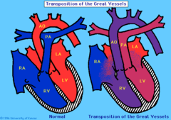
A shunt (e.g., PDA, ASD, VSD).
What is required for survival in Transposition of the Great Arteries (TGA)?

RV hypertrophy and LV hypoplasia.
What ventricular changes occur in Transposition of the Great Arteries (TGA)? (2)

Heaped-up calcified masses on the outflow surface.
What is the characteristic gross finding in calcific aortic stenosis?

Age-related degeneration or congenital bicuspid valve.
What are the two main causes of calcific aortic stenosis?

Concentric LV hypertrophy.
What does calcific aortic stenosis lead to in the left ventricle?

Aschoff bodies with Anitschkow cells.
What is the hallmark of the acute phase of rheumatic heart disease?

Fish-mouth (buttonhole) stenosis and commissural fusion.
What are the chronic valvular changes seen in rheumatic disease?
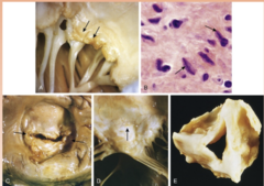
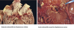
Friable, bulky vegetations.
What is the gross appearance of infective endocarditis?

Septic emboli and ring abscess.
What complications can arise from infective endocarditis?

Streptococcus viridans (subacute) and Staphylococcus aureus (acute).
Which pathogens commonly cause infective endocarditis? (2)
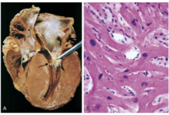
Myocyte disarray and interstitial fibrosis.
What microscopic findings are characteristic of hypertrophic cardiomyopathy (HCM)?
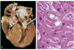
Asymmetric septal hypertrophy.
What pattern of hypertrophy is typical in hypertrophic cardiomyopathy (HCM)?

Sudden death
What major clinical risk is associated with hypertrophic cardiomyopathy (HCM)?
Cystic medial degeneration.
What is the most common underlying lesion in aortic dissection?

Hypertension and Marfan syndrome.
What are major risk factors for aortic dissection? (2)
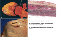
Rupture leading to cardiac tamponade.
What is a major complication of aortic dissection?
Portal hypertension
What causes esophageal varices?

Tortuous, dilated submucosal veins.
What is the gross appearance of esophageal varices?

Rupture leading to fatal hematemesis.
What is the major risk associated with esophageal varices?

Gray-white pseudomembranes with fungal hyphae.
What are the characteristic findings of Candida esophagitis?

Punched-out ulcers and multinucleated squamous cells with nuclear inclusions.
What are the characteristic findings of HSV esophagitis?

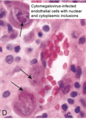
Endothelial and stromal cells with nuclear and cytoplasmic inclusions.
What are the characteristic findings of CMV esophagitis?
Eosinophils (followed by neutrophils).
Which inflammatory cells appear first in GERD?

Basal cell hyperplasia
What epithelial change is seen in GERD?

Stricture, ulcer, and Barrett esophagus.
What are complications of GERD? (3)

Intestinal metaplasia
What type of metaplasia occurs in Barrett esophagus?

Goblet cells
What finding is diagnostic of Barrett esophagus?

Adenocarcinoma
Barrett esophagus is a precursor to which cancer?

Barrett esophagus
What condition is the main origin of esophageal adenocarcinoma?
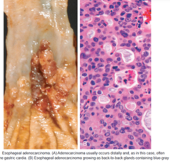
The distal esophagus
Where in the esophagus does adenocarcinoma typically occur?

Gland formation.
What is the histologic pattern of adenocarcinoma?

Alcohol and tobacco use
What strong risk factors are associated with esophageal squamous cell carcinoma?

The middle third
Where in the esophagus does squamous cell carcinoma most commonly arise?

As gray-white plaques that progress to polypoid masses.
How does squamous cell carcinoma appear early in its development?

Neutrophils above the basement membrane.
What microscopic finding indicates active inflammation in acute gastritis?
Erosions and hemorrhage.
What are severe manifestations of acute gastritis?
The antrum.
Where in the stomach is H. pylori gastritis typically located?

Neutrophils forming pit abscesses.
What acute inflammatory cells are seen in H. pylori gastritis, and what structure do they form?
Lymphocytes and plasma cells.
What chronic inflammatory cells are present in H. pylori gastritis? (2)
Intestinal metaplasia
What metaplastic change can H. pylori gastritis progress to?

Sharply punched-out ulcers with clean bases.
What is the gross appearance of peptic ulcers?
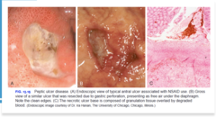
Duodenum
Which location is more commonly affected in peptic ulcer disease?
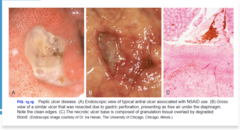
Scarring
What feature is seen in chronic peptic ulcer lesions?

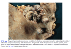
Gland-forming tumors
What is the histologic appearance of intestinal-type gastric adenocarcinoma?
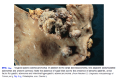
Inflammatory and metaplastic changes.
What background is associated with the intestinal type of gastric adenocarcinoma?
Signet ring cells
What cells are characteristic of diffuse-type gastric adenocarcinoma?

Thickened "leather bottle" stomach (linitis plastica).
What gross appearance is associated with diffuse-type gastric adenocarcinoma?

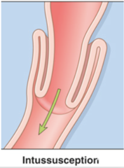
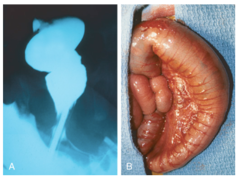
Telescoping of bowel leading to obstruction and ischemia.
What happens in intussusception?

Peyer patch hyperplasia.
What is a common cause of intussusception in children?
Absence of ganglion cells in the rectum.
What is the defining microscopic feature of Hirschsprung disease?
Dilation (megacolon).
What structural change occurs proximal to the aganglionic segment in Hirschsprung disease?

Hemorrhagic, ulcerated mucosa and transmural necrosis.
What are acute morphological findings in ischemic bowel disease? (3)

The splenic flexure.
What bowel region is a classic watershed zone susceptible to ischemia?
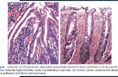
Reperfusion injury
What is a major source of damage in ischemic bowel disease?
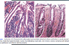
Villous atrophy, crypt hyperplasia, and increased intraepithelial lymphocytes.
What are the key microscopic features of celiac disease? (3)

Cryptitis and crypt abscesses.
What histologic findings are seen in acute self-limited colitis?
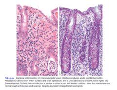
Crypt architecture is preserved (unlike in IBD).
How is crypt architecture in acute self-limited colitis different from IBD?

Pseudomembranes composed of inflammatory debris.
What is the characteristic gross/microscopic finding in C. difficile colitis?

"Volcano lesions"
What lesion type is typical of C. difficile colitis?
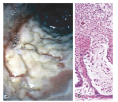
Skip lesions
What pattern of bowel involvement is seen in Crohn disease?
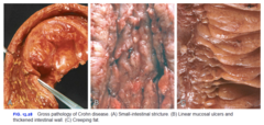
Transmural inflammation.
What layers of the bowel are affected in Crohn disease?

Cobblestone mucosa
What is the gross appearance of the mucosa in Crohn disease?
