Advanced Diagnostic Imaging FULL SUBJECT
 Call Kai
 Learn
Learn Practice Test
 Spaced Repetition
 Match
Match Flashcards
 Knowt Play
1/117

There's no tags or description
Looks like no tags are added yet.
Name | Mastery | Learn | Test | Matching | Spaced | Call with Kai | Chat |
|---|
No analytics yet
Send a link to your students to track their progress
118 Terms
Categories of Bone Tumours
Benign
Quasimalignant
Malignant
Primary
Secondary (metastatic)
Types of Benign Bone Tumours
Osteochondroma - Heriditary Multiple Exostoses (HME)
Haemangioma
Bone Island (enostoma)
Secondary Metastasis
Appearance | Lytic - 75% | Blastic - 15% | Mixed - 10% |
Male | Lung | Prostate | Prostate |
Female | Breast | Breast | Breast |
Primary malignant bone tumours
Multiple Myeloma - 43% - older
Osteosarcoma - 20% - young
Chondrosarcoma - 10% - middle age
Ewing’s Sarcoma - 7% - young
Multiple Myeloma - Radiological Features
Bone Scans - Cold spots
Early Signs = Gross osteoporosis
Hallmark feature = Punched out lesions (rain drop skull)
Vertebra Plana = Wrinkled vertebra
Pedicles will be preserved
Rarely, sclerotic lesions occur (ivory vertebra)

Solitary myeloma - Radiological Features
Localised form of plasma cell proliferation
70% of solitary plasmacytoma lesions develop into diffuse Multiple Myeloma
Typically appears as:
Geographic
Soap bubbled
Highly expansile

Solitary myeloma - Common sites
Mandible
Ilium
Vertebrae
Ribs
Proximal Femur
Scapula
Osteosarcoma - Radiological Features
Classic lesions presents as a permeative or ivory medullary lesion in the metaphysis of a long tubular bone
Sunburst or sunray periosteal response is characteristic
Often, Codman’s triangle associated with destructive lesion
Cortical disruption with soft tissue mass formation - often larger
peripheral edge of eccentric lobulated mass appear roughened and irregular may be referred to as cumulus cloud

Ewing’s Sarcoma - clinical features
Metastasis:
Frequently
Early
To lung
To Bone
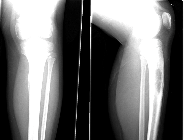
Ewing’s Sarcoma - Radiological Features
Diaphyseal
Laminated Periostitis
Short spiculated periostitis
Saucerisation (erosion on outer surface)
Chordoma - Clinical Features
Arises from notochord remnant tissue
1% of primary malignant bone tumours
Crosses disc spaces
Slow growing
Peak in 40-70 y/o
M 2:1 F
More calcification = better prognosis
Chordoma - Involved sites
Axial (majority)
Sacrum 50%
Sphenoid 35%
Spine 15%
Extra-axial (insanely rare. 7 Reported cases)
Pubic ramus
Femur
Tibia
Metatarsal
Lung
Rib
Ulna

Chordoma - Spread
15% metastasise (including to bone)``
Non-Hodgkin’s Lymphoma - Clinical Features
No prior lymph node involvement
Round cell neoplasm
4% of primary malignant bone tumors
20-40 y/o
M 2:1 F
Dull pain not relieved by rest (vs multiple myeloma which is)
Patient generally looks surprisingly well
Non-Hodgkin’s Lymphoma - Sites
Sites:
Knee 40%
Femur
Pelvis
Humerus
Tibia
Ribs, scapulae, vertebrae
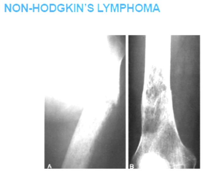
Non-Hodgkin’s Lymphoma - Radiographic Features
Diaphyseal
Lytic
If present (uncommon periostitis)
Pathological fractures are common
Rarely the lesion is sclerotic and mimics blastic lesions
Giant Cell tumour - characteristics
Quasimalignant - 20% malignant
Highly vascular connective tissue tumour
5% of all primary malignant
15% of all benign bone tumours
Giant Cell tumour - Clinical Features
20-40
M:F equal
Benign - M 2:3F
Malignant - M 3:1 F
May get large before symptomatic
Pathological fracture precipitates symptoms
Giant Cell tumour - Sites
Femur - distal
Tibia - proximal
Radius - distal
Humerus - proximal
Sacrum is most common spinal site for GCT

Giant Cell tumour - Radiological features
Epiphyseal
Lytic
Expansile
Subchondral
40% soap-bubbly
Eccentricly located (off to one side of tubule bone)

Osteoma - Characteristics
Arises in intramembranous bones
Rarely multiple osteomas are associated with colonic polyposis and soft tissue tumours = Gardner’s syndrome
Osteoma - Clinical features
Usually asymptomatic
Male 1:3 Female
No malignant degeneration
Excise if symptomatic
Osteoma - Sites
Skull
Mandible

Osteoma - radiological features
Radiopaque
Skull tables
Paranasal sinuses
Mandible

Osteoid Osteoma - Characteristics
Differentiate from Brodie’s Abscess
Osteoid Osteoma - Clinical Features
10-25 y/o
M 2:1 F
Painful!!!
Aching worse with inactivity
Dramatically relieved by aspirin
Osteoid Osteoma - Sites
50% femur and tibia
Proximal end of femur
Spine (posterior neural arch)
Cervical 27%
Thoracic 12%
Lumbar 60%
Sacral 2%
Osteoid Osteoma - Radiological Features
Nidus <1cm diameter
Sclerosis - reactive
Periostitis depends on location in bone
Cortical (most common)
Cancellous (in medullary cavity)
Subperiosteal (creates strongest periosteal reaction)

Osteoblastoma - characteristics
1% of benign bone tumours
Giant osteoid osteomas
Histologically similar to osteoid osteoma
Nidus >1cm (larger than osteoid osteoma)
Osteoblastoma - Clinical Features
70% <20 y/o
M 2:1 F
Progressive onset of pain
Spinal lesions produce painful scoliosis
May cause spinal stenosis
Osteoblastoma - Sites
Spine - posterior neural arch
Femur
Foot
Ankle
Osteoblastoma - Radiological features
Scoliosis
Spinal stenosis
Expansile
Radiolucent
Metaphyseal/diaphyseal in tubular bones
Mild reactive sclerosis
Enchondroma - Characteristics
10% of benign tumors
Occur in any enchondrally formed bone
Cartilage cell rests in metaphysis growth
Most common benign tumour in hand
Enchondroma - Clinical Features
10-30 y/o
M=F
Usually asymptomatic
Pathological fracture leads to X-ray leading to discovery
Enchondroma - Sites
Hands 50%
Femur
Humerus
Ribs
Enchondroma - Radiological Features
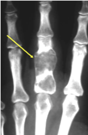
Geography
Radiolucent
Expansile
Metaphyseal
Endosteal scalloping
Calcification 50%

Enchondroma - DDx
Fibrous dysplasia
Simple Bone Cyst
Giant Cell Tumour
Bone infarct (when calcified)
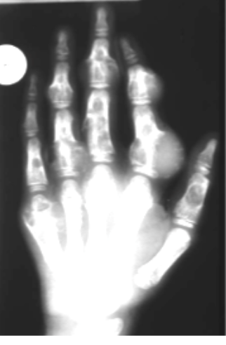
Enchondromatosis - Characteristics
Ollier’s Disease
Identical lesions to those of solitary endochondroma
May see a streaky “celery stick” form involving the metaphyses
If cavernous soft tissue haemangiomata are present, this constitutes maffucci's syndrome
Malignant degeneration has been reported in up to 50%
Closer lesion is to the spine, the more likely it will undergo malignant change
Enchondromatosis - Clinical Features
Usually asymptomatic
May have associated deformities
Enchondromatosis - Sites
Hands
Feet
Knee
Pelvis
Ribs
Enchondromatosis - Radiology
As for solitary enchondroma
Celery stick form is seen in metaphysis
Non-Ossifying Fibroma - Clinical Features
Non cancerous tumour of connective tissue
>2cm
8-20 year olds
Asymptomatic unless pathological fracture
M:F 2:1
Non-Ossifying Fibroma - Common Sites
Lower extrem
Distal tibia
Knee
Prox humerus
Fibula
Non-Ossifying Fibroma - Radiographic Features
Solitary lesion
Eccentric
Soap bubbly
Arises from cortex and expands into medulla
Metaphyseal
Sclerotic border
Narrow zone of transition

Simple Bone Cyst - Clinical Features
Not really a tumour - fluid filled cyst in a chamber lined with fibrous tissue
3% of benign bone tumours
Unicameral (one room)
It is usually asymptomatic unless pathological fracture (occurs in ⅔)
75% are prox femur or humerus
Can also be in pelvic and calcaneous
3-14
M 2;1 F
Simple Bone Cyst - Common Sites
Proximal humerus
Proximal femur
Humerus 2:1
Calcaneus
Pelvis
Centrally located
Simple Bone Cyst - Radiographic Features
Single or multichambered (soap bubbly)
Geographic
Radiolucent
Broad at metaphyseal
Narrow at diaphyseal
Expansile
Endosteal scalloping but not usually wider than metaphysis
No periosteal response
No cortex breach
No soft tissue mass
Fallen fragment sign in 10% of cases - especially with fracture, a piece of the wall of the cyst falls into the most gravitationally dependent part of the cyst

Aneurysmal Bone Cyst - Clinical Features
Vessels within not actually aneurysmal, just blood filled cavity - tumour like
1% of biopsied tumours
Most common benign tumour of clavicle
Young 5-20
M 2:3 F
Sudden onset of pain and rapid increase in severity
Pathological fracture common
If in spine, can cause paraplegia
Often hx of trauma
80% in long tubular bones and spine
Femur, tibia, thoracic and lumbar most common
When spinal - often neural arch (may mimic osteoblastoma)
Aneurysmal Bone Cyst - Sites
80% long tubular bones and in the spine
Femur and tibia most common
Tx and Lx most common spinal
Predilection for the neural arch
metaphyseal, eccentric and can be diaphyseal
Aneurysmal Bone Cyst - Radiographic Features

Varied presentation
Eccentric
Metaphyseal - expands to epiphysis after growth plate closure (opposite to sbc which grows away) (however ABCs can be diaphyseal)
Expansile, radiolucent and rapidly growing (mimic malignancy)
Cortex thins but usually intact
Periosteal buttressing at edges (like codmans)
May be soap bubbly, show trabeculae
Can cross growth plate (rare)

Paget’s Disease - Clinical Features
focal disorder of bone metabolism causing accelerated bone remodelling
Disease occurs when osteoclasts break down faster than osteoblasts replace it. Might be inflammatory, endocrine autoimmune etc.
M:F 2:1
Rare <40, most>55
90% asymptomatic
Pain can be present if paget’s causes bone deformities or fractures
Increasing hat size may be noted due to enlargement of calvaria
Pelvis
Vertebral body
Clavicle
Humerus
Ribs
Paget’s is subarticular and extends to involve the entire bone
High incidence in the UK
Paget’s Disease - Sites
Pelvis
Vertebrae
Clavicle
Humerus
Ribs
Fibula LEAST LIKELY
Paget’s Disease - Complications
Deformities due to bone softening
Proximal femur- shepherds crook (coxa vara)
Tibia - sabre shin deformity (anterior bowing)
Protrusio acetabili
Frontal and patietla bossing
Basilar invagination
Lentiasis ossei (lyon face)
Fractures
Vertebral body expansion leading to stenosis
Transverse banana - Like fractures of long bones
Pseudo fractures - lines of demineralisation replaced by fibrous tissue
Anaemia due to fibrous replacement of bone marrow
Secondary degenerative changes due to destruction of articular cartilages
Hypercalcuria (from bone resorption) - urinary calculi - uteric colic pain
High output cardiac failure (if ⅓ skeleton or more involved)
Malignant degeneration to osteosarcoma (new pain, increase size) poor prognosis
Leontiasis ossea of the pelvis
high output of cardiac failure
Malignant degeneration relatively rare but possible
Paget’s Sarcoma - Femur, humerus, innominate, skull, tibia

Paget’s Disease - Pathological Features
Replacement of normal bone (patternless)
Pagetic vascular and fibrotic bone
Been called mosaic structure of Paget’s disease

Paget’s Disease - Radiographic features
Hot on bone scan
Skull - early lytic (osteoporosis circumscripta), later cotton wool
Vertebral involvement: thickened enlarged end plates (squared off appearance) picture frame vertebra
Ivory vertebra - homogenous increase in radiopacity
Increased coarsening of trabeculae
Pelvis - cortical thickening, bone expansion causing thickening of the iliopectineal line which obliterates Kohler’s tear drop + brim or rim sign

Paget’s Disease - Stages
Osteolytic
Mixed
Blastic
Malignant
Fibrous Dysplasia - Clinical features
Fibrous dysplasia is a rare genetic disorder that causes abnormal bone growth
Replaced by fibrous tissue
Poorly formed trabeculae
Begins in medullary
Monostotic or polyostotic (* McCune-Albright syndrome - associated with cafe au lait spots and endocrine issues - growth hormone excess affects FD bone more than normal bone)
Asymptomatic so incidence is unknown
Bowing deformities (shepherds crook) and pathological fractures
Cafe au lait spots in cost of maigne in 30% polyostotic
Fibrous Dysplasia - Types
Monostotic - one bone involved
Polyostotic - two or more involved
Fibrous Dysplasia - Sites
Monostotic - typically the ribs, tibia, skull
Polyostotic - femur, skull, tibia,. Humerus, ribs, fibula, radius and ulna
Vertebral involvement is rare

Fibrous Dysplasia - Radiographic Features
Skeletal lesions NOT present at birth
Normal bone replaced by abnormal fibrous tissue
Cause widening of the medullary cavity, thinning of cortex and endosteal scalloping
Rarely malignant degeneration to fibrosarcoma or osteosarcoma
Ground glass appearance (a characteristics hazy, indistinct, or speckled pattern seen on X-rays and CT scans, indicating abnormal boe formation within the affected area)
Cherubism - cheeks appear bigger due to FD of mandible
Soap bubbly appearance
Endosteal scalloping
Mildly expansile lesion
Geographic - encapsulated by sclerotic border
Pathological fracture
Most radiolucent, soap bubbly, located or trabeculated
Ground glass or smokey - trabecular patterns wiped out
Geographic, encapsulated by sclerotic border
Bone expansion with widening of medullary canal, endosteal scalloping
Bone softening
Expansile rib lesion
Pseudoarthrosis can occur as a result of pathological fracture and non union

Neurofibromatosis Type 1 - Clinical Features
Cafe au lait spots (smooth margins - coast of california, not raised)
Fibroma molluscum (elevated cutaneous fibrous nodule)
Osseous deeformities and lesions
(Neurofibroma also present)
Elephantiasis neuromatosa - thick large soft tissue folds
Pulsating exophthalmos (temporal lobe herniation through deficient/absent posterior wall of orbit)
Spinal involvement (includes paraplegia, kyphoscoliosis, altantoaxial subluxation)
Neurofibromata
Malignant degeneration to neurofibrosarcoma
Lesions biopsied multiple times more likely to degenerate sarcoma
Neurofibromatosis Type 1 - Radiographic Features

50% have skeletal lesions
50% have kyphoscoliosis (acute angle usually lower thoracic 5-7 vertebrae)
Posterior vertebral scalloping (intraspinal masses - pressure effect)
Two types of scalloping
Posterocentral scalloping along multiple levels (due to dural extasia ballooning of the dural sheath and trapped CSF associated with herniated nerve root sheaths
Eccentric and unilateral - dumbbell shape, half in spinal canal half in IVF (enlarges IVF) see image C1/2
Twist ribbon appearance (scalloping of rib margins)
Pseudoarthrosis of the tibia in 50%
Multiple large noon ossifying fibromas
Focal giantism
Renal artery stenosis
1% get adrenal tumour pheochromcytoma and 20% get neurofibromatosis
Osteomalacia may occur

Battered Child Syndrome - Other names
Child abuse
Non-accidental injury
Parent infant traumatic stress syndrome
Caffey-Kempe syndrome
Battered Child Syndrome - Clinical Features
A child with multiple fractures of the long tubular bones and subdural haematoma
Trauma is usually inflicted by parent, guardian or de facto partner
Children usually less than 6 years old when brought in for medical attention, and are often less than 2 years old when exposed to trauma
After age 4, children can either escape the abuser or talk about what is happening
Any evidence of repeated trauma in a child including bruising, burns should arouse suspicion
Repeated episodes of multiple fractures are highly suggestive
Battered Child Syndrome - Radiographic features

Multiple fractures (especially at rubs) at different stages of healing is a hall-mark feature
Particularly significant are fractures at the corner of metaphyses (especially medial side of distal femoral metaphysis)
Periosteum loosely attached at the shaft of long bones but more firmly attached to the metaphysis
Lifting of the periosteum of the shaft (exuberant callous), especially tubular long bones
Periostitis is seen within 1-2 weeks of injury (helps date the injury)

Battered child Syndrome - Shaking a child sequelae
Brain damage - intracranial and subdural hematoma
Retinal detachment
Spinous process fracture of thoracic and lumbar spine
Humeral and rib fractures
Dystrophic Calcification (myositis ossificans) - Clinical Features
Most calcification 95-98% is dystrophic (lacking in form) = small to large amorphous calcification in damaged soft tissue
Calcification can occur as part of non-specific inflammatory response to tissue damage (trauma, infection, chronic disease, response to medical devices)
Calcification can progress to ossification - meaning a cortex and medullary space are formed
Should be called heterotopic ossification

Dystrophic Calcification - Sites

Elbow - brachialis anterior
Thigh - quadriceps femoris and adductor muscle
Knee - medial collateral ligament (pelligrini-Stieda Disease)
Shoulder - AC ligament rupture

Dystrophic Calcifications - Differentials
Vascular
Venous insufficiency
Infection
Parasitic infestation
Cysticercosis
Dracunculiasis
Armillifer armillatus
Neoplasm
Osteoma
Osteosarcoma
Tumour
Drugs
Vitamin D
Autoimmune
Dermatomysitis
Scleroderma
Trauma
Heterotopic ossification
Injection granulomas
Necrosis
Thoracic Aorta Aneurysm - Clinical Features
M 3:1 F
Peak age 65
Many asymptomatic
Found incidentally on chest images
Symptomatic presentations most commonly are pain in the:
Substernal region
Back (mid-thoracic)
Shoulder
Hoarseness (recurrent laryngeal nerve compression)
Stridor
Dyspnea
Dysphagia
Facial engorgement (superior vena caval compression)
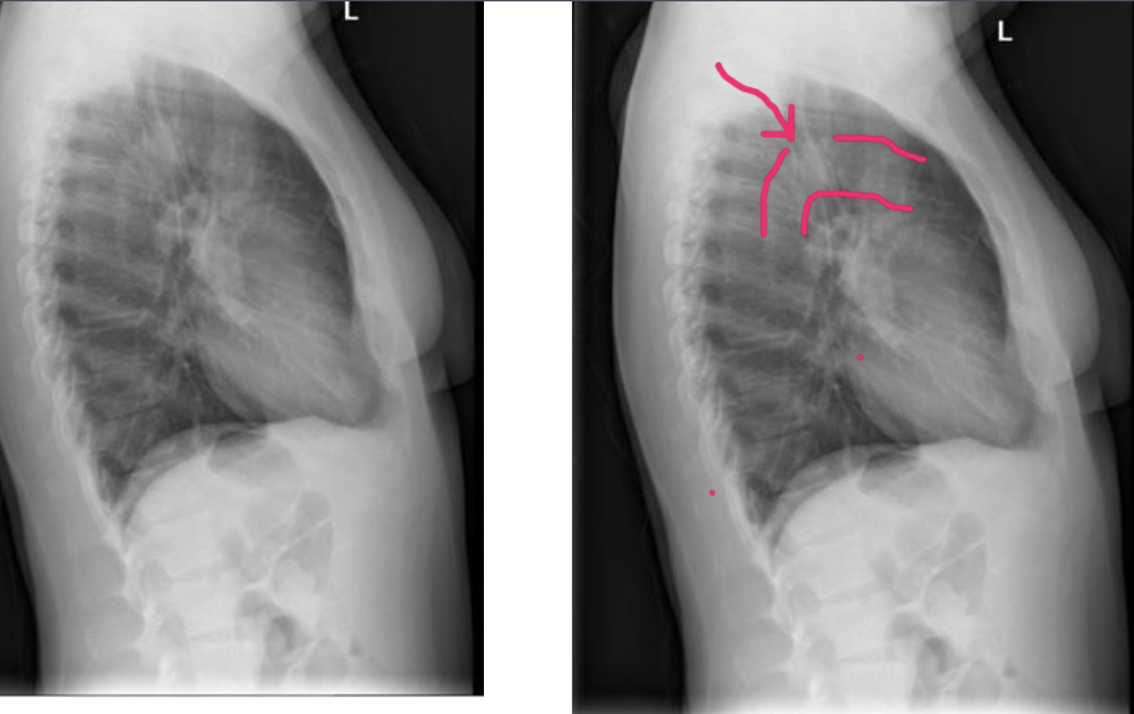
Thoracic Aorta Aneurysm - Differentiate from age related dilation and tortuosity
Typical senile changes are usually made up of five features:
Ascending aorta sweeps to the right
Aortic arch lies close to the clavicle
Tracheal indentation is more prominent
Diameter is enlarged between 3 and 4 cm
Descending aorta is tortuous in its course and projects more laterally into the thoracic cavity

Thoracic Aorta Aneurysm - Location suggestions
Ascending aorta suggests syphilis, Marfan’s Syndrome and AS
Aortic arch location is associated with atherosclerosis and trauma
Descending aorta is more commonly involved with trauma and collagen disease

Abdominal Aorta Aneurysm - Clinical Features
Abdominal aorta most common vessel for aneurysm below the diaphragm
AAA is probably the most common extraspinal, potentially life-threatening disorder encountered in patients presenting with back pain
2-6% of general population
For every 5 years after the age of 65, the risk increases by 40%
Males are more frequently as M 5:1 F
Abdominal Aorta Aneurysm - Sites
Sites of involvement in descending order of incidence are:
Abdominal aorta
Aortic arch
Descending thoracic aorta
Ascending aorta

Abdominal Aorta Aneurysm - Risk Factors/incidence
Known risk factors:
Male
>60
Hypertension
Heart disease
Hyperlipidemia
Peripheral vascular disease
Positive family hx of AAA
Smoking is the most important risk factor that can be avoided
40% of patients have hypertension
30% have heart disease
Almost 30% incidence in male siblings of patients with AAA

Abdominal Aorta Aneurysm - Rupturing
At least 25% of AAA > 5cm will rupture within 5 years
Those AAAs under 5cm have a rupture rate of only 5%
AAA on average expands by 4-5mm each year
Can cause anterior scalloping in 5%
Vertebral Artery Tortuosity - Clinical Features
Age-related vertebral arterial wall thinning and elongation precipitates dilation and ectasia
When symptoms do occur from this altered blood flow to the hindbrain they are often bizarre and clinically confusing
Called the barre-lieou syndrome, symptoms include headaches, dizziness, nystagmus and suboccipital pain as well as nausea and vomiting
Occasionally leads to pulsatile transmission to adjacent bony structures which may produce extrinsic bony erosion (Hadley’s erosive defect)
An Astute diagnostician may recognize the erosive defunct on cervical plain films
Majority unilateral
Symptoms occur if radicular compression or rarely if path fracture results from extensive erosion
Pregnancy may precipitate radicular symptoms
Bilateral atherosclerotic occlusion of the carotid arteries may precipitate vertebral artery dilation as a compensatory mechanism for maintaining cerebral perfusion

Vascular Calcification

Ricket’s (hypovitaminosis) - General features
Childhood equivalent of osteomalacia
Systemic disease of infancy and childhood
Premature birth is a predisposing factor
Calcification of bone matrix is deficient
Ricket’s - Clinical Features
6 months - 1yo
Maternal vitamin D prevents manifestations for up to 6 months
Infant has softened skull bones
Costochondral junction is enlarged - rachitic rosary
Limb deformities with epiphyseal swellings are noted
Infant is irritable and shows increased sweating
Ricket’s - Laboratory
Serum calcium is normal to low
Serum phosphorus is slightly low
Alkaline phosphatase is increased
Ricket’s - Radiographic Features (active)

Changes at end of long bones - knee, ankle and wrist
Metaphysis is flared, widened or trumpeted
Physis is widened
Metaphysis appears frayed - brush border
Bowing deformities
Greenstick fractures
Pseudofractures - rare
Costochondral junction is enlarged

Ricket’s - Radiographic Features (healing)
Mineralisation of zone of provisional calcification
Dense metaphyseal band
Deminerralisation of subperiosteal osteoid tissue
Mimics periosteal new bone formation
Calcification of ossification centres appears as a marginal ring
Changes mimic active scurvy and heavy metal toxicity

Scurvy (Hypovitaminosis D) - General
Humans are unable to synthesize vitamin C so must get it in their diet
Ascorbic acid (vitamin C) is much higher in human milk than in cow’s
Incidence of infantile scurvy low with breast feeding
In infants = barlow’s disease
Scurvy - Clinical Features
6–9 months
Malaise and weakness
Perifollicular haemorrhage
Hyperkerototic papules
Bleeding
Gums
Joints
Subperiosteally
Anaemia
Delayed wound healing
Loose teeth and mouth infections
Fever
Scurvy - Laboratory
Normal plasma ascorbic acid = 0.6 mg%
<0.2 mg% is a deficiency state
Deficiency state 3–6 months must be maintained before symptoms occur
Scurvy - Pathology
Both chondroblastic and osteoblastic activity is disordered
Changes prevalent where growth is rapid
Devvelopment and calcification of new cartilage cells occurs normally
Removal of the calcified cartilage matric is abnormal
No new inter-cellular matrix is laid down between these new cells
Ossification fails to occur
Bones may fracture at metaphyseal sites and not heal
Blood vessels wall fragility - lack of cementing between endothelial cells
Scurvy - Sites
Anterior ribs
Distal femur
Proximal humerus
Both ends of the tib and Fib
Distal radius and Ulna
Scurvy - Radiology
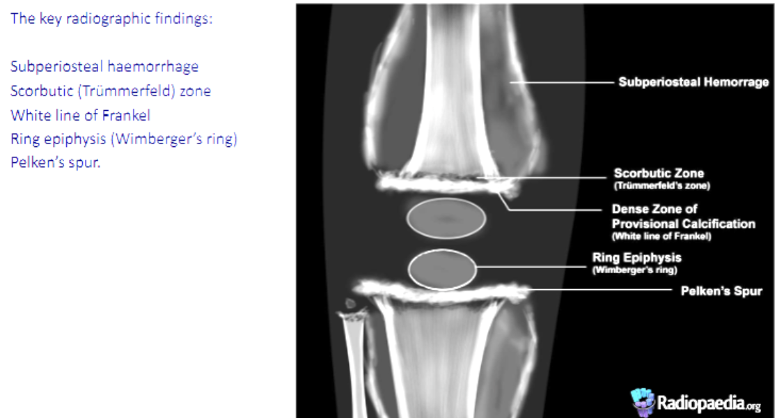
Dense metaphyseal bands
Ground glass appearance
Halo ossification centre
Cortical thinning
Corner sign
Lateral spurs (pelkan’s spurs)
Subperiosteal haematomas
Metaphyseal fractures
Atrophic scurvy line (trummerfeld zone)
Soft tissue oedema
Spinal changes
Healing
Heavy Metal Poisoning (lead) - Clinical Features
Infants and children
History of eating flakes of paint
Can lead to cerebral edema
Can be fatal if meninggoencephalitis occurs
Mortality is 25%
Can see radiographic changes within 1 month
Lead poisoning - radiology
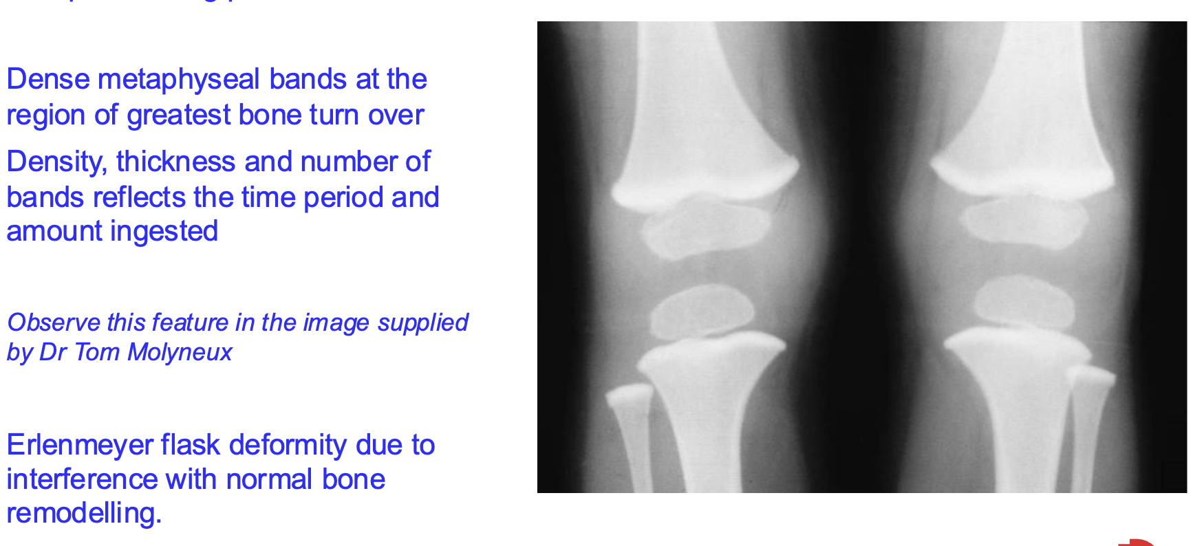
Lead lines - dense metaphyseal bands seen at the site of greatest bone turnover
Lead is metabolised similarly to calcium
Interferes with normal remodelling to result in the erlenmeyer flask deformity of the femur
Lead lines reflect:
Amount of ingested lead (density of band)
Time period of ingestion (thickness of band)
Number of periods of ingestion (number of bands)
Dense metaphyseal bands can also be seen in:
Normal children under 4
Scurvy
Healing rickets
Cretinism (juvenile hypothyroidism)
Metal intoxications such as:
Bismuth
Phosporus
Mercury (administered as an injection to treat certain disorders

Phosphorus poisoning - General
Rare nowadays but formerly common as a result of administration of phosphorylated cod liver oil for rickets and tuberculosis
Produces dense metaphyseal bands
Bands fade after ingestion ceases
Radium Poisoning - General
Caused malignancies arising in bone such as bone sarcomas
Ossified Posterior Longitudinal Ligament Syndrome (OPLLS) - General
Condition may be associated with compression of the spinal cord
Definition of ossified posterior longitudinal ligament syndrome diffuse idiopathic skeletal hyperostosis and for this to occur in Japan (Japanese disease)
Occurs most commonly is Cx and least commonly in Lx
Ossified Posterior Longitudinal Ligament Syndrome - Clinical Features
Symptoms if present are mostly associated with cervical spine involvement and least frequently in Tx and Lx
Findings may include insidious onset of motor and sensory deficits mainly of lower limb cord myelopathy is heralded by progressive difficulty walking
S/S are likely with ossified ligament occupies more than 60% of the sagittal diameter of the cervical spinal canal
Ossified Posterior Longitudinal Ligament Syndrome - Pathology
Ossified ligament usually spans 3 to 4 adjacent spinal segments
Microscopically the affected ligament demonstrates superficial well0formed cortical bone but poorly formed marrow cavities
Normal ligamentous tissue is found in the deeper layers
Spinal cord changes in OPLL may include flattening of the cord, grey matter infarction and demyelination of the lateral and posterior white columns
Ossified Posterior Longitudinal Ligament Syndrome - Radiology
Findings best detected on lateral plain film
Most common in mid and upper Cx
Dense ligament may be up to 5mm in thickness and parallels to the posterior body margins
OPLL may be separated from the underlying vertebrae by a radiolucent zone representing normal ligament
Underlying discs and apophyseal joints are usually unaffected by degenerative change
85% of patients with OPLL also have DISH. 50% of DISH have OPLL
Progressive myelopathy requires decompression laminectomy

Neurotrophic Arthropathy - General
Advanced DJD
Seen in patients with impaired proprioception or diminished pain sensation
Relationship noted between DJD and tabes dorsalis
Neurotrophic Arthropathy - Clinical Features
Neurotrophic arthropathy was formerly most common due to syphilis but now patients with diabetes mellitus
Neurotrophic changes in approximately 5% of diabetics
Changes usually affect ankle and foot in diabetes
In syphilis 20% of patients develop change affecting lumbar, knee and ankle
Syringomyelia 25% show neurotrophic change of the upper extremity shoulder wrist and elbow
Neurological manifestation associated include wider gait, loss of reflexes and pain insensitivity
Affected joints may be painless, unstable and or produce crepitus
Recurrent and painless joint effusion seen early in the condition
Charcot’s joint may take weeks to years to develop
Neurotrophic Arthropathy - Pathological
Thought to be as a result of ineffectual neurological protection from mechanical trauma
Neurovascular theory exists which estimates neurologically mediated increased intraosseous blood flow leads to stimulation of osteaclastic bone resorption
Resorption leads to fracture and joint damage
In weight bearing regions, secondary hypertrophic changes are superimposed. Intra-articular cartilage fibrillation and flaking has been found together with cartilaginous and osseous loose bodies