Skull (Part III)
 Call Kai
 Learn
 Practice Test
 Spaced Repetition
 Match
 Flashcards
 Knowt Play
1/100

There's no tags or description
Looks like no tags are added yet.
101 Terms
zygomatic arch and infratemporal crest divide
temporal fossa from infratemporal fossa
zygomatic arch is made of
temporal process of zygomatic bone and zygomatic process of temporal bone
infratemporal crest divides
greater wing of sphenoid bone from lateral pterygoid process of sphenoid bone
bones that form pterion
frontal bone
parietal bone
greater wing of sphenoid bone
squamous part of temporal bone
parts of bones that form pterion form the
medial boundary of temporal fossa
superior temporal line marks
superior boundary of temporal fossa
superior temporal line is attachment site for
temporal fascia
inferior temporal line is attachment site for
temporalis m.
infratemporal fossa is bounded laterally by
ramus of mandible
infratemporal fossa is bounded anteriorly by
infratemporal surface of maxilla

posterior superior alveolar foramina
small hole(s) on infratemporal surface of maxilla that transmits posterior superior alveolar nerves and arteries
infratemporal fossa is bounded medially by
lateral pterygoid plate of sphenoid bone
infratemporal surface of maxilla is limited superiorly by
inferior orbital fissure
infratemporal surface of maxilla is limited medially by
pterygomaxillary fissure

pterygomaxillary fissure is continuous with
inferior orbital fissure
pterygomaxillary fissure is passageway between
pterygopalatine and infratemporal fossa
pterygomaxillary fissure forms lateral wall of
pterygopalatine fossa

pterygopalatine fossa serves as
major neurovascular crossroad between oral cavity, nasal cavity, nasopharynx, orbit, masticator space, and middle cranial fossa
infratemporal fossa is bounded posteriorly by
styloid process of temporal bone
infratemporal fossa is bounded superiorly by
greater wing of sphenoid bone and associated features
mandibular division of trigeminal nerve (V3) enters cranial cavity via
foramen ovale
middle meningeal artery enters cranial cavity through
foramen spinosum

petrotympanic fissure
narrow crevice between petrous and tympanic parts of temporal bone that provides passageway for chorda tympani nerve

articular tubercle
lateral bony prominence on root of zygomatic process of temporal bone

articular eminence
anterior slope of mandibular fossa

sphenoid spine
small downward projection from greater wing of sphenoid bone that is attachment site for sphenomandibular ligament
bony nasal septum is formed by
vomer and perpendicular plate of ethmoid bone


conchae
lateral walls of nasal cavities that project into nasal cavity
superior and middle nasal concha are features of
ethmoid bone
lateral to middle nasal concha are
uncinate process and bulla of ethmoid bone

ethmoidal bulla
elevation produced by middle ethmoidal air cells
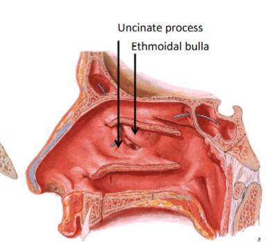
uncinate process
curved, hook-like process

perpendicular plate of palatine bone
contributes to lateral nasal wall

sphenoethmoidal recess
space located superior to superior nasal concha and anterior to sphenoid bone

sphenopalatine foramen
hole between perpendicular plate of palatine bone and medial pterygoid plate on lateral nasal wall
sphenopalatine foramen is passageway between
pterygopalatine fossa and nasal cavity

ethmoid sinuses
extensive permeations of air spaces in ethmoid bone

vaginal process of temporal bone
inferior projection from temporal bone that is immediately anterior to styloid process
vaginal process of temporal bone is attachment site for
muscles associated with tongue and larynx
sphenoid bone has two bilateral pterygoid processes that project downward
medial and lateral pterygoid processes

inferior head of lateral pterygoid muscle attaches to
lateral surface of lateral pterygoid process
deep head of medial pterygoid muscle attaches to
medial surface of lateral pterygoid process

hamulus of medial pterygoid plate
hook-like process that acts as pulley for tensor veli palatine tendon to glide around

pterygoid fossa (purple star on diagram)
V-shaped divergence of medial and lateral pterygoid plates

pterygoid notch (red arrow on diagram)
inferior bony deficiency between medial and lateral pterygoid plates
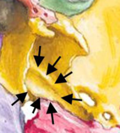
scaphoid fossa
shallow, boat-shaped depression posterior to root of medial pterygoid plate
scaphoid fossa is muscle attachment site for
tensor veli palatini m.

pterygoid canal
horizontal channel that transmits nerve of pterygoid canal

palatovaginal canal (yellow arrow on diagram)
horizontal channel between vaginal process of sphenoid bone and perpendicular plate of palatine bone
palatovaginal canal transmits
pharyngeal nerve and pharyngeal branch of sphenopalatine artery

ala of vomer
superior, thick, horizontal projections that articulate with sphenoid bone

incisive canal
opening in anterior part of intermaxillary suture, posterior to maxillary central incisors
incisive foramen is located at
junction between primary and secondary palates

posterior nasal spine
median projection from posterior border of hard palate

greater palatine foramen
hole in palatomaxillary suture that transmits nerves and vessels onto hard palate

pyramidal process of palatine bone
posterior and lateral projection from junction of horizontal and perpendicular plates of palatine bone
pyramidal process of palatine bone serves as
attachment site for deep head of medial pterygoid m.
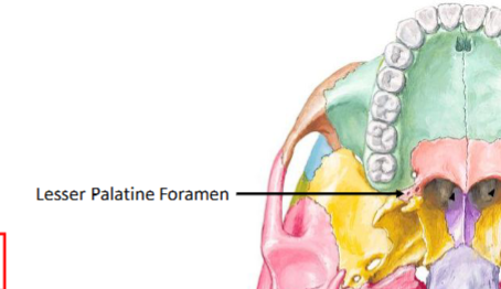
lesser palatine foramen
small holes in pyramidal process of palatine bone that transmit nerves and vessels onto soft palate
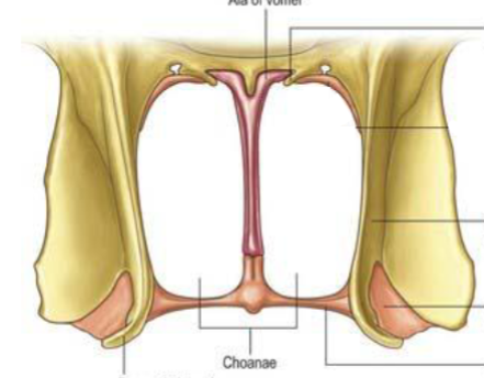
choana
paired openings that provide communication between nasopharynx and nasal cavities
choana is bound medially by
vomer
choana is bound superiorly by
ala of vomer and vaginal process of sphenoid bone
choana is bound laterally by
medial pterygoid plate
choana is bound inferiorly by
horizontal plate of palatine bone

maxillary tuberosity
rounded eminence on inferior part of infratemporal surface of maxilla; located posterior to last molar
maxillary tuberosity serves as attachment site for
superficial head of medial pterygoid m.

interalveolar septum
thin plate of bone separating two adjacent alveoli

alveolar jugum
vertical grooves or pits on outer surface of alveolar process formed by roots of two adjacent teeth

interradicular septum
thin plate of bone separating roots within a single alveoli

gomphosis
fibrous joint in skull that joins tooth and alveolar sockets

piriform aperture (dashed outline on diagram)
anterior bony opening of nasal cavity formed by nasal bones, nasal notches of maxilla, and anterior nasal spine
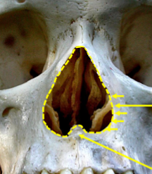
nasal notch (shorter arrow on diagram)
lateral and inferior portion of maxillary contour
anterior nasal spine (longer arrow on diagram)
sharp anterior projection of maxilla at midline inferior border of piriform aperture

maxillary sinus
air-filled cavity in central part (body) of maxilla
petrous part of temporal bone is embryologically derived from
otic capsule

groove for lesser petrosal nerve leads to
foramen ovale

groove for greater petrosal nerve leads to
internal opening of carotid canal

portion
central point of superior border of external acoustic meatus
mandible
only movable bone in skull that consists of tooth-bearing body and vertical ramus
mandibular ramus divides superiorly into
coronoid and condylar processes, separated by mandibular notch


coronoid notch
gentle concave curve on anterior surface of mandibular ramus
coronoid notch is palpable featured used in
administering inferior alveolar nerve block

temporal crest
vertical ridge from coronoid process to behind 3rd molar
temporal crest is attachment site of
temporalis muscle

pterygoid fovea
roughened depression on anterior surface of mandibular neck
pterygoid fovea is insertion site of
inferior head of lateral pterygoid m.

masseteric tuberosity
roughened area at lateral aspect of mandibular angle
masseteric tuberosity is attachment site for
some lower fibers of masseter m.

pterygoid tuberosity
roughened area on medial aspect of mandibular angle
pterygoid tuberosity is insertion site for
medial pterygoid muscle

retromolar triangle
small, depressed area posterior to last mandibular molar; bound by bony ridges of alveolar bone

mylohyoid line serves as
attachment site for mylohyoid muscle and delineates sublingual from submandibular fossa

mandibular foramen
on inner surface of mandibular ramus
inferior alveolar nerve enters mandible via
mandibular foramen to innervate mandibular teeth

lingula
small, tongue-like projection of bone near anterior margin of mandibular foramen
lingula is ligamentous attachment site for
sphenomandibular ligament

mandibular sulcus
shallow groove sloping from condylar process toward mandibular foramen

mylohyoid groove
shallow furrow extending from margin of mandibular foramen; where mylohyoid nerve lies

retromylohyoid area
smooth surface on mandibular ramus between mylohyoid groove and back of mylohyoid line

digastric fossae serve as attachment sites for
anterior bellies of digastric muscle

mental spines
bony projections near posterior aspect of mandibular symphysis; site of muscle attachment, usually more than one