A&P lecture 9A-9B
 Call Kai
 Learn
 Practice Test
 Spaced Repetition
 Match
 Flashcards
 Knowt Play
1/97

There's no tags or description
Looks like no tags are added yet.
98 Terms
What is RICE?
Rest, Ice, Compression, Elevation
Terminologies
Myo, mys, and sarco (flesh) are prefixes for muscle
Skeletal muscle tissue is
Organs that are attached to bones and skin
Skeletal muscle fibers are
Longest of all muscle and have striations (stripes)
Smooth muscle tissue is found in
Walls of hollow organs
Excitability (responsiveness)
ability to receive and respond to stimuli by changing its membrane potential; neurotransmitter
Contractility
Ability to shorten forcibly when stimulated; sets muscle apart from other tissue
Extensibility
Ability to be extended or stretched; can be stretched beyond resting length when relaxed
Elasticity
Ability to recoil to resting length after stretching
Four important functions of muscle
Produce movement: responsible for all locomotion and manipulation
Maintain posture and body position
Stabilize joints (even as they pull on bones)
Generate heat as they contract (plays a role in maintaining normal body temperature)
Skeletal muscle is an organ made up of different tissues with three features
nerve and blood supply
connective tissue sheaths
attachments
In general each muscle receives a
Nerve, artery, and one or more veins
Contracting muscle fibers require
Huge amounts of oxygen and nutrients
Support cells will
Hold together the muscle, preventing the bulging muscles from bursting during strong contractions
Epimysium
Dense irregular connective tissue surrounding entire muscle; may blend with fascia
Perimysium
Fibrous connective tissue surrounding fascicles (groups of muscle fibers)
Endomysium
Fine areolar connective tissue surrounding each muscle fiber

What is A covering
Epimysium

What is B covering
Fascicle

What is C covering
Perimysium

What is D covering
Endomysium

What is E covering
Muscle fiber

What is F covering
Perimysium
Direct (fleshy)
Epimysium fused to periosteum of bone or perichondrium of cartilage
Indirect
Connective tissue wrappings extend beyond muscle as ropelike tendon or sheetlike aponeurosis
Sarcolemma
Muscle fiber plasma membrane
Sarcoplasm
Muscle fiber cytoplasm
Sarcoplasm contains
glycosomes for glycogen storage, as well as myoglobin for O2 storage
Myofibrils are
Densely packed, rodlike elements can contain 1000s; accounts for ~80% of muscle cell volume

What is A covering
Sarcolemma
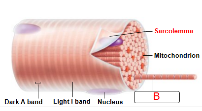
What is B covering
Myofibril
Striations are
Stripes formed from repeating series of dark and light bands along length of each myofibril
Myofilaments are
Orderly arrangement of actin and myosin myofilaments within sarcomere
Actin myofilaments are
Thin filaments
Extend across I band and partway in A band
Anchored to Z discs
Myosin myofilaments
Thick filaments
Extend length of A band
Connected at M line
Tropomyosin and troponin are
Regulatory proteins bound to actin
Structures that help from the myofibril are
Protein titin, dystrophin
Duchenne muscular dystrophy (DMD) is the
Most common and serious form of muscular dystrophies, muscle-destroying diseases that generally appear during childhood
Sarcoplasmic reticulum
Network of smooth endoplasmic reticulum tubules surrounding each myofibril
SR functions in regulation of
Intracellular Ca2+ levels, stores and releases Ca2+
T tubules
Tube formed by protrusion of sarcolemma deep into cell interior
Triad
Area formed from terminal cistern of one sarcomere, T tubule, and terminal cistern of neighboring sarcomere
Triad relationships
When an electrical impulse passes by, T tubule proteins change shape, causing SR proteins to change shape, causing release of calcium into cytoplasm
Contraction
The activation of cross bridges to generate force
Sliding filament model of contraction
States that during contraction, thin filaments slide past thick filaments, causing actin and myosin to overlap more
When nervous system stimulates muscle fiber, myosin heads are
Allowed to bind to actin, forming cross bridges, which cause sliding (contraction) process to begin
Four steps must occur for skeletal muscle to contract
Nerve stimulation
Action potential, an electrical current, must
be generated in sarcolemmaAction potential must be propagated along
sarcolemmaIntracellular Ca2+ levels must rise briefly

What is step 1 of the NMJ
AP arrives at axon terminal of motor neuron.
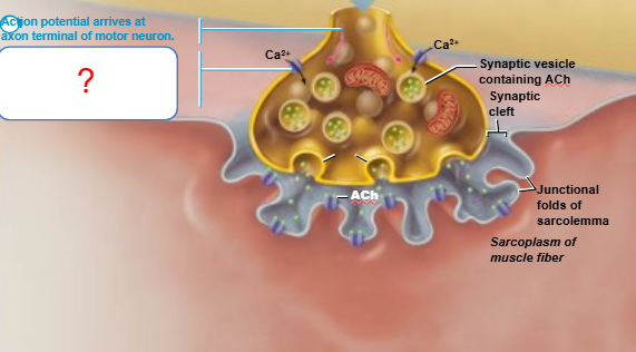
What is step 2 of the NMJ
Voltage-gated channels open. Ca2+ enters the axon terminal

What is step 3 of the NMJ
Influx of Ca2+ causes synaptic vesicles to release ACh via exocytosis
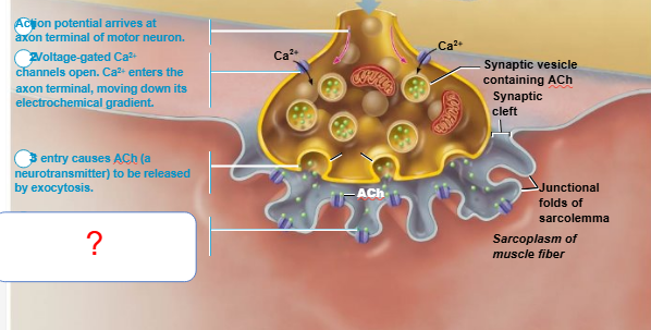
What is step 4 of the NMJ
ACh diffuses through the synaptic cleft and binds to receptors on sarcolema

What is step 5 of the NMJ
Sodium influx and K+ (potassium) influx causes the resting membrane potential to the change. This continues until muscle reaches end plate potential
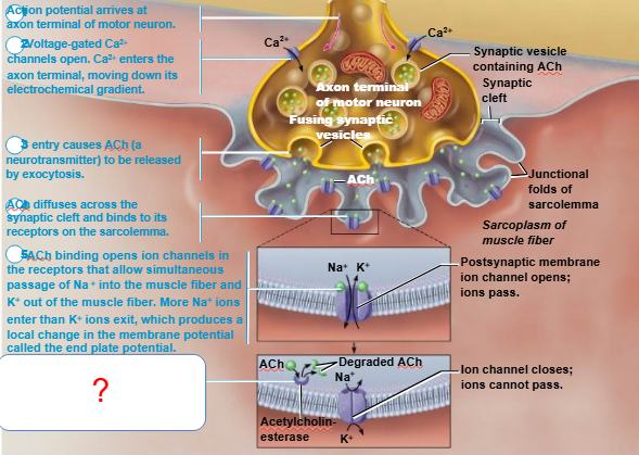
What is step 6 of the NMJ
Effects of Ach are terminal 1-diffusion 2-ACh breaks it apart
Myasthenia gravis is characaterized by
Drooping upper eyelids, difficulty swallowing and talking, and generalized muscle weakness it also an autoimmune disease
Generation of AP occurs in three steps
End plate potential
Depolarization
Repolarization
End plate potential
Results in local depolarization called end plate potential
Depolarization
Generation and propagation
of an action potential (AP)
Repolarization
Restoration of resting conditions
Refractory period
Muscle fiber cannot be stimulated for a specific amount of time, until repolarization is complete
Excitation-contraction (E-C) coupling
Events that transmit AP along sarcolemma (excitation) are coupled to sliding of myofilaments (contraction)

What is step 1 of ECC
The AP propagates down the T-tubule and sarcolemma
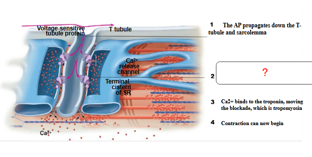
What is step 2 of ECC
Ca2+ is released into the sarcoplasm
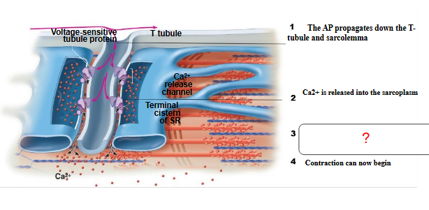
What is step 3 of ECC
Ca2+ binds to the troponin, moving the blockade, which is tropomyosin

What is step 4 of ECC
Contraction can now begin
At low intracellular Ca2+ concentration:
Tropomyosin blocks active sites on actin
Myosin heads cannot attach to actin
Muscle fiber remains relaxed

What is occurring in stage 1 of CBF
Formation of crossbridge the myosin head binds to actin

What is occurring in stage 2 of CBF
The power stroke (contraction); myosin head pivots

What is occurring in stage 3 of CBF
Cross-bridge detachment requires ATP

What is occurring in stage 4 of CBF
Cocking the myosin head
Rigor mortis
3–4 hours after death, muscles begin to stiffen
ATP is also needed for cross bridge detachment
Isometric contraction
no shortening; muscle tension increases but does not exceed load
Isotonic contraction
Muscle shortens because muscle tension exceeds load
Motor unit consists of the
Motor neuron and all muscle fibers (four to several hundred) it supplies
Muscle twitch
Simplest contraction resulting from a muscle fiber’s response to a single action potential from motor neuron
Latent period
Events of excitation-contraction coupling
Period of contraction
Cross bridge formation Tension increases
Period of relaxation
Ca2+ reentry into SR Tension declines to zero
Muscle responses are graded by
Changing frequency of stimulation
Changing strength of stimulation

Low stimulation frequency
unfused (incomplete) tetanus (partial relxation)

Fused tetanus.
At even higher stimulus frequencies, there is no
relaxation at all between stimuli.

High stimulation frequency
Fused (complete) tetanus.
At higher stimulus frequencies, there is no relaxation at all between stimuli. This is fused (complete) tetanus.
Recruitment works on
Size principle
Isotonic contractions
Muscle changes in length and moves load

Isotonic contraction (concentric) on stimulation muscle
Develops enough tension (force) to lift the load (weight).

Isometric contraction when stimulated
The tension increases to the muscle’s peak tension-developing capability, but the muscle does not shorten.
ATP is regenerated quickly by three mechanisms:
Direct phosphorylation of ADP by creatine phosphate (CP)
Anaerobic pathway: glycolysis and lactic acid formation
Aerobic respiration
Creatine phosphate
Is a unique molecule located in muscle fibers that donates a phosphate to ADP to instantly form ATP
Direct phosphorylation: enzyme & duration?
Enzyme: Creatine kinase / Duration: ~15 seconds
In the absence of oxygen
Anaerobic glycolysis, pyruvic acid is converted to lactic acid
Anaerobic pathway
Glycolysis and lactic acid formation
Aerobic respiration
Produces 95% of ATP during rest and light-to-moderate exercise

Within the 6 seconds of short-duration exercise
ATP is stored in muscles is used first

Within the 10 seconds of short-duration exercise
ATP is formed from creatine phosphate and ADP (direct phosphorylation).

Within the 30-40 seconds of high-intensity exercise
Glycogen stored in muscles is broken down to
glucose, which is oxidized to generate ATP
(anaerobic pathway).

What occurs within the hours of prolonged duration exercise
ATP is generated by breakdown of several nutrient
energy fuels by aerobic pathway.
Muscle fatigue is
Physiological inability to contract despite continued stimulation
For a muscle to return to its pre-exercise state:
Oxygen reserves are replenished
Lactic acid is reconverted to pyruvic acid
Glycogen stores are replaced
ATP and creatine phosphate reserves are resynthesized