Psych 1000-001, Biological Basis of Behaviour
 Call Kai
 Learn
Learn Practice Test
 Spaced Repetition
 Match
Match Flashcards
 Knowt Play
1/82

Earn XP
Description and Tags
Chapter 3
Name | Mastery | Learn | Test | Matching | Spaced | Call with Kai | Chat |
|---|
No analytics yet
Send a link to your students to track their progress
83 Terms
Neurons
Neurons are specialized cells that transmit electrical impulses and convert them into chemical signals. Each neuron’s structure is uniquely adapted to its function, reflecting the specific connections it forms with other cells. The human brain contains about 86 billion neurons on average.
Parts of a Neuron
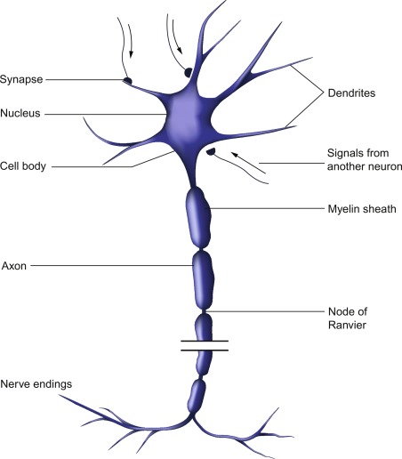
Cell body, dendrites, axon hillock, Schwann cells, nodes of Ranvier, myelin sheath, axon, nerve terminals.
Cell Body / Soma
The control center of a neuron.
Processes information:
It receives signals from the dendrites, integrates them, and determines whether to generate an action potential.Contains the nucleus:
The nucleus holds the neuron’s DNA and controls the production of proteins and other molecules needed for cell function.Supports cell health:
The cell body contains organelles (like mitochondria, ribosomes, and the endoplasmic reticulum) that provide energy and synthesize neurotransmitters and structural proteins.
The cell body keeps the neuron alive, energized, and functionally coordinated — it’s like the neuron’s brain and engine room.
Dendrites
The branch-like extensions of a neuron that receive signals from other neurons.
Receive incoming signals: Dendrites pick up electrical or chemical messages (neurotransmitters) from the axon terminals of other neurons across synapses.
Transmit information to the cell body (soma): These signals are then sent toward the neuron's cell body, where they are processed and integrated.
Determine neuron response: If the total incoming signals are strong enough, the neuron may generate an action potential.
Dendrites = antennae of the neuron — they detect and collect information from neighbouring neurons.
Axon Hillock (anatomy)
A critical region of the neuron located at the junction between the cell body (soma) and the axon.
Integrates signals:
It sums all excitatory and inhibitory inputs coming from the dendrites and cell body.
Decision point for firing:
If the combined signals reach the threshold potential (usually around –55 mV), the axon hillock initiates an action potential.Triggers action potentials:
Once the threshold is reached, voltage-gated sodium channels open here, starting the electrical impulse that travels down the axon.
Think of the axon hillock as a neuron’s “gatekeeper” — it decides whether the incoming signals are strong enough to fire off a message.
Axon
The long, slender projection of a neuron that carries electrical signals away from the cell body toward other neurons, muscles, or glands.
Conducts action potentials:
The axon transmits the electrical impulse generated at the axon hillock along its length.Transmits signals to other cells:
At its axon terminals, the axon triggers the release of neurotransmitters into the synapse, communicating with the next neuron or target cell.Insulated for speed (in myelinated neurons):
Many axons are wrapped in myelin, which speeds up signal transmission through saltatory conduction(jumping from node to node).
The axon is like a highway for electrical signals, carrying messages from the neuron’s “control center” (cell body) to other cells.
Myelin Sheath
A fatty layer that wraps around the axons of many neurons.
Insulates the axon:
Prevents electrical signals from leaking out, keeping the impulse strong.
Speeds up signal transmission:
Myelin allows action potentials to jump between nodes of Ranvier in a process called saltatory conduction, making signal travel much faster.Protects the axon:
Acts like a protective covering, preventing damage to the axon.
The myelin sheath is like insulation around an electrical wire — it protects the wire and helps the electricity travel faster.
Nerve Terminal
The end part of an axon that communicates with other neurons, muscles, or glands. Also called the axon terminal or synaptic terminal.
Stores neurotransmitters:
Tiny sacs called synaptic vesicles in the terminal hold chemical messengers (neurotransmitters).Releases neurotransmitters:
When an action potential reaches the terminal, it triggers the release of neurotransmitters into the synapse (the gap between neurons).Transmits signals to the next cell:
Neurotransmitters bind to receptors on the next neuron or target cell, passing the signal along.
The nerve terminal is like the delivery dock of a factory — it releases packages (neurotransmitters) to send messages to the next “station” (neuron, muscle, or gland).
Nodes of Ranvier
The small gaps in the myelin sheath along a myelinated axon.
Enable saltatory conduction:
The action potential “jumps” from node to node rather than traveling continuously along the axon. This speeds up signal transmission.Allow ion exchange:
These gaps contain voltage-gated sodium (Na⁺) and potassium (K⁺) channels, which regenerate the action potential as it moves along the axon.Maintain signal strength:
By refreshing the electrical impulse at each node, the action potential doesn’t lose strength over long distances.
Nodes of Ranvier are like charging stations along a highway — the signal “recharges” at each node, allowing it to travel faster and farther.
Synaptic Cleft
The tiny gap between the axon terminal of one neuron and the dendrite or cell body of the next neuron (or a target cell like a muscle or gland).
Separates neurons:
Neurons don’t physically touch each other; the cleft keeps them slightly apart.
Allows chemical communication:
When an action potential reaches the axon terminal, neurotransmitters are released into the cleft. These chemicals cross the gap and bind to receptors on the next cell to transmit the signal.Controls signal flow:
The cleft ensures that communication is directional and regulated, preventing unwanted or continuous activation.
The synaptic cleft is like a tiny river between two islands — messages (neurotransmitters) float across the gap to deliver instructions to the other side.
Synapse
The junction between two neurons (or a neuron and a target cell, like a muscle or gland) where information is transmitted.
Presynaptic neuron: The neuron sending the signal, ending in an axon terminal.
Synaptic cleft: The tiny gap between the two cells where neurotransmitters are released.
Postsynaptic neuron (or cell): The receiving neuron or target cell that has receptors for the neurotransmitters.
Converts the electrical signal (action potential) in the presynaptic neuron into a chemical signal via neurotransmitters.
The chemical signal crosses the synaptic cleft and is detected by the postsynaptic cell, potentially triggering a new action potential.
A synapse is like a relay station — one neuron passes the message across a small gap to the next “station” to continue the signal.
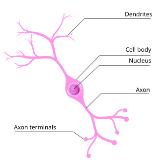
Multipolar Neuron
A type of neuron that has one axon and multiple dendrites extending from its cell body. This is the most common type of neuron in the human nervous system.
One axon: Carries signals away from the cell body.
Many dendrites: Receive signals from multiple other neurons, allowing for integration of information.
Cell body (soma): Contains the nucleus and organelles, maintaining cell function.
Multipolar neurons are mainly involved in motor control and integration of information within the brain and spinal cord.
They process and transmit information from many inputs to a single output.
Think of a multipolar neuron as a hub with many antennas (dendrites) receiving signals and one outgoing cable (axon) sending the message.
Bipolar Neuron
A type of neuron that has one axon and one dendrite extending from opposite ends of the cell body.
One axon: Sends signals away from the cell body.
One dendrite: Receives signals toward the cell body.
Cell body (soma): Contains the nucleus and organelles for cell maintenance.
Bipolar neurons are specialized for sensory functions, such as vision, smell, and hearing.
They relay information from sensory receptors to other neurons in the central nervous system.
A bipolar neuron is like a straight two-way street — one “lane” bringing signals in (dendrite) and one “lane” sending signals out (axon).
Unipolar Neuron
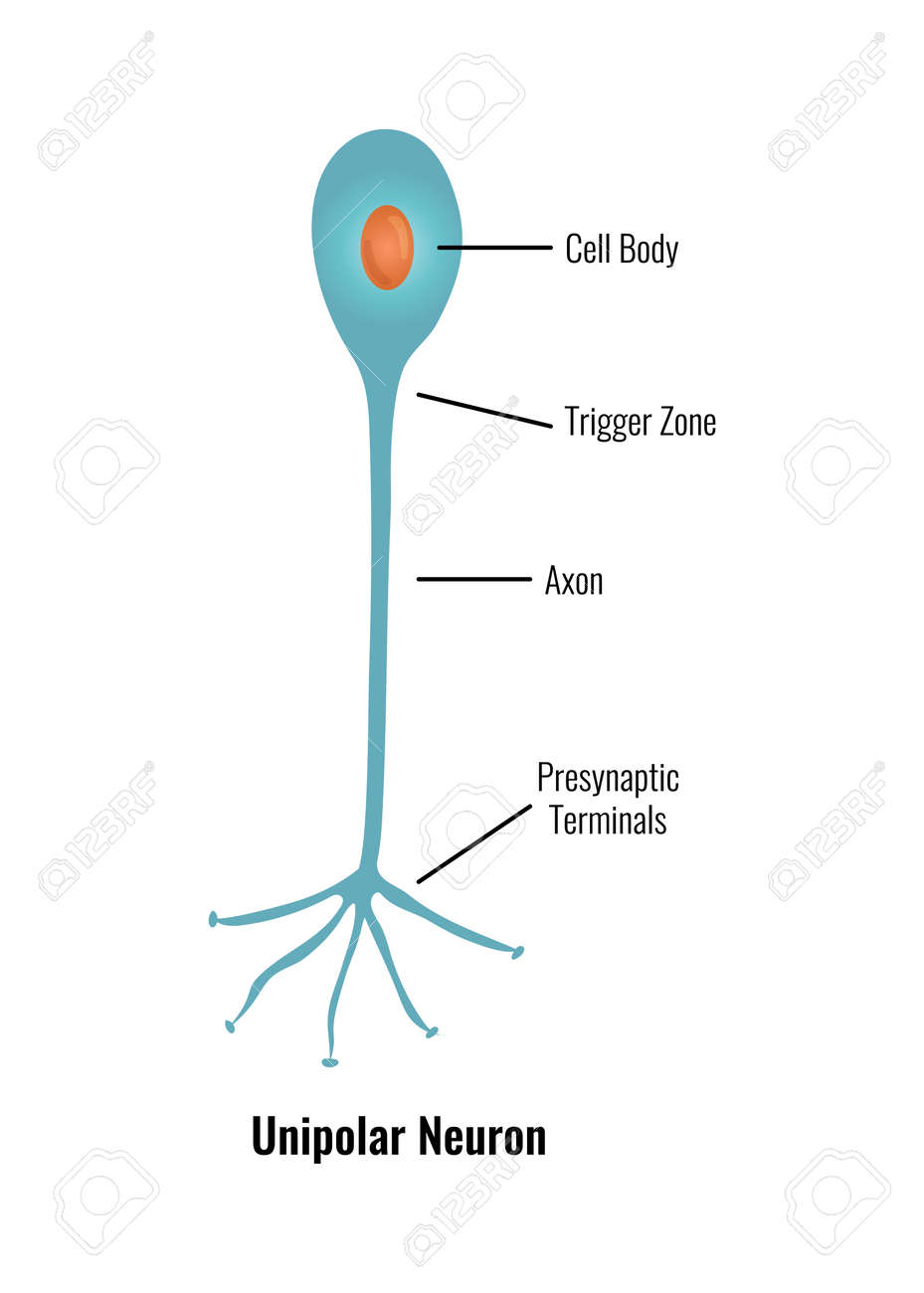
A type of neuron in which a single process extends from the cell body and then splits into two branches: one that acts like a dendrite to receive signals and one that acts like an axon to send signals. (also called a pseudo-unipolar neuron)
Single process from the cell body: Splits into two branches.
Peripheral branch: Receives sensory information from receptors in the body.
Central branch: Sends the signal into the central nervous system (CNS).
Unipolar neurons are mainly sensory neurons that carry information from the body to the CNS.
They are common in dorsal root ganglia of the spinal cord.
A unipolar neuron is like a T-shaped road — the stem (cell body) connects to a road that splits into two lanes: one bringing information in and one sending it out.
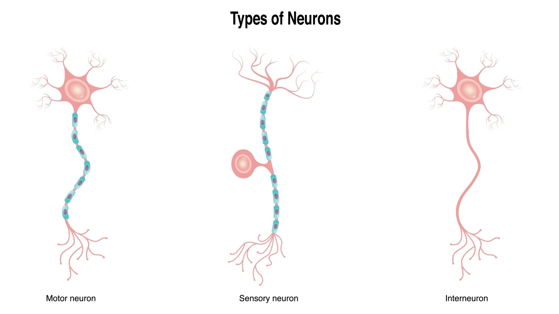
Sensory Neuron
A neuron that carries information from sensory receptors in the body toward the central nervous system (CNS). (also called an afferent neuron)
Receives stimuli: Detects changes in the environment (like touch, temperature, pain, light, or sound) through sensory receptors.
Transmits signals to CNS: Sends electrical impulses from the body to the brain or spinal cord.
Structure: Many sensory neurons are unipolar, but some can be bipolar (e.g., in the eye or ear).
Converts physical or chemical stimuli into electrical signals.
Allows the body to perceive sensations and respond appropriately.
Sensory neurons are like messenger wires that report information from the body to the control center (brain/spinal cord).
Motor Neuron
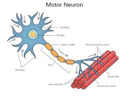
A neuron that carries signals away from the central nervous system (CNS) to muscles or glands, enabling movement or secretion. (also called an efferent neuron)
Receives commands from CNS: The cell body is usually located in the spinal cord or brain.
Sends signals to effectors: The axon transmits impulses to muscles (for movement) or glands (for secretion).
Structure: Most motor neurons are multipolar, with one axon and multiple dendrites.
Controls voluntary and involuntary movements.
Triggers muscle contraction or glandular activity in response to CNS signals.
Motor Neurons are like outgoing electrical wires that carry commands from the control centre (CNS) to machines (muscles/glands) to make them act.
Interneurons
Neurons that connect other neurons within the central nervous system (CNS). They process information and coordinate responses between sensory and motor neurons. (also called association neurons)
Located in CNS: Found mainly in the brain and spinal cord.
Connect neurons: Link sensory neurons (afferent) to motor neurons (efferent).
Structure: Typically multipolar, with multiple dendrites and one axon for integrating information.
Integrate sensory input from sensory neurons.
Process information and determine the appropriate response.
Send commands to motor neurons to trigger actions.
Interneurons are like middle managers — they receive reports from sensors (sensory neurons), decide on a course of action, and pass instructions to workers (motor neurons).
Glial Cells
The supporting cells of the nervous system. Unlike neurons, they do not generate electrical impulses, but they play essential roles in maintaining neuron function, protection, and overall brain health. (neuroglia)
Support and structure: Provide physical scaffolding for neurons.
Insulation: Produce myelin (e.g., oligodendrocytes in CNS, Schwann cells in PNS) to speed up signal transmission.
Protection: Act as immune defense in the CNS (e.g., microglia remove debris and pathogens).
Nourishment: Supply nutrients and oxygen to neurons (e.g., astrocytes regulate blood flow and maintain the blood-brain barrier).
Regulate environment: Maintain ion balance and remove excess neurotransmitters from synapses.
Types: Astrocytes, Oligodendrocytes, Microglia, Ependymal cells
Glial cells are like the support staff in a company — they maintain the environment, provide resources, protect the team, and ensure smooth operations, while neurons do the “main work” of transmitting messages.
Astrocytes
Star-shaped glial cells that support and nourish neurons by supplying essential nutrients and maintaining the proper chemical environment. They also play a key role in forming and maintaining the blood-brain barrier, which protects the brain from harmful substances in the blood while allowing necessary nutrients to pass through.
Oligodendroglia
Glial cells in the central nervous system (CNS) that produce myelin sheaths, which wrap around axons of neurons. These myelin sheaths insulate the axons, allowing electrical signals (action potentials) to travel more quickly and efficientlyalong the neuron. By supporting rapid signal transmission, oligodendrocytes play a critical role in the proper functioning of neural networks within the brain and spinal cord.
Ependymal Cells
Specialized glial cells that line the ventricles of the brain and the central canal of the spinal cord. They are responsible for producing and circulating cerebrospinal fluid (CSF), which cushions the brain and spinal cord, providing protection against mechanical shocks, and also helps transport nutrients and remove waste productswithin the central nervous system.
Microglia
Specialized glial cells in the central nervous system (CNS) that function as the primary immune defense of the brain and spinal cord. They are phagocytic cells, meaning they ingest and break down cellular debris, waste products, and harmful pathogens. By removing damaged neurons and infectious agents, microglia help maintain a healthy neural environment, protect the CNS from infection, and support overall brain function.
Schwann Cells
Glial cells in the peripheral nervous system (PNS) that produce myelin sheaths around the axons of neurons. These myelin sheaths insulate the axons, allowing faster and more efficient transmission of electrical impulses along the nerve fibers. In addition to supporting rapid signal conduction, Schwann cells also aid in the repair and regeneration of damaged peripheral nerves, making them essential for maintaining proper function in the PNS.
Resting potential
A cell’s resting membrane potential refers to the difference in electrical charge between the inside (intracellular fluid) and the outside (extracellular fluid) of the cell.
Because the neuronal membrane is semi-permeable, certain charged particles (ions) become more concentrated on one side of the membrane than the other. The resting potential of a neuron is about –70 mV. It is maintained mainly by the ions sodium (Na⁺) and potassium (K⁺). Potassium tends to drive the membrane potential toward –90 mV, while sodium pushes it toward +60 mV. The balance between these opposing forces results in the neuron’s resting potential of –70 mV.
Axon Hillock (transmission)
Neurons receive both excitatory and inhibitory signals.
Excitatory inputs cause depolarization, raising the membrane potential above its resting level.
Inhibitory inputs cause hyperpolarization, lowering the membrane potential below its resting level.
If the axon hillock receives enough excitatory input to reach the threshold potential (typically between –55 mV and –40 mV), an action potential is triggered. This opens voltage-gated sodium channels, allowing Na⁺ ions to enter the cell and make the membrane potential more positive.
When the potential reaches around +35 mV, the sodium channels become inactivated, and voltage-gated potassium channels open. The resulting efflux of K⁺ ions restores the negative charge inside the cell — a process known as repolarization.
Action Potential
When a neuron receives a stimulus:
If the stimulus is strong enough to reach the threshold (around –55 mV), the neuron fires an action potential.
If the stimulus is too weak (below threshold), no action potential occurs.
Once triggered, the action potential always has the same size and strength — it doesn’t get bigger with a stronger stimulus.
In other words, a weak stimulus doesn’t create a small action potential, and a strong one doesn’t create a bigger one.
Think of it like flipping a light switch:
If you press the switch enough, the light turns on completely.
If you don’t press hard enough, it stays off.
There’s no “dim” version — it’s either on or off.
A neuron either fires a full action potential or doesn’t fire at all — there’s no in-between or partial firing.
Refractory Period
Right after a neuron fires an action potential, it temporarily cannot fire again.
Absolute refractory period: The neuron is completely incapable of generating another action potential, no matter how strong the stimulus.
Relative refractory period: The neuron can fire again, but only if it receives a stronger-than-normal stimulusbecause the membrane potential hasn’t fully returned to its resting level
Impulse Propogation
For a neuron to transmit a signal to another cell, the action potential must travel along the axon and trigger the release of neurotransmitters at the axon terminal.
In myelinated neurons, the electrical impulse jumps from one node of Ranvier to the next, a process known as saltatory conduction.
Neurotransmitters
Chemical messengers stored in tiny sacs called vesicles within nerve cells (neurons). When a neuron is activated, these chemicals are released into the space between neurons (called the synaptic cleft) to transmit signals to other neurons, muscles, or glands. Neurotransmitters are how neurons “talk” to each other. When a nerve impulse reaches the end of a neuron, it causes vesicles (small storage bubbles) to release neurotransmitters into the synaptic cleft. These chemicals then bind to receptors on the next neuron, continuing the communication chain throughout the nervous system. Ex: dopamine, seratonin
Acetylcholine (ACh)
A neurotransmitter that plays a key role in muscle movement, learning, and memory. It helps transmit signals from nerve cells to muscles, allowing the body to move. When a person has Alzheimer’s disease, the neurons that produce acetylcholine gradually break down. As a result, the levels of this neurotransmitter decrease, which contributes to problems with memory and thinking.
Dopamine
A neurotransmitter that helps control movement, learning, attention, and emotions. It plays a major role in the brain’s reward and pleasure system, influencing how we feel motivation and satisfaction. An oversupply of dopamine is linked to schizophrenia, a mental disorder that can cause hallucinations and disorganized thinking. An undersupply of dopamine is associated with Parkinson’s disease, leading to tremors, stiffness, and difficulty with movement.
Serotonin
A neurotransmitter that helps regulate mood, hunger, sleep, and arousal (alertness).
An undersupply of serotonin is linked to depression and other mood disorders.
Certain antidepressant medications, such as SSRIs (Selective Serotonin Reuptake Inhibitors), increase serotonin levels in the brain to help improve mood and relieve symptoms of depression.
Norepinephrine
A neurotransmitter that helps control alertness, arousal, and the body’s stress response. It prepares the body to take action by increasing heart rate, blood flow, and focus. An undersupply of norepinephrine can lead to depressed mood, low energy, and difficulty concentrating.
Gamma-aminobutyric acid
GABA is the brain’s main inhibitory neurotransmitter, meaning it helps calm the activity of neurons and prevents overstimulation.
An undersupply of GABA is linked to seizures, tremors, and insomnia, since the brain becomes overly active without enough of this calming chemical.
Glutamate
The brain’s main excitatory neurotransmitter, meaning it stimulates neurons and helps them send signals. It plays an important role in learning, memory, and overall brain function. An oversupply of glutamate can overstimulate the brain, which may lead to migraines or seizures.
Endorphins
Neurotransmitters that help reduce pain and increase feelings of pleasure or well-being. They act as the body’s natural painkillers, released during activities such as exercise, laughter, or moments of stress. An oversupply caused by opioid drug use (such as morphine or heroin) can suppress the body’s natural endorphin production, making it harder for the body to manage pain or feel pleasure without the drug.
Agonist
A molecule that enhances or mimics the action of a neurotransmitter. It can increase the effect of the neurotransmitter by either stimulating its receptor or boosting its release.
Antagonist
A molecule that blocks or inhibits a neurotransmitter’s action. It prevents the neurotransmitter from binding to its receptor, reducing or stopping its effect.
Central Nervous System (CNS)
This nervous system consists of the brain and spinal cord. It acts as the body’s control center, processing information, making decisions, and directing the body’s responses. The brain interprets information from the senses, plans actions, and controls thoughts, emotions, and memory. The spinal cord acts as a communication highway, sending messages between the brain and the rest of the body. Together, they coordinate everything the body does, from moving muscles to regulating vital functions like breathing and heart rate.
Peripheral Nervous System (PNS)
This nervous system is made up of sensory and motor neurons that connect the CNS (brain and spinal cord) to the rest of the body. It gathers information from the environment and body, and transmits commands from the CNS to muscles and glands. The PNS acts like the body’s communication network: Sensory neurons carry information from the senses (like touch, sight, and sound) to the CNS. Motor neurons carry instructions from the CNS to muscles and glands, telling the body how to respond. This system allows the brain and spinal cord to sense the world, react to changes, and control movement.
Functions of nervous system
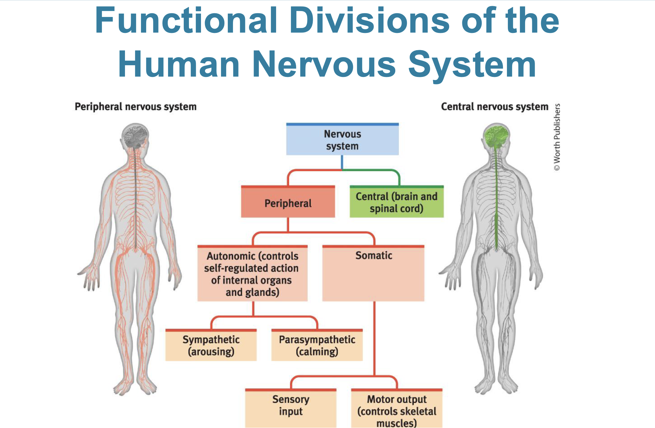
Somatic Nervous System (SNS)
Controls voluntary movements of the body’s skeletal muscles. Allows you to consciously move, walk, grab objects, or type on a keyboard.
Autonomic Nervous System (ANS)
Regulates involuntary functions such as heartbeat, digestion, and breathing. Has two subdivisions: sympathetic and parasympathetic.
Sympathetic Nervous System
This subdivision of the autonomic nervous system arouses the body and expends energy. It prepares the body for action, increasing alertness, heart rate, and blood flow to muscles. It also supports voluntary control of skeletal muscles during stressful or emergency situations.
Parasympathetic Nervous System
This subdivision of the autonomic nervous system calms the body and conserves energy. It supports routine maintenance activities such as digestion, repair, and rest. It also controls involuntary muscles and glands, helping the body maintain balance and recovery after stress.
Feedback System
Involves communication between the brain, glands, hormones, and body:
The brain sends signals to the pituitary gland.
The pituitary gland signals other endocrine glands to release hormones.
Hormones affect the body and send feedback to the brain, which adjusts responses as needed.
This system shows the close interplay between the nervous system and the endocrine system, coordinating bodily functions and maintaining balance.
Endocrine System
A network of glands that secrete hormones directly into the bloodstream. These hormones travel throughout the body and influence various tissues, including the brain, to regulate processes like growth, metabolism, mood, and stress response.
Pituitary Gland
This is often called the “master gland” because it regulates the hormone release of other endocrine glands, including the adrenal glands.
It operates as part of a feedback system:
The hypothalamus in the brain sends signals to the pituitary.
The pituitary gland releases hormones that stimulate other glands.
Those glands release their own hormones, which affect the body and send feedback to the brain, allowing the system to self-regulate.
Stress
When a stressful event occurs, the hypothalamus signals the pituitary gland to release hormones that stimulate the adrenal glands. The adrenal glands then release cortisol, a stress hormone that increases blood sugar and prepares the body for a “fight or flight” response. Stress doesn’t only come from physical threats—experiencing prejudice or social stress can also trigger cortisol release. Chronic or sustained stress can have long-term effects:
It increases the risk of depression in adolescents and adults.
Essentially, a stressed body can contribute to a depressed mind.
Brain and Spinal Cord
The adult brain contains about 86 billion neurons, which communicate through complex neural networks and pathways. These networks allow the nervous system to send and receive information efficiently using electrochemical signals, controlling everything from reflexes to complex thoughts. The spinal cord serves as the main communication link between the peripheral nervous system (PNS) and the brain:
Ascending neural fibers carry sensory information from the body to the brain.
Descending neural fibers carry motor-control signals from the brain back to the body.
An example of this communication is the pain reflex: when you touch something hot, sensory neurons send a signal to the spinal cord, which triggers an immediate reflex to pull your hand away before the brain even processes the pain.
Brain
Weight: ~3 pounds (about 1.4 kg)
Proportion of body weight: ~2% in an average adult
Size: Roughly the size of two fists put together
Energy use: Consumes about 20% of the body’s energy, despite its small size
Hemispheres: The left and right hemispheres are largely symmetrical, with a few functional exceptions
Folds and grooves: The gyri (ridges) and sulci (grooves) increase surface area; without them, the brain wouldn’t fit inside the skull
Brainstem
The central core of the brain, located where the spinal cord swells as it enters the skull. It controls many automatic survival functions, such as heartbeat, breathing, and swallowing.
Medula
Located at the base of the brainstem and controls vital automatic functions such as heartbeat and breathing.
Pons
This sits above the medulla in the brainstem. It helps coordinate movement and plays a key role in controlling sleep and arousal.
Cerebellum
This, often called the “little brain”, is located at the rear of the brainstem in the hindbrain.
Functions:
Processes sensory input to help understand the body’s position in space
Coordinates movement and balance, ensuring smooth and precise motions
Supports nonverbal learning and skill memory, such as riding a bike or playing an instrument
Reticular Formation
A network of nerves that runs through the brainstem and extends into the thalamus.
Functions:
Filters incoming sensory information, helping the brain focus on what’s important
Regulates arousal and alertness, determining wakefulness and attention
Note: For categorization, it is associated with both the midbrain and hindbrain, as its nerve network spans across these regions.
Midbrain
This connects the hindbrain to the forebrain and contains structures located deep within the brain.
Key Structures:
Substantia nigra – Produces dopamine; critical for movement control
Ventral tegumental area (VTA) – Produces dopamine; involved in reward, motivation, and reinforcement learning
Thalamus
Located on top of the brainstem and acts as the brain’s sensory relay station.
Functions:
Directs incoming sensory messages (except smell) to the appropriate sensory areas of the cerebral cortex
Transmits outgoing messages from the cortex to the cerebellum and medulla, helping coordinate movement and responses
Limbic System
A set of structures located mostly in the forebrain. It is closely linked with the hypothalamus, which controls the nearby pituitary gland to regulate hormones.
Functions:
Regulates emotions such as fear, anger, and pleasure
Drives basic behaviors like hunger, thirst, and sex
Supports memory formation
Key Structures:
Amygdala – Processes emotions, especially fear and aggression
Hypothalamus – Regulates hormones, body temperature, hunger, thirst, and sexual behavior
Hippocampus – Essential for forming new memories and connecting them to emotions
Amygdala
Consists of two lima bean-sized neural clusters located within the limbic system.
Functions:
Processes emotions, especially fear, aggression, and rage
Helps detect threats and triggers appropriate emotional and physiological responses
Hypothalamus
A neural structure located below the thalamus. It plays a central role in maintaining the body’s homeostasis.
Functions:
Regulates maintenance activities such as hunger, thirst, body temperature, and sexual behavior
Controls the endocrine system by directing the pituitary gland to release hormones
Links to emotion and reward: contains pleasure/reward centers that influence motivation and behavior
Related Concepts:
Reward deficiency syndrome – A condition where natural brain reward pathways are underactive, leading to cravings or addictive behaviors
Hippocampus
A neural centre located within the limbic system.
Functions:
Processes conscious, explicit memories of facts and events, preparing them for long-term storage
Some neural clusters influence hunger, while others help regulate thirst, body temperature, and sexual behavior
Decreases in size and function with age, contributing to cognitive decline and memory difficulties
Cerebral Cortex
A thin layer of interconnected neurons that covers the cerebral hemispheres. It is the brain’s ultimate control and information-processing center.
Functions:
Governs thinking, perceiving, planning, and decision-making
Processes sensory input and coordinates voluntary movement
Supports higher-level cognitive functions, such as language, reasoning, and problem-solving
Cerebral Hemispheres
The brain is divided into two hemispheres, left and right, which are largely symmetrical but have specialized functions.
Each hemisphere contains four lobes: Frontal, Parietal, Temporal, Occipital.
Frontal Lobe
These lobes are the portion of the cerebral cortex located just behind the forehead.
Functions:
Controls voluntary muscle movements
Involved in speaking and language production (Broca’s area)
Supports higher cognitive functions, such as planning, decision-making, and judgment
Parietial Lobe
Lobes located on the top and toward the rear of the head, forming part of the cerebral cortex.
Functions:
Processes sensory information from the body, including touch, pressure, temperature, and pain
Integrates spatial awareness and helps coordinate body position and movement
Occipital Lobe
Lobes located at the back of the head and form part of the cerebral cortex.
Functions:
Processes visual information from the eyes
Contains the primary visual cortex, which receives input from the visual fields and interprets shapes, colors, and motion
Temporal Lobe
Lobes located roughly above the ears and form part of the cerebral cortex.
Functions:
Processes auditory information from the ears
Supports language comprehension (Wernicke’s area)
Plays a key role in memory formation and emotional association
Somatosensory Cortex
Located at the front of the parietal lobes, just behind the motor cortex.
Functions:
Receives and processes sensory information from the skin and body, including touch, pressure, temperature, and pain
Registers and interprets sensations from different body parts, allowing precise perception of texture, shape, and movement
Visual Cortex
Located in the occipital lobes at the rear of the brain.
Functions:
Receives visual input directly from the eyes
Processes and interprets visual information, such as color, shape, motion, and depth
Sends processed data to other brain areas for object recognition and spatial understanding
Auditory Cortex
Located in the temporal lobes, positioned just above the ears.
Functions:
Receives and processes auditory information from the ears
Interprets pitch, volume, tone, and sound location
Plays a key role in understanding speech and language
Association Areas
Regions of the cerebral cortex found in all four lobes of the brain. Unlike sensory or motor areas, they are not involved in primary motor or sensory functions.
Functions:
Integrate and interpret information from various brain regions
Prefrontal lobes: enable judgment, planning, decision-making, personality expression, and memory processing
Other areas help with language comprehension, spatial awareness, and recognizing faces or objects
Damage:
Can cause different types of losses depending on the location:
Damage to the prefrontal cortex may impair planning, personality, and social behavior
Damage to parietal association areas may disrupt spatial awareness
Damage to temporal association areas may affect language or object recognition
Hemispheric Laterization
The two hemispheres of the cerebral cortex differ in their motor, cognitive, and emotional functions — a concept known as lateralization, or specialization of the hemispheres.
Functions of Each Hemisphere:
Left Hemisphere:
In most people, it controls language, speech, reading, and writing
Handles logical reasoning, analytical thinking, and mathematical processing
Associated with positive emotions and approach-related behaviors
Right Hemisphere:
Specializes in spatial abilities, such as visualizing shapes and recognizing faces
Plays a role in creativity, music, and emotional expression
Associated with negative emotions and withdrawal-related behaviors
Lateralization
Refers to the way specific functions are more dominant in one hemisphere than the other.
Handedness
The left hemisphere controls the right hand, and the right hemisphere controls the left hand.
Most right-handed individuals show stronger left-hemisphere dominance for language and logic.
Brain Plasticity
The brain’s ability to reorganize and form new connections depends on when lateralization occurs.
In early development, the brain is more plastic (flexible), allowing other areas to take over functions if one side is damaged.
As lateralization becomes more established with age, plasticity decreases.
Electroencephalograms and Event-Related Potentials
EEG:
A non-invasive technique that measures electrical activity in the brain using electrodes placed on the scalp.
Detects brain waves produced by groups of firing neurons.
ERP:
A specific application of EEG that records brain responses to particular sensory, cognitive, or motor events.
ERPs are used to study how the brain processes stimuli over time.
Limitations:
ERPs cannot precisely identify where in the brain activity is occurring.
They have excellent temporal resolution (can measure timing of brain activity very accurately) but poor spatial resolution (can’t show exact brain location).
Animal Studies
Researchers often use this to explore the relationship between brain function and behavior.
Methods:
Scientists can temporarily activate or deactivate specific brain areas to observe resulting changes in behavior.
Two common techniques include:
Lesioning: Intentionally damaging or removing a small part of the brain to study how behavior changes when that area is no longer functional.
Transcranial Magnetic Stimulation (TMS): A non-invasive method that uses magnetic fields to temporarily disrupt or stimulate brain activity in targeted regions.
Neuroimaging Techniques: CAT Scan
Purpose:
Neuroimaging techniques allow scientists to observe brain structure and activity safely and non-invasively.
Computerized Axial Tomography (CAT or CT) Scan:
Uses X-ray technology to create clear, detailed two-dimensional (2D) images of the brain.
Combines multiple X-ray images taken from different angles to form a cross-sectional view of brain structures.
Commonly used to detect brain injuries, tumors, bleeding, or structural abnormalities.
Diffusion Tensor Imaging (DTI)
Purpose:
DTI is a type of MRI-based neuroimaging that measures the orientation and integrity of white matter in the brain.
Functions and Applications:
Maps neural pathways and connections between brain regions.
Evaluates the health and structure of white matter, which is crucial for efficient communication between neurons.
Used to study brain disorders, including traumatic brain injury (TBI), multiple sclerosis, and neurodegenerative diseases.
Positron Emission Tomography (PET) Scan
Purpose:
PET scans are functional neuroimaging techniques used to study brain activity by tracking blood flow and metabolic processes.
How It Works:
A harmless radioactive substance (tracer) is injected into the bloodstream.
Active brain areas consume more energy, so the tracer accumulates in those regions.
The PET scanner detects the tracer, creating a color-coded map of brain activity.
Applications:
Identifying active brain regions during tasks
Studying brain disorders, including Alzheimer’s disease, epilepsy, and tumors
Researching cognition, emotions, and neural pathways
Functional Magnetic Resonance Imaging (fMRI)
Purpose:
fMRI is a non-invasive functional neuroimaging technique that measures brain activity by tracking changes in blood flow.
How It Works:
fMRI detects oxygenated blood in the brain, which increases in areas with higher neural activity.
Unlike PET scans, no radioactive injection is required.
Produces high-resolution, real-time images of which brain regions are active during specific tasks.
Applications:
Studying cognition, perception, emotion, and motor functions
Mapping brain activity before surgery
Researching brain disorders such as stroke, Alzheimer’s disease, and schizophrenia