Function and combinatorial action of transcription factors
 Call Kai
 Learn
 Practice Test
 Spaced Repetition
 Match
 Flashcards
 Knowt Play
1/11

There's no tags or description
Looks like no tags are added yet.
12 Terms
Structure of a transcription factor, and evidence
A DNA binding domain (e.g. helix-turn-helix) and an activation domain (acidic domains, glutamate and proline rich). Domain swap experiments provided evidence for this, where activation domains from one transcription factor were joined to binding domains of another - the binding domains did not facilitate transcription on their own, and were shown to be necessary to bring the activating domains close.
How does the Lac repressor bind the Lac operator sequence
Lac operator is a palindrome - this allows binding of a transcription factor as a dimer, so facilitates cooperativity
There are multiple binding sites in the Lac operon - each dimer binds to one. The dimers then tetramerise, so each tetramer binds two operator sequences.
The binding site of the Lac repressor contains a helix-turn-helix motif - this binds to the major groove of the DNA, and there are specific interactions between the bases and the amino acids
DNA binding domain
Small structural domains, often containing a helix-turn-helix motif, that bind to specific short DNA sequences, through interactions between specific amino acid side chains and specific nitrogenous bases, e.g. Gln - A and Arg - G
Similarities between eukaryotic and bacterial transcription factors
They both often contain helix-turn-helix domains.
Homeotic transformation
Transcription factors expressed in the wrong place, so the wrong tissue type is expressed in a certain place, e.g. Drosophila expressing legs where their antennae should be
Eukaryotic DNA binding domains that aren’t helix-turn-helix motifs
Zinc fingers - alpha helices coordinated by Zn2+ ions
Leucine zipper - dimer of two helices, forming a coil-coil. Basic residues in the extended alpha-helical regions interact with the DNA backbone at adjacent sites in the major groove. The coiled-coil dimerisation domain is stabilised by hydrophobic interactions between the monomers
Both of these, and HTH motifs, contact the bases in a direct manner
How do transcription factors promote transcription?
RNA polymerase (the sigma factor in the case of bacteria) binds to the promoter with a low affinity, so it has a short residence time
The transcription factor also has a short residence time by itself (DNA binding domain is small, there are very few residues, so it has a low affinity) and binds to the promoter
The activating domain binds to the RNA polymerase, and stabilises it - they bind cooperatively
This increases the residence time of RNA polymerase, so makes transcription initiation more likely
Evidence for the model of cooperative binding between TFs and RNA polymerase
CAP TF (Lac operon): activating region binds to the alpha subunit of RNA polymerase: the sole role of the activating region is to bind RNA polymerase
Genetic trickery can be used to replace the activating domain with a protein, and fuse another protein to the RNA polymerase - these two proteins bind each other and that is sufficient to activate transcription
Transcription is also activated if CAP is fused directly to RNA polymerase
Cooperativity between DNA-binding proteins
Each DNA-binding protein has a relatively weak interaction with DNA, which allows binding sites for each protein to be small
The protein has simultaneous interactions with DNA and with each other
This does not require the utilisation of energy - no ATP is used; does not require conformational changes in the proteins - they only need to bind each other
Makes binding to DNA stronger, as if one protein dissociates from the DNA, its interaction with the other protein, still bound to DNA, increases its local concentration and allows it to efficiently rebind to DNA
Example of feedback in transcription regulation: maintaining a lysogenic state
Bacteriophage lambda repressor
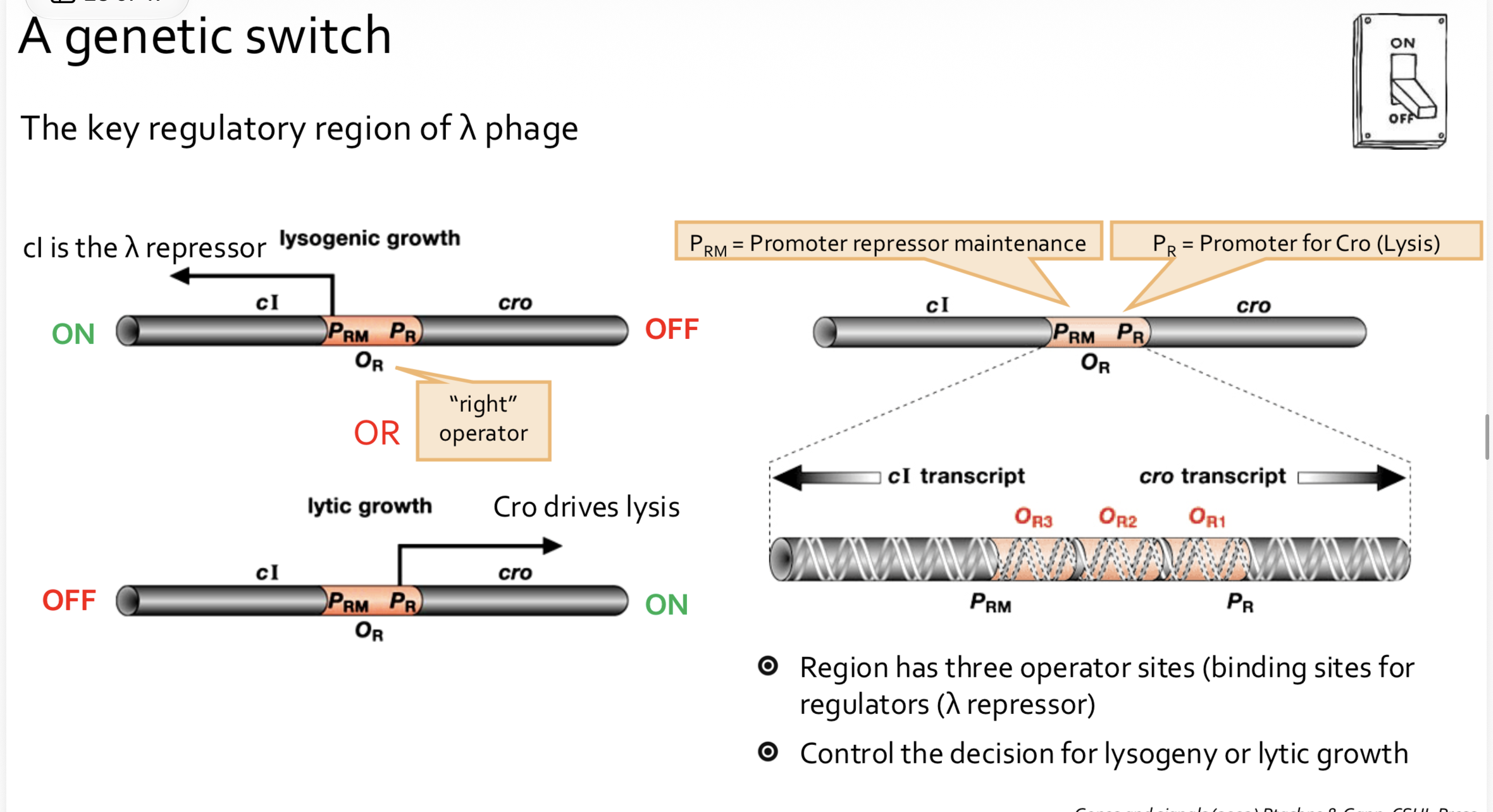
The operator region has three operator sites/binding sites ofr regulators. Upstream of the operator region is the gene encoding the lambda repressor (this causes lysogenic growth, which is when the bacteria divide with the bacteriophage in a dormant state) and downstream is the Cro gene, which causes lytic growth. The OR3 site is next to the promoter for lambda, and the OR1 site is next to the promoter for Cro.
The lambda repressor has the highest affinity (via cooperative binding as dimers, then as a tetramer) for OR1 and OR2 - this allows it to repress transcription from the PRM (Cro) promoter, and stabilise RNA polymerase on the PR (lambda) promotor to promote transcription of its own gene - this is positive feedback
At high concentrations of lambda repressor, it binds at its low-affinity site, OR3, and blocks its own transcription - negative feedback
This leads to auto-regulation of expression, keeping lambda repressor levels within range
Strong feedback and strong cooperativity makes this system very long-lasting
Switching from lysogenic state to lytic state
Stress signal (e.g. UV irradiation) stimulates the production of activated RecA, which cleaves the lambda repressor - this separates the DNA binding domain and activating domain (N terminal part) from the dimerisation and tetramerisation domains (in the C-terminal part)
Lambda repressors can no longer bind cooperatively to each other, so affinity is reduced, so they dissociate from the OR1 and OR2 regions
This means that the PR (Cro) promoter is now available for transcription, leading to Cro synthesis, which binds to the OR3 site with high affinity, blocking expression of the lambda gene
This breaks the lysogenic state and leads to lytic growth
How is the diversity of responses necessary for the development of complex multicellular organisms created
Genes that control development tend to be transcription factors that have binding sites in many different genes, so can activate many different genes
Transcription factors can combine to make heterodimers as well as homodimers - the combinatorial effect of different short DNA binding domains allows specificity and builds complexity