Neurobio Unit 2 Diagrams
 Call Kai
 Learn
 Practice Test
 Spaced Repetition
 Match
 Flashcards
 Knowt Play
1/92

There's no tags or description
Looks like no tags are added yet.
93 Terms

rubbery thick protective covering over brain. has blood vessels in it that if damaged —> subdural hematoma
Dura mater

web like appearance, below the dura mater
Arachnoid layer

creates fluid spaces that creates buffer
subarachnoid space

delicate innermost layer that clings directly to the outer surface of the brain and spinal chord, dipping deeply into every groove
Pia mater

where CSF is produced in the lateral ventricles
choroid plexus

Part of the meninges. Where CSF flows after leaving the ventricles
subarachnoid space

name the region
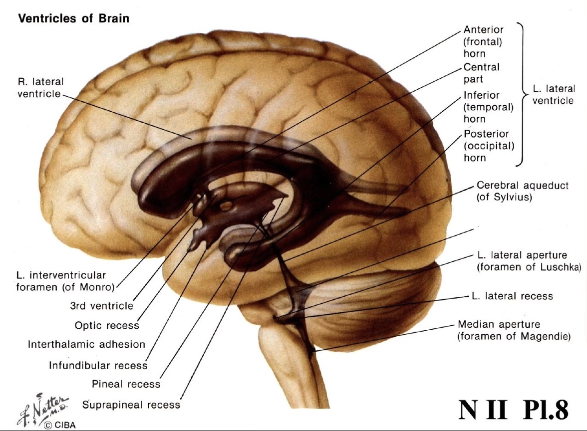
anterior (frontal) horn of left lateral ventricle

name the structure
inferior (temporal) horn of left lateral ventricle

name the structure
posterior (occipital) horn of left lateral ventricle

name the structure
cerebral aquiduct
name the structure
4th ventricle

name the structure
3rd ventricle

name the structure
telencephalon

name the structure
diencephalon

name the structure
retina

name the structure
mesencephalon (midbrain)

name the structure
metencephalon (pons and cerebellum)

name the structure
myelencephalon (medulla)
name the structure
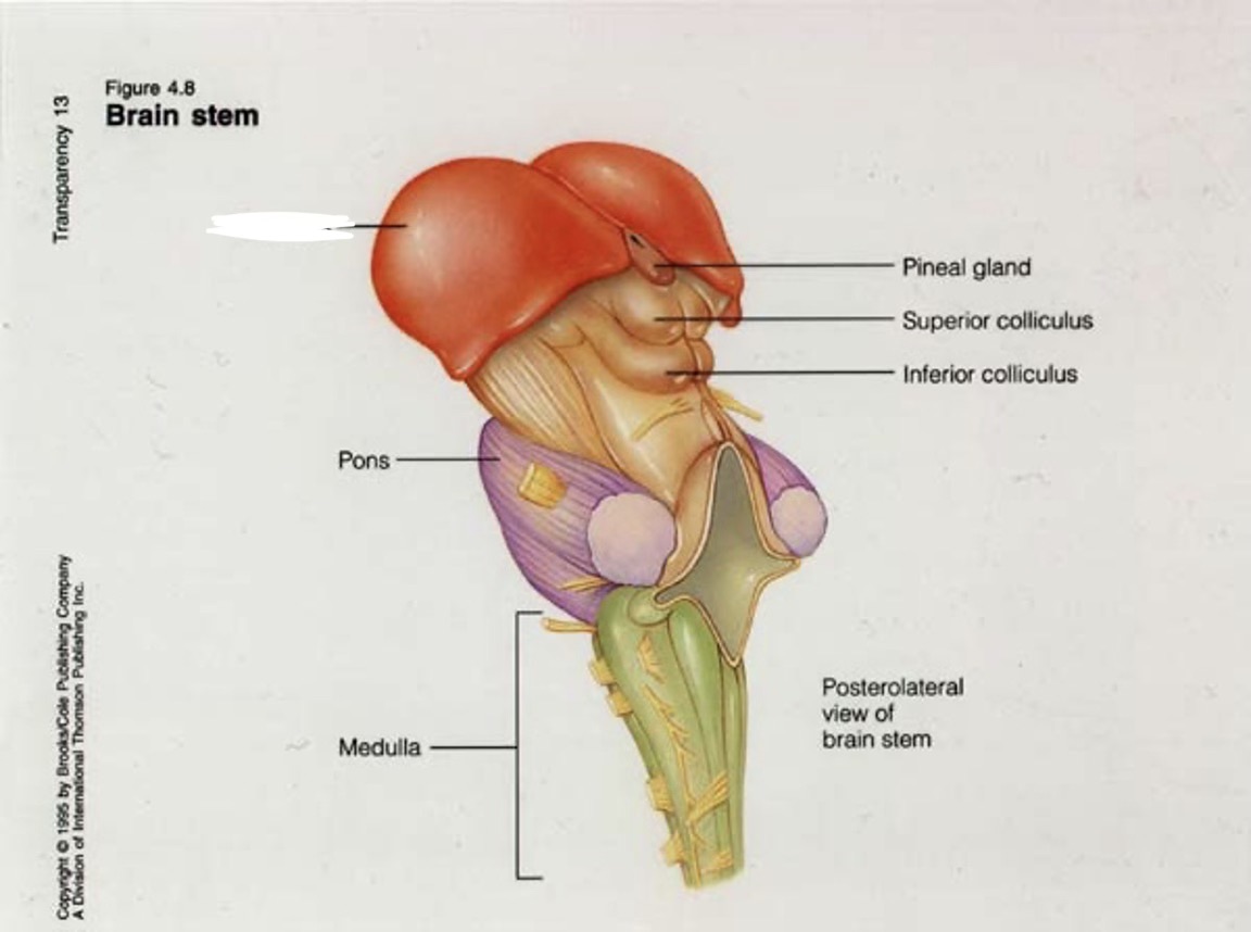
thalumus

name the structure
pineal gland

name the structure
superior colliculus

name the structure
inferior colliculus

name the structure
pons

name the structure
medulla
name the structure
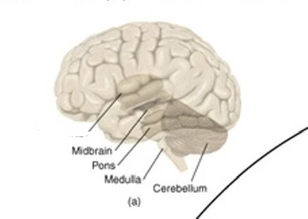
thalamus

name the structure
midbrain

name the structure
pons

name the structure
medulla

name the structure
cerebellum

name the structure
cerebral peduncles

name the structure in the MIDBRAIN
superior colliculus

name the structure in the MIDBRAIN
periaqueductal gray matter

name the structure in the MIDBRAIN
cerebral aqueduct

name the structure in the MIDBRAIN
reticular formation

name the structure in the MIDBRAIN
red nucleus

name the structure in the MIDBRAIN
substantial nigra

name the structure
medulla

name the structure
cerebellum

name the structure
pons

name the structure
3rd ventricle

name the structure
4th ventricle

name the lobe
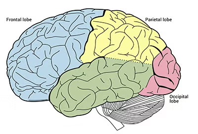
frontal lobe

name the lobe
parietal lobe
name the lobe
temporal lobe

name the lobe
occipital lobe

name the structure
hypothalamus

name the structure
bands of myelinated axons

name the structure
right thalamus

name the structure
cerebellum

name the structure(s) of the BASAL GANGLIA
caudate nucleus and putamen

name the structure(s) of the BASAL GANGLIA
tail of caudate nucleus

name the structure(s) of the BASAL GANGLIA
globus pallidus

name the structure of the BASAL GANGLIA
caudate nucleus (body)

name the structure of the BASAL GANGLIA
globus pallidus (external segment)

name the structure of the BASAL GANGLIA
globus pallidus (internal segment)

name the structure of the BASAL GANGLIA
caudate nucleus (tail)

name the structure of the BASAL GANGLIA
putamen

name the structure of the LIMBIC SYSTEM (error monitering)

name the structure of the LIMBIC SYSTEM (myelinated axons carrying info from left to right hemisphere)
corpus callosum

name the structure of the LIMBIC SYSTEM (new memories)
fornix

name the structure of the LIMBIC SYSTEM (new memories)
mammillary body

name the structure of the LIMBIC SYSTEM (processing fear + negative affect)
amygdala

name the structure of the LIMBIC SYSTEM (consolidating new memory into long term memory)
hippocampus

name the structure (separates precentral gyrus containing primary motor cortex from postcentral gyrus containing somatosensory cortex)
central sulcus

name the structure
lateral fissure

name the structure in the frontal lobe
primary motor cortex

name the structure in the parietal lobe
primary somatosensory cortex

name the structure in the occipital lobe
primary visual cortex

name the structure in the temporal lobe
auditory association cortex

name the structure in the temporal lobe
visual association cortex

name the structure (formed by retinal ganglion cells)
optic nerve

name the structure (where optic nerves meet)
optic chiasm

name the structure (wraps around dyencephalon (hypothalamus and thalamus)
optic tract

name the structure (part of what optic tract wraps around)
thalamus

name the structure
midbrain

name the structure (10% projects here for retinal reflexes. called the retino tectal projection)
superior colliculus

name the structure in the Macack brain
dorsal stream

name the structure in the Macack brain
ventral stream

name the structure in the Macack brain
MST

name the structure in the Macack brain
MT (V5)
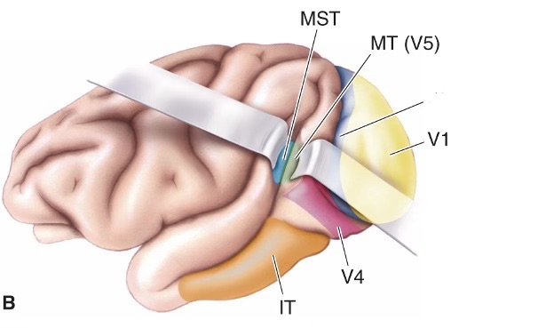
name the structure in the Macack brain
V2

name the structure in the Macack brain
V1

name the structure in the Macack brain
V4

name the structure in the Macack brain
IT

name the structure in the human brain
V3A

name the structure in the human brain
V3

name the structure in the human brain
V1

name the structure in the human brain
V2

name the structure in the human brain
V4

name the structure in the human brain
face and object recognition areas

name the structure in the human brain
V5 (MT)

name the structure in the macaque and human brains
area 17 (V1)

name the structure in the human brain
calcarine fissure