brain and cn

0.0(0)
Studied by 0 people
call kaiCall Kai
learnLearn
examPractice Test
spaced repetitionSpaced Repetition
heart puzzleMatch
flashcardsFlashcards
GameKnowt Play
Card Sorting

1/77

encourage image

There's no tags or description

Looks like no tags are added yet.

78 Terms

1
New cards
What are the different parts of the brain?

Cerebrum

Diencephalon

Brainstem

Cerebellum

2
New cards
What components make up the Diencephalon?

Epithalamus

Thalamus

Hypothalamus

Neurohypophysis

3
New cards

What components make up the Brainstem?

Midbrain

Pons

Medulla Oblongata

4
New cards
What is rostral vs caudal?

towards the nose vs. towards the tail

5
New cards

How is the gray vs white matter composed within the brain and what is within these areas?

• Gray matter: contains little myelin, but mostly somas, dendrites, and proximal portions of axons; location where information is processed (synapses)

– Cerebral cortex: the outer layer of the brain

– Nuclei: deeper portions of the brain surrounded by white matter

White: highly myelinated axons called tracts, carrying information to different parts of the CNS that is deep to the cerebral cortex

6
New cards

What structures help support and protect the brain?

Cranium

Meninges

Cerebrospinal Fluid

Blood-brain barrier

7
New cards

What are meninges?

fibrous connective tissue that surrounds and protects the CNS
(include dura mater, arachnoid mater, pia mater)

<p><span style="background-color: transparent;">fibrous connective tissue that surrounds and protects the CNS</span><br><span style="background-color: transparent;">(include dura mater, arachnoid mater, pia mater)</span></p>
8
New cards
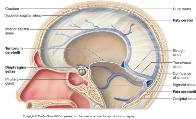
What are dural sinuses? Name one dural sinus.

Veins that pools blood to be emptied out from the brain into the internal jugular veins

Superior sagittal sinus
Inferior sagittal sinus
Straight sinus
Transverse sinus
Sigmoid sinuses
Cavernous sinuses

<p><span style="background-color: transparent;">Veins that pools blood to be emptied out from the brain into the&nbsp;internal jugular veins</span></p><p>Superior sagittal sinus<br>Inferior sagittal sinus<br>Straight sinus<br>Transverse sinus<br>Sigmoid sinuses<br>Cavernous sinuses</p>
9
New cards
What is the falx cerebri?

separates the right and left hemisphere within the longitudinal fissure
<p><span style="background-color: transparent;">separates the right and left hemisphere within the&nbsp;longitudinal fissure</span></p>
10
New cards
What are arachnoid villi and what does it do?

extensions of the arachnoid mater that passes CSF from  subarachnoid space into the dural venous sinuses

<p><span style="background-color: transparent;">extensions of the arachnoid mater that passes CSF from&nbsp; subarachnoid space into the dural venous sinuses</span></p>
11
New cards
What are the different ventricles of the brain and the order that CSF flow through them?

Right and left lateral ventricles

Third ventricle

Cerebral aqueduct

Fourth ventricle

<p>Right and left lateral ventricles </p><p>Third ventricle </p><p>Cerebral aqueduct </p><p>Fourth ventricle</p>
12
New cards
What is the central canal?

Space that runs from brain through spine to dispose of CSF.

13
New cards
What is the CSF’s function?

Buoyancy: reducing the weight of the brain

Protection: prevents brain from hitting cranium

Chemical stability: regulates chemical environment

14
New cards

How is CSF produced?

• Choroid plexus: within each ventricle has ependymal cells with capillaries to produce 30% CSF (other 30% from the lining of ventricles; 40% at subarachnoid space)

• Ependymal cells: produces CSF and also line the ventricles and central canal causing  CSF flow

• CSF from subarachnoid space goes to venous sinuses through arachnoid villi

– Superior sagittal sinus: dural venous sinus that is above the falx cerebri

15
New cards
How does CSF flow?

CSF from subarachnoid space goes to venous sinuses through arachnoid villi

16
New cards

What is the blood brain barrier?

protects brain from toxins from blood, allows water, glucose and lipid soluble substances to pass through

formed by Astrocytes

17
New cards
What are the structures that make up the Cerebrum?

Cerebral Hemispheres

Corpus Callosum

Cerebral Cortex

Gyri Fissure

Sulci Fissure

Longitudinal Fissure

Transversal Fissure

18
New cards
What is the corpus callosum?

nerve fibers (commissure tracts) that connects the right and left hemisphere

<p><span style="background-color: transparent;">nerve fibers (commissure tracts) that connects the right and&nbsp;left hemisphere</span></p>
19
New cards
What is the cerebral cortex and what type of matter is it made of?

The outer gray matter of the brain

20
New cards
What are gyri vs sulci vs fissures?

Gyri: folds to increase the surface area

Sulci: shallow grooves

Fissures: deeper grooves

<p><span style="background-color: transparent;">Gyri: folds to increase the surface area</span></p><p><span style="background-color: transparent;">Sulci: shallow grooves</span></p><p><span style="background-color: transparent;">Fissures: deeper grooves</span></p>
21
New cards
What is the longitudinal vs transverse fissure?

Longitudinal fissure: deep groove that separates the right and left cerebral hemisphere

Transverse fissure: separates cerebrum and cerebellum

<p><span style="background-color: transparent;">Longitudinal fissure: deep groove that separates the right and left cerebral hemisphere</span></p><p><span style="background-color: transparent;">Transverse fissure: separates cerebrum and cerebellum</span></p>
22
New cards
What is made of gray matter in the brain?

Cerebral cortex

Basal nuclei

Limbic system

23
New cards
Where is the white matter located at?

Inside the brain, inside the cerebral cortex

24
New cards
What are the cerebral lobes?

Frontal lobe

Parietal lobe

Occipital lobe

Temporal lobe

Insula

25
New cards

What is the function of the Frontal lobe? (243)

principal area for

  • motor functions

    • voluntary motor function

    • planning

  • mental functions

    • motivation

    • foresight

    • memory

    • mood

  • social functions

    • emotion

    • social judgment

    • aggression


26
New cards

What is the function of the Temporal Lobe?

principal area for

  • hearing

  • smell

    Also contains area for

  • learning

  • memory

  • some aspects of vision and emotion


27
New cards

What is the function of the Parietal Lobe?

principal area that

  • receives and interpreting general senses

  • taste

  • some visual processing


28
New cards

What is the function of the Occipital Lobe?

principal visual center of the brain

29
New cards

What are the sulci that divide the Frontal and Parietal lobes?

Central sulcus

30
New cards
What is motor control?

voluntary motor functions

31
New cards
What are the general senses?

Touch

Nociception

Pressure

Vibration

Temperature

Proprioception

32
New cards
What is nociception?

pain sense

33
New cards
What is proprioception?

positional sense

34
New cards
What are the special senses?

Vision

Hearing

Equilibrium

Taste

Smell

35
New cards
What are association areas?

integrate and store information

36
New cards

Which areas of the brain are for motor control?

Premotor Cortex / Somatic Motor Association

Primary Motor Cortex / Somatic Motor Area

<p><span style="background-color: transparent;">Premotor Cortex / Somatic Motor Association</span></p><p><span style="background-color: transparent;">Primary Motor Cortex / Somatic Motor Area</span></p>
37
New cards

What is the premotor cortex for?

rostral of precentral gyrus and where we

  • plan our actions

  • regulates eye movement for reading


<p><span style="background-color: transparent;">rostral of precentral gyrus and where we </span></p><ul><li><p><span style="background-color: transparent;">plan our actions </span></p></li><li><p><span style="background-color: transparent;">regulates eye movement for reading</span></p></li></ul><p></p>
38
New cards

What is the primary motor cortex?

precentral gyrus of frontal lobe that

  • sends signals to brainstem and spinal cord for skeletal muscle contraction on the contralateral side


<p><span style="background-color: transparent;">precentral gyrus of frontal lobe that </span></p><ul><li><p><span style="background-color: transparent;">sends signals to brainstem and spinal cord for skeletal muscle contraction on the </span><span style="background-color: transparent; color: rgb(255, 0, 0);"><strong>contralateral </strong></span><span style="background-color: transparent;">side</span></p></li></ul><p></p>
39
New cards

What does the basal nuclei and cerebellum do?

Refinement of motor control

<p>Refinement of motor control</p>
40
New cards

Which areas of the brain are for general senses?

Primary somatosensory cortex, Somatosensory association area

<p><span style="background-color: transparent;">Primary somatosensory cortex, Somatosensory association area</span></p>
41
New cards

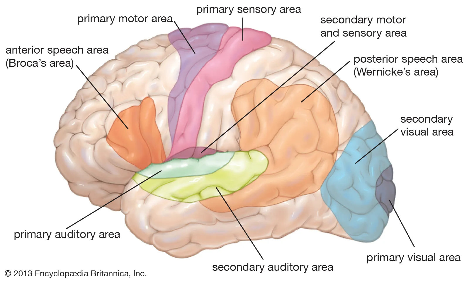
What is the primary somatosensory cortex?

postcentral gyrus that detect general senses on contralateral side
<p><span style="background-color: transparent;">postcentral gyrus that detect general senses on contralateral side</span></p>
42
New cards

What is the somatosensory association area?

caudal of postcentral gyrus of the parietal lobe; makes sense of sensation

<p><span style="background-color: transparent;">caudal of postcentral gyrus of the parietal lobe; makes sense of sensation</span></p>
43
New cards

Which part of the brain is for vision?

Primary visual cortex

Visual association area

<p><span style="background-color: transparent;">Primary visual cortex</span></p><p><span style="background-color: transparent;">Visual association area</span></p>
44
New cards

Which lobe is primarily for vision?

Occipital

<p>Occipital</p>
45
New cards

What is the visual association area?

anterior of occipital,
posterior part of the parietal lobe,
inferior temporal lobe

recognize faces and familiar objects

<p>anterior of occipital, <br><span style="background-color: transparent;">posterior part of the parietal lobe,</span><br><span style="background-color: transparent;">inferior temporal lobe  </span></p><p><span style="background-color: transparent;">recognize faces and familiar objects</span></p>
46
New cards

Which part of the brain is for hearing?

Primary auditory cortex

Auditory association area

<p><span style="background-color: transparent;">Primary auditory cortex</span></p><p><span style="background-color: transparent;">Auditory association area</span></p>
47
New cards

Which lobe is primarily for hearing?

Temporal lobe

<p>Temporal lobe</p>
48
New cards

What is the auditory association area?

inferior to temporal lobe to recognize spoken words, familiar music and voice

<p><span style="background-color: transparent;">inferior to temporal lobe to recognize spoken words, familiar music and voice</span></p>
49
New cards

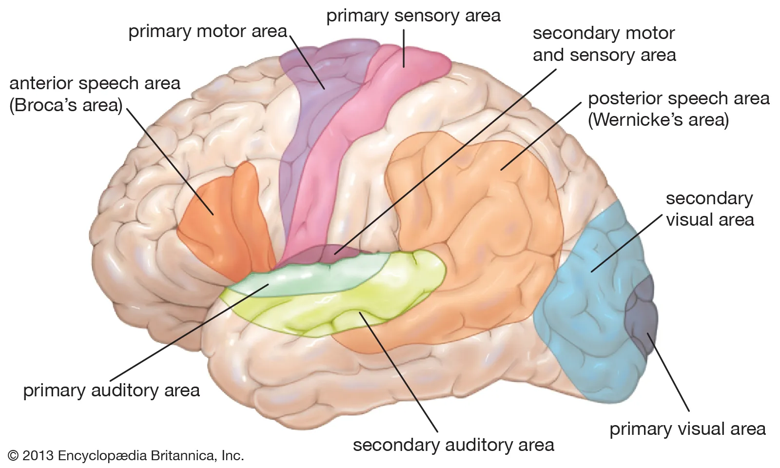
Which part of the brain is for equilibrium?

Cerebellum and brainstem nuclei
<p><span style="background-color: transparent;">Cerebellum and brainstem nuclei</span></p>
50
New cards

Which part of the brain is for taste?

inferior part of the postcentral gyrus of parietal lobe and anterior region of insula
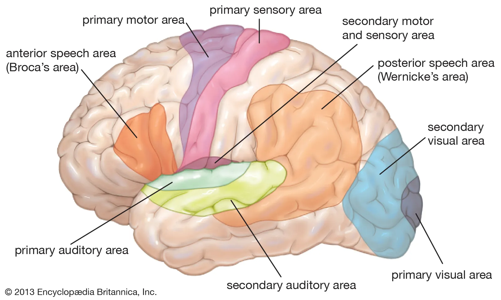
<p><span style="background-color: transparent;">inferior part of the <strong>postcentral gyrus </strong>of parietal lobe and <strong>anterior region of insula</strong></span></p>
51
New cards

Which lobe is primarily for taste?

Insula
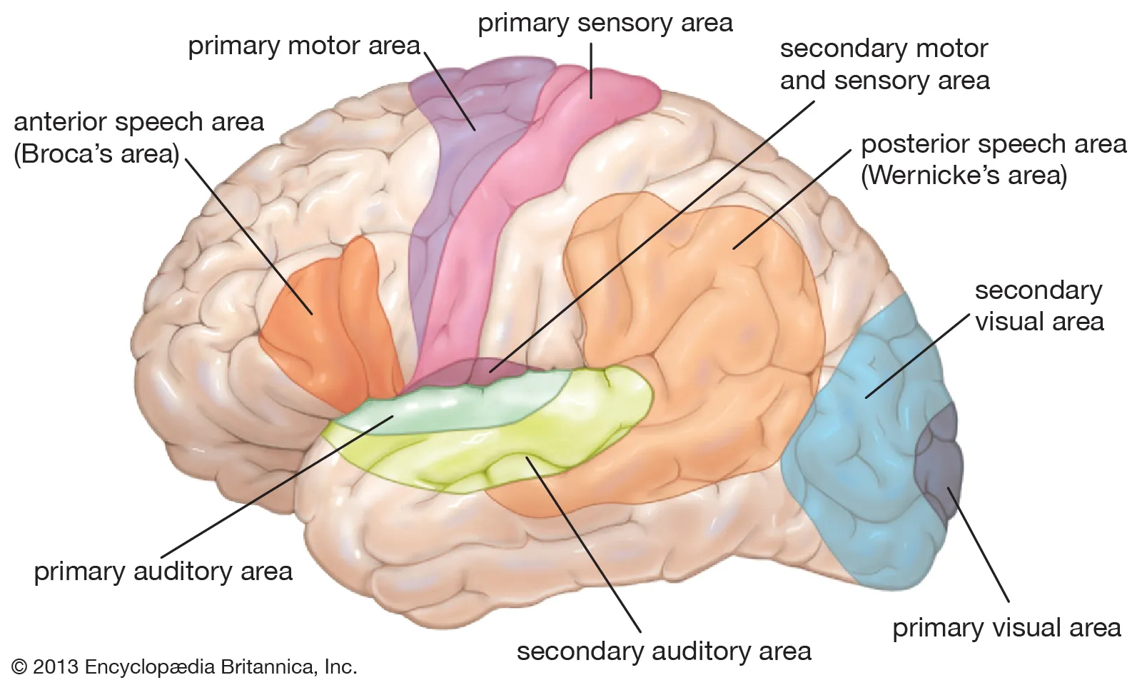
<p>Insula</p>
52
New cards

Which part of the brain is for smell?

medial surface of temporal lobe and inferior surface of frontal lobe

<p><span style="background-color: transparent;">medial surface of temporal lobe and inferior surface of frontal lobe</span></p>
53
New cards

What lobe is primarily for smell?

Temporal Lobe

<p>Temporal Lobe</p>
54
New cards

Functions of the Wernicke area and Broca area?

recognizes spoken and written language

inferolateral part of frontal lobe, which transmit signals to the precentral gyrus to eventually cause muscles to help us speak

<p><span style="background-color: transparent;">recognizes spoken and written language</span></p><p><span style="background-color: transparent;">inferolateral part of frontal lobe, which transmit signals to the precentral gyrus to eventually cause muscles to help us speak</span></p>
55
New cards

Which hemisphere are the Wernicke and Broca area usually found?

left cerebral hemisphere

<p>left cerebral hemisphere</p>
56
New cards

What is the function of the opposing hemisphere?

emotional aspect of language

<p><span style="background-color: transparent;">emotional aspect of language</span></p>
57
New cards

What is the function of the basal nuclei?

lateral to the thalami contain many brain centers for motor control

58
New cards

What is the function of the limbic system?

  • center of emotion and learning

  • center for gratification and aversion


59
New cards

What are the functions of the components in the limbic system?

Hippocampus: memory role

Amygdala / Amygdaloid Body: emotion and mood role, especially fear

Olfactory bulbs, tracts, and cortex: odors producing emotions/memories

Mammillary bodies: nuclei in diencephalon that interconnect other parts of the limbic system, may have a memory role

<p><strong>Hippocampus</strong>: <span style="background-color: transparent;">memory role</span></p><p><strong>Amygdala / Amygdaloid Body</strong>: <span style="background-color: transparent;">emotion and mood role, especially fear</span></p><p><strong>Olfactory bulbs, tracts, and cortex</strong>: <span style="background-color: transparent;">odors producing emotions/memories</span></p><p><span style="background-color: transparent;"><strong>Mammillary </strong></span><strong>bodies</strong>: nuclei in diencephalon that interconnect other parts of the limbic system, may have a memory role</p>
60
New cards

What are the functions of the components of the diencephalon? (- thalamus)

Epithalamus: hosts pineal gland, endocrine gland that secretes melatonin which regulates circadian rhythm

Thalamus: processes most information (except olfaction) and relay them to cerebral cortex; also for motor control and memory

Hypothalamus

Neurohypophysis: stores hormones

61
New cards
What is the function of the hypothalamus?

Hormone secretion
Autonomic effects
Thermoregulation
Food and water intake
Sleep and circadian rhythms
Memory
Emotional

62
New cards

What structure is also related to the hypothalamus?

Infundibulum / Neurohypophysis

63
New cards
What is the infundibulum?

stalk that connects the hypothalamus to pituitary gland

64
New cards
What is the neurohypophysis?

Posterior pituitary gland; stores hormones made by the hypothalamus

65
New cards
What hormones does the posterior pituitary gland secrete?

oxytocin and ADH

66
New cards
Where do the hormones from the posterior pituitary come form?

Hypothalamus

67
New cards
Why isn’t the anterior pituitary part of the neurohypophysis?

anterior pituitary gland is not derived from nervous tissue

68
New cards

What is the function of the midbrain?

connects hindbrain to forebrain, connects third and fourth ventricles

<p><span style="background-color: transparent;">connects hindbrain to forebrain, connects third and fourth ventricles</span></p>
69
New cards

What is the function of the pons?

Transmit signals from forebrain to cerebellum

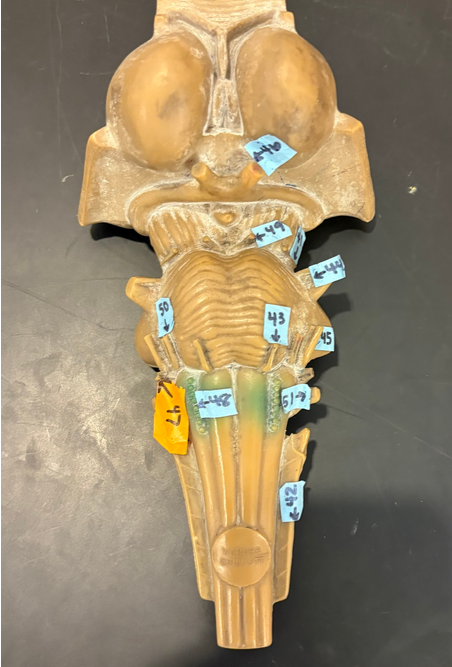
  • Trigeminal Nerve (V) (44)

  • Abducens Nerve (VI) (43)

  • Facial Nerve (VII) (50)

Autonomic respiratory centers

<p>Transmit signals from forebrain to cerebellum</p><ul><li><p>Trigeminal Nerve (V) (44)</p></li><li><p>Abducens Nerve (VI) (43)</p></li><li><p>Facial Nerve (VII) (50)</p></li></ul><p>Autonomic respiratory centers</p>
70
New cards

What is the function of the medulla oblongata?

Manages heart, circulation and breathing

Pyramid body (swaps sides of control)

Cardiac center

Vasomotor center

Respiratory centers

  • Vestibulocochlear Nerve (VIII) (45)

  • Glossopharyngeal Nerve (IX) (47)

  • Vagus Nerve (X) (51)

  • Accessory Nerve (XI) (42)

  • Hypoglossal Nerve (XII) (Hypoglossal)


<p>Manages heart, circulation and breathing</p><p>Pyramid body (swaps sides of control)</p><p>Cardiac center</p><p>Vasomotor center</p><p>Respiratory centers</p><ul><li><p><span style="background-color: transparent;">Vestibulocochlear Nerve (VIII) (45)</span></p></li><li><p><span style="background-color: transparent;">Glossopharyngeal Nerve (IX) (47)</span></p></li><li><p><span style="background-color: transparent;">Vagus Nerve (X) (51)</span></p></li><li><p><span style="background-color: transparent;">Accessory Nerve (XI) (42)</span></p></li><li><p><span style="background-color: transparent;">Hypoglossal Nerve (XII) (Hypoglossal)</span></p></li></ul><p></p>
71
New cards
What structures form the corpora quadrigemina?

Superior colliculi

Inferior colliculi

72
New cards

What are the functions of the colliculi?

Superior colliculi:

  • for visual attention

  • tracking moving objects

  • reflexes (blinking, focusing, pupillary dilation and constriction)

Inferior colliculi:

  • receive signal from inner ear and relay to other parts of brain like the thalamus

  • reflexive turning to sound


<p><span style="background-color: transparent;"><strong>Superior colliculi</strong>: </span></p><ul><li><p><span style="background-color: transparent;">for visual attention</span></p></li><li><p><span style="background-color: transparent;">tracking moving objects</span></p></li><li><p><span style="background-color: transparent;">reflexes (blinking, focusing, pupillary dilation and constriction)</span></p></li></ul><p><span style="background-color: transparent;"><strong>Inferior colliculi</strong>: </span></p><ul><li><p><span style="background-color: transparent;">receive signal from inner ear and relay to other parts of brain like the thalamus </span></p></li><li><p><span style="background-color: transparent;">reflexive turning to sound</span></p></li></ul><p></p>
73
New cards
What space divides the cerebrum from the cerebellum?

Transverse Fissure

74
New cards

What are the different structures of the cerebellum?

Cerebellar hemispheres

Vermis: motor control, balance, and emotional regulation, connects cerebellum hemispheres

Folia: small folds on cerebellum, increase surface area

Arbor vitae: white branches on the cerebellum, Signal Conduit, Connection and Communication with brainstem


75
New cards

Are cranial nerves part of the CNS?

No!

76
New cards

List the Cranial Special Sensory Nerves

special sensory nerves

CN1 olfactory: smell

CN2 optic: vision

CN8 vestibulocochlear: hearing and equilibrium

77
New cards

List the Cranial Motor Nerves

motor nerves

CN3 oculomotor: eyeball and eyelid movement, accomidation

CN4 trochlear: superior eyeball movement

CN6 abducens: lateral eye movement

CN11 accessory: head and shoulder rotation

CN12: hypoglossal: tongue movement

78
New cards

List the Cranial Multiple Use Nerves

multiple use nerves

CN5 trigeminal: face and chewing

CN7 facial: face expressions and 2/3rd of the tongue taste, tears

CN9 glossopharyngeal taste 1/3rd of tongue, swallowing and salivation

CN10 vagus: heart rate reduction and digestion