Anatomy 4 (Muscles)
 Call Kai
 Learn
 Practice Test
 Spaced Repetition
 Match
 Flashcards
 Knowt Play
1/169

There's no tags or description
Looks like no tags are added yet.
170 Terms
Muscle Characteristics
Contractile
Excitable
Elastic
Extensible
Muscle Functions
Movement
Stability
Storage
Guard entrance/exit
Thermogenesis
Sarcolemma
Muscle cell membrane
Sarcoplasm
Muscle cell cytoplasm
Epimysium
Fibrous connective outer layer holding the entire muscle together
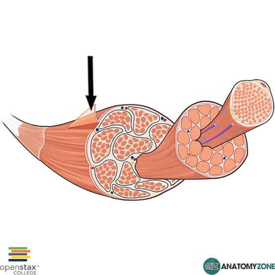
Perimysium
Areolar layer that surrounds bundles of muscle fibers

Endomysium
Areolar layer surrounding each muscle fiber

Sarcolemma
Plasma membrane of individual muscle fibers
Myofibrils
Rod-shaped organ in muscle cells made of sarcomeres that makes muscle contract

Sarcoplasmic Reticulum
Modified ER in muscle cells of fluid-filled sacs that encircle each myofibril
Stores and releases Ca
Contain terminal cisterns
Terminal Cisterns
Part of the SR and release Ca
Traid
T-Tubules
Small invagination of the sarcolemma filled with fluid that helps move electrical signals down the fiber
Triad
Triad
2 terminal cisterns and 1 T-Tubule
Sarcomere
Functional unit of myofibrils
Thick and thin filaments between z disks

I Band
Thin filaments only
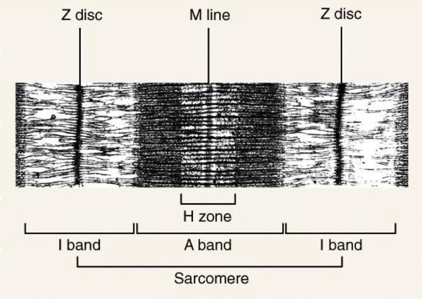
A Band
Thick and thin filaments

H Zone
Thick filaments only

Contractile Muscle Proteins
Actin (thin) and myosin (thick)
Regulatory Muscle Proteins
Troponin and tropomyosin
Act as an on/off switch for muscle contraction by controlling contact between actin and myosin
Structural Muscle Proteins
Titin
Myomesin
Nebulin
Dustrophin
Titin
Coiled between Z-lines for elasticity

Myomesin
Found in the M-line to cross-link myosin

Nebulin
Stiffens/stabilizes actin

Dystrophin
Aligns actin with sarcolema
Muscular Dystrophy
Flexion
Decrease angles between bones
Extension
Increase angles between bones
Lateral Flexion
Flexion in the frontal plane
Abduction
Away from midline
Adduction
Towards midline
Supination
Turns palms anteriorally
Pronation
Turns palms posteriorally
Inversion
Turning sole medially
Eversion
Turning sole laterally
Dorsiflexion
Top of the foot moves towards tibia
Plantar Flexion
Sole of the foot moves posteriorly
Protraction
Forward from horizontal plane
Retraction
Backward from horizontal plane
Origin
Attachment to a stable, less movable bone
Insertion
Moveable end of the muscle
Parallel Arrangment
Fibers run parallel along axis
Converging Arrangment
Fibers come to together on one end
Unipennate Arrangment
Attach to 1 side of a tendon
Bipennate Arrangment
Attach to 2 sides of tendon
Multipennate Arrangment
Attach to central tendon on all sides
Circular Arrangement
Rings of fibers
Frontalis
Creases forehead
O: Aponeuosis
I: Skin
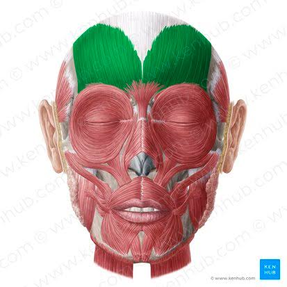
Aponeurosis
Tendon sheet
Orbicularis Oculi
Around the eyes to squint

Orbicularis Oris
Around the mouth to purse lips

Levator Palpebrae
On eyelids to open eyes

Zygomaticus Major and Minor
Smile
O: Zygomatic arch
I: Skin

Platysma
Tightens skin
O: Fascia on clavicle
I: Mandible

Depressor Anguli Oris
Side of mouth going down

Nasalis
On nose

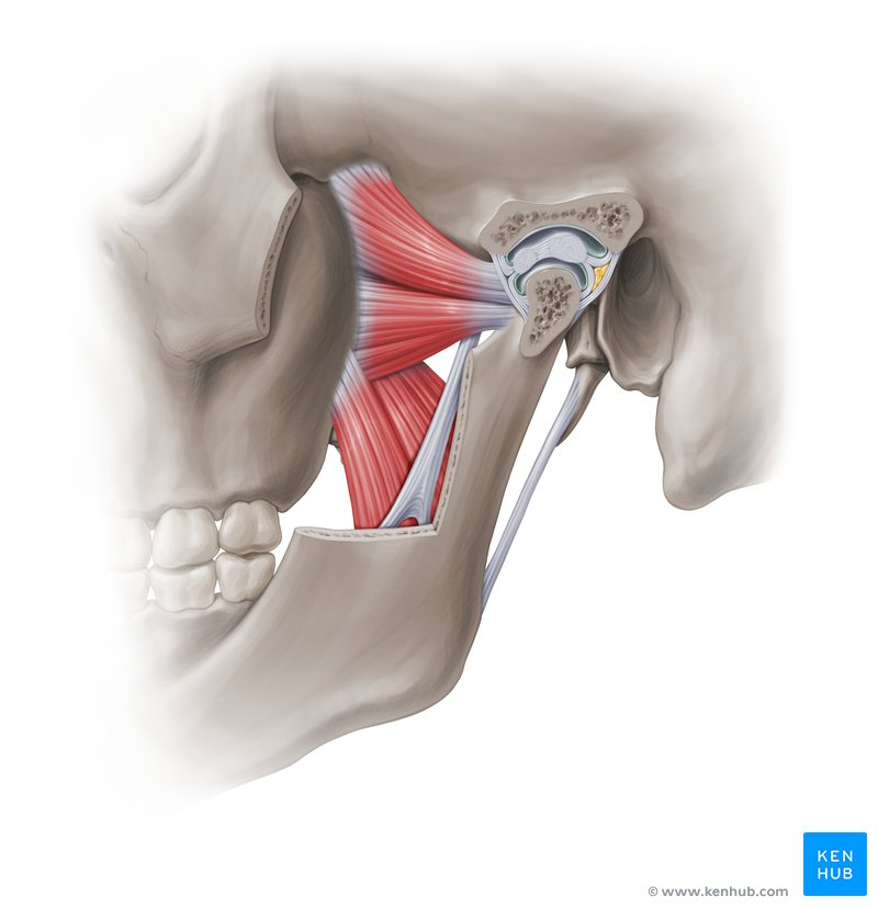
Masseter
Closes jaw
O: Zygomatic arch
I: Mandibular ramus

Buccinator
In cheeks

Mentalis
On chin

Temporalis
Closes the jaw
O: Temporal fossa
I: Coronoid process

Pterygoids
Medial: On bottom
Lateral: On top, opens the jaw
Mylohyoid
Opens the jaw
O: Mylohyoid line of mandible
I: Hyoid

Rectus Muscles
4 side of the eyes
Medial
Lateral
Superior
Inferior

Superior Oblique
Move eye down and out

Inferior Oblique
Move eye up and out

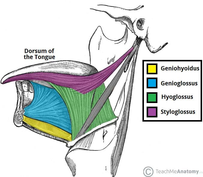
Genioglossus
Anterior/outwards and rolls tongue
O: Mandible
I: Tongue

Styloglossus
Tongue up and back
O: Styloid process
I: Tongue
Palatoglossus
Tongue up and back
O: Soft palate
I: Tongue

Hyoglossus
Tongue sides down
O: Hyoid
I: Tongue
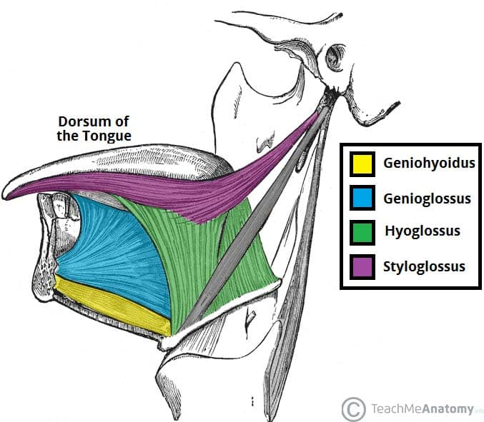
Geniohyoid
Tongue muscle that depresses mandible
O: Mandible
I: Hyoid

Diagastric
Moves mandible down
Mastoid to chin

Stylohyoid
Stabilize hyoid for tongue movement
O: Styloid
I: Hyoid

Geniohyoid
Mandible down
O: Mandible
I: Hyoid

Omohyoid
Stabilize hyoid for tongue movement
O: Hyoid
I: Clavicle

Sternohyoid
Stabilize hyoid for tongue movement
O: Sternum
I: Hyoid

Sternothyroid
Stabilize hyoid for tongue movement
O: Sternum
I: Thyroid

Thyrohyoid
Stabilize hyoid for tongue movement
O: Thyroid
I: Hyoid

Sternocleidomastoid
Head movement
O: Manubrium and clavicle
I: Mastoid

Other muscles of head movement
Semispinalis capitis
Splenius capitis
Longissimus capitis
3 scalenes
Rectus Sheath
Tendon sheath over abs

Linea Alba
Midline between abs

Linea Semilunaris
Lateral edge of abs

Inguinal Ligament
ASIS to pubic tubercle; marked by V line

External Oblique
Rotation, trunk + lat flex
O: Lower ribs
I: Linea alba
Runs diagonally downward

Internal Oblique
Trunk + lat flex
O: Lumbodorsal fascia + iliac crest
I: Linea alba
Run upward and inward perpendicular to external obliques

Transversus Abdominus
Tension to support the spine and pelvis
O: Lumbodorsal fascia iliac crest, lower ribs
I: Linea alba
Runs horizontally

Tranversalis Fascia
Under transversus abdominus
Rectus Abdominus
Trunk flexion and forced exhalation
O: Pubic crest and symphysis
I: Xiphoid and lower ribs

Quadratus Lumborum
Ext, lat flex
O: Iliac crest
I: Lower ribs and lumbar vert

Inguinal Canal
Canal formed by 3 arches of muscle (2 obliques and transversus abdominus)
Holds nerves, tendons, spermatic cord
Natural Respiration Muscles
Ext and Int Intercostals
Transversus Thoracis
Rectus Abdominus
Diaphragm
External Intercostal
Elevate rib below for inhale
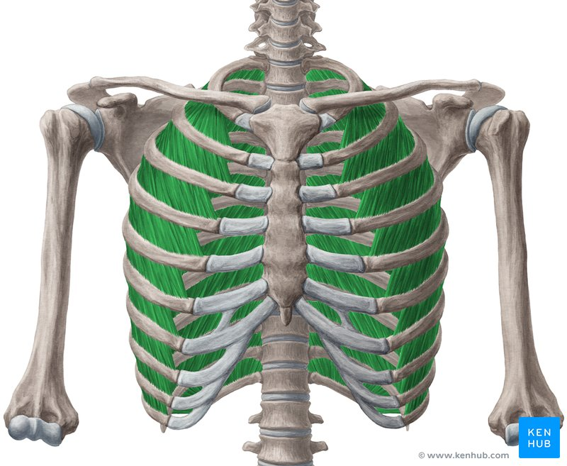
Internal Intercostals
Depress rib above during exhale

Transversus Thoracis
Ribs to sternum

Diaphragm
O: Everywhere (ribs and L3)
I: Central tendon and connect with pleura and pericardium

Diaphragm Landmarks + Vertebrae
IVC: T8
Esophagus: T10
Aorta: T12
Accessory Respiration Muscles
Sternocleidomastoid
Scalenes
Pec Minor
Internal Intercostals
Rectus Abdominus
Superficial Back Muscles
Trap
Lat
Rhomboid Maj/Min
Levator Scapulae
Deep Back Muscles
Erector Spinae Group
Multifidus
Quadtratus Lumborum
Erector Spinae Group
Along the vertebral column for ext, lat flex, rot

Multifidus
Spinal extension and rotation
O: Transverse processes
I: Spinous processes

Subclavius
Elevates 1st rib, depresses 2nd rib
O: 1st rib
I: Subclavian groove
