Objective 16
 Call Kai
 Learn
 Practice Test
 Spaced Repetition
 Match
 Flashcards
 Knowt Play
1/35

There's no tags or description
Looks like no tags are added yet.
36 Terms
M0
AML with minimal differentiation
M1
AML without maturation
M2
AML with maturation
M3
Acute promyelocytic leukemia
M4
Acute Myelomonocytic leukemia
M5
Acute monoblastic and monocytic leukemia
M6
Pure erythroid leukemia
M7
Acute megakaryoblastic Leukemia
What are the FAB classifications for Acute Myeloid Leukaemia?
M0: AML with minimal differentiation
M1: AML without maturation
M2: AML with maturation
M3: acute promyelocytic leukaemia
M4: Acute myelomonocytic leukemia
M5: Acute monoblastic and monocytic leukaemia
M6: Pure erythroid leukaemia
M7: Acute megakaryoblastic leukemia
What is the WHO classification for Myeloid Leukemia?
AML with recurrent genetic abnormalities
AML with meylodysplasia-related changes
Therapy-related myeloid neoplasms
AML-NOS
Myeloid sarcoma
Myeloid proliferation related to Down Syndrome
Acute Leukemias of ambiguous lineage

AML with Recurrent Genetic Abnormalities
t(8;21) (q22;q22);RUNX1/RUNX1T1 (fusion)
5% of all AML cases
children and young adults
dysplastic granular cytoplasm
Auer rods
Pseudo Pelger-Huet cells
Hypogranulation
Eosinophilia
Monosomy 7
> 10% blasts
FAB M2

AML with Recurrent Genetic Abnormalities
inv(16)(p13.1;q22.1) or t(16;16)(p13.1;q22); CBFB-MYH11
accounts for 5-8% of all AML cases
Myeloblasts, monoblasts, AMML, promyelocytes
eosinophilia with dysplastic changes
extramedullary disease
CNS
> 10% blasts
FAB 4

AML with Recurrent Genetic Abnormalities
AML with t(15;17)(q22;q12); PML-RARA
known as acute promyelocytic leukemia (FAB M3)
5-10% of AML cases
differentiation block at promyelocytic stage
Dx: detection of t(15;17) mutation
auer rods, DIC
microgranular variant - 30-40% APL
> 10% blasts
treatment
ATRA
Arsenic trioxide
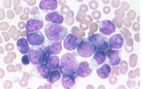
AML with Recurrent Genetic Abnormalities
t(9;11)(p22;q23);MLLT3-MLL
6% of AML cases
monoblasts
immature monocytes
large blasts, abundant cytoplasm
> 10% blasts
gingival, skin involvement
DIC

AML with Recurrent Genetic Abnormalities
t(6;9)(p23;q34);DEK-NUP214 rearrangement
> 10% peripheral blood or BM blasts with/without monocytic features
associated with basophilia / multilineage dysplasia
poor prognosis
low WBC count
Describe the AMLs with Recurrent Genetic Abnormalities
7 types involving a translocation or inversion
variable frequency
DIC with t(15,17) , less frequent with t(9;11)
favorable prognosis in:
AML t(8;21)
Inv(16)
t(15;17)
t(9;11)

Describe AML with Myelodysplasia related changes
in older adults (poor prognosis)
> 20% blasts w’ multilineage dysplasia
history of: MDS, MPN, MDS associated abnormality
neutrophil hypergranulation
Pseudo Pelger-Huet
7q-, 5q-
dysplastic megakaryocytes
ringed sideroblast
vacuoles in erythroblasts
pancytopenia
Describe therapy-related Myeloid Neoplasms (TR-MN)
comprise 10-20% AML, MDS, MPN
median age: 61 yrs
treatment:
alkylating agents, radiation, topoisomerase II inhibitor
poor prognosis
t(9;22)
11q23(MLL)
11p15(NUP98)

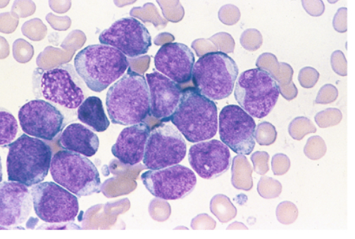
Describe AML with Minimal Differentiation
Immunophenotype
CD13+, CD33+, CD34+, CD17+
Auer rods absent
lack of cellular maturation
negative results with MPO/ SBB
comprises < 5% of AML
Describe AML without Maturation
blasts comprise 90% of nonerythroid cells in BM
fewer than 10% of leukocytes show maturation to promyelocyte stage
Auer rods present
Positive MPO and SBB
Immunophenotype
CD13+,CD33+, CD117+,CD34+
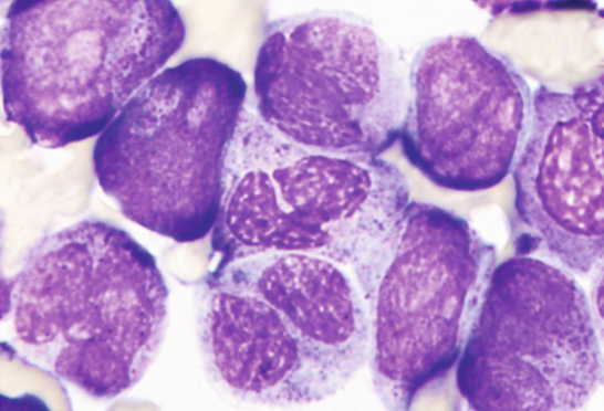
Describe AML with Maturation
> 20% blasts
at least 10% maturing cells of neutrophil lineage
fewer than 20% monocytic lineage
auer rods present
Which classification system uses M0-M7 for AML?
FAB
What WHO subtype is associated with this.
Philadelphia chromosome. Associated with a poorer prognosis.
t(9;22) - BCR-ABL1
What WHO subtype is associated with this.
Often occurs in infants; associated with CNS involvement and organomegaly.
t(v;11q23.3) – KMT2A
What WHO subtype is associated with this.
Common in childhood ALL; generally favorable prognosis.
t(12;21) – ETV6-RUNX1
What WHO subtype is associated with this.
Cells have TOO MANY chromosomes (more than 50); generally favorable prognosis in children.
Hyperdiploidy
What WHO subtype is associated with this.
Cells have TOO FEW chromosomes (fewer than 46); generally poor prognosis.
Hypodiploidy
What WHO subtype is associated with this.
Rare subtype; may be associated with eosinophilia.
t(5;14) – IL3-IGH
What WHO subtype is associated with this.
Genetic abnormality involving chromosomes 1 and 19.
t(1;19) – TCF3-PBX1
What are the B-cell ALL WHO subtypes?
t(9;22) – BCR-ABL1
t(v;11q23.3) – KMT2A
t(12;21) – ETV6-RUNX1
Hyperdiploidy
Hypodiploidy
t(5;14) – IL3-IGH
t(1;19) – TCF3-PBX1
Describe this WHO category of AML.
AML with recurrent genetic abnormalities
AML caused by specific, recognizable genetic changes or chromosome abnormalities.
Describe this WHO category of AML.
AML with myelodysplasia-related changes
AML associated with abnormal-looking blood cell development, often with a history of MDS or MPN
Describe this WHO category of AML.
Therapy-related myeloid neoplasms (TR-MN)
AML or another myeloid disease that develops after previous chemotherapy or radiation treatment.
Describe this WHO category of AML.
AML-NOS
AML that doesn't fit into one of the more specifically defined categories
Describe this WHO category of AML.
Myeloid sarcoma
A collection of cancerous myeloid cells forming a tumor outside the bone marrow.
Describe this WHO category of AML.
Acute leukemias of ambiguous lineage
Leukemia cells that cannot be clearly classified as either myeloid or lymphoid, or show characteristics of both.