Spinal Cord - Microscopic / Cross-Section Anatomy
 Call Kai
 Learn
Learn Practice Test
 Spaced Repetition
 Match
Match Flashcards
 Knowt Play
1/17

There's no tags or description
Looks like no tags are added yet.
Name | Mastery | Learn | Test | Matching | Spaced | Call with Kai | Chat |
|---|
No analytics yet
Send a link to your students to track their progress
18 Terms
White Matter
Outer region of a spinal cord cross section. Has 3 columns: Dorsal Funiculus, Lateral Funiculus, and Ventral Funiculus. Lighter outer area surrounding the butterfly-shaped gray matter.

Dorsal Funiculus
White matter on the posterior side of the spinal cord. White matter between the two dorsal horns. Posterior = Dorsal

Ventral Funiculus
White matter on the anterior side. White matter anterior to the ventral horns. Anterior = Ventral

Lateral Funiculus
White matter on the sides of the spinal cord. White matter lateral to the gray horns. Side = Lateral

Dorsal Median Sulcus
Shallow groove along the posterior/dorsal midline of the spinal cord. Small/shallow indentation on the dorsal side.
(Top Middle)

Ventral Median Fissure
Deep groove along the anterior/ventral midline. The large, deep indentation.
(Bottom Middle)

Dorsal Horn
Posterior/dorsal projection of gray matter. The thin, pointed upper wings of the butterfly-shaped gray matter.

Ventral Horn
Anterior/ventral projection of gray matter. Usually the large, rounder/broader horns. Toward deep ventral fissure.

Lateral Horn
Lateral projection of gray matter. Small gray-matter projection sticking outward between the dorsal and ventral horns.
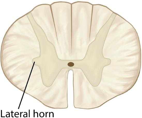
Gray Commissure
Gray matter connecting the left and right sides of the spinal cord. Center of the butterfly-shaped gray matter. Gray bridge surrounding the central canal.

Central Canal
Small space in the center of the spinal cord. Within the gray commisure. Tiny hole/dot in the middle of the gray matter.

Dorsal Root
Enters the spinal cord on the dorsal/posterior side. Root associated with the dorsal root ganglion.

Ventral Root
Connected to the ventral/anterior side of the spinal cord. Root on the ventral side that does NOT have a ganglion.

Dorsal Root Ganglion
Plural = Ganglia. Enlargement associated with the dorsal root. Obvious bulge/swelling along the dorsal root.

Multipolar Neuron
Cell body with many processes extending from it/

Unipolar Neuron
Rounded cell body with a single main process.

Glial Cell/ Neuroglia
Typically appears much smaller than a neuron, often seen as small dark nuclei around neurons.
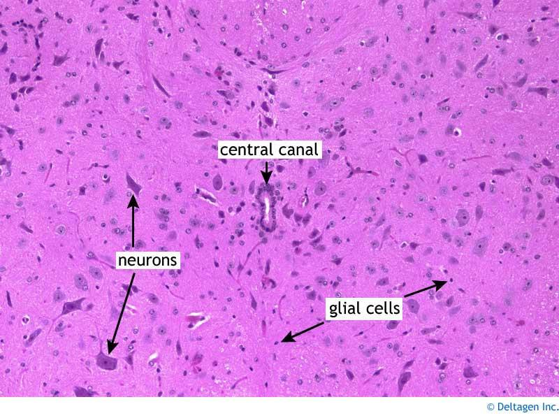
Gray Matter vs. White Matter
Gray matter: Primarily neuron cell bodies, dendrites, synapses, and unmyelinated axons.
White matter: Primarily bundles of myelinated axons.
Spinal cord: Gray matter is internal; white matter is external.
Cerebrum: Gray matter forms the outer cortex; white matter is deeper.