Genetics Exam 3
 Call Kai
 Learn
 Practice Test
 Spaced Repetition
 Match
 Flashcards
 Knowt Play
1/53

There's no tags or description
Looks like no tags are added yet.
54 Terms
Types of viral genomes
DNA/RNA viruses: single or double stranded
RNA ←→ DNA viruses:
retroviruses: ssRNA → DSDNA
hepaDNAviruses: partially dsDNA
*are diverse: diff sizes, linearity, size, etc.
Supercoiling
coils in DNA that occur during replication and transcription
facilitations compacting of DNA into bacterial chromosomes
done by topoisomerases
enzymes that cut 1/both DNA strands
winds/unwinds helix before resealing ends
found in both prokaryotes and eukaryotes
Polytene chromosomes vs lampbrush
giant chromosomes that form due to repeated replication w/o division
puff regions: areas of unwound chromatin
due to high activity/high gene expression
type of huge chromosome found in vertebrate oocytes (immature eggs) and some spermatocytes of insects
contain large amounts of looping and uncondensing
occur as DNA is actively being transcribed (into RNA)
can be isolated/removed to be studied during diplotene stage
of prophase I
Chromatin, Chromatin remodeling
decondensed, uncoiled chromosomes in interphase…
Euchromatin
Uncoiled and active
Appears unstained during interphase
Heterochromatin
Condensed areas are mostly inactive
Appears stained during interphase
*chromosomal banding techniques: Differential staining along longitudinal axis of mitotic chromosome (Staining patterns resemble polytene chromosome bands)
Remodeling:
structure changes to accommodate DNA interactions
relax the compact structure
expose DNA regions to regulatory proteins
has a reversal mechanism for inactivity
used in replication and gene expression
Histones
protiens made of lysine and argenine that DNA is wrapped around to make nuleosomes
Epigenetics
chemical after-modifications to DNA → usually occur along histone tails (have targets along the chromatin fiber for chemical modifications)
acetylation: adding acetyl group for gene activation
done by: Enzyme: histone acetyltransferase (HAT)
removed by: Histone: deacetylase
added to lysine (+ charge) - it becomes neutralized
Methylation: Adds methyl groups to arginine and lysine residues in histones
done by: Enzyme methyltransferase
DOESNT change charge, just acts as a marker
sometimes has positive correlation with gene activity
if base C methylated: negatively correlates w/ gene activity
forms 5-methyl cytosine
can occur at CpG island: Region of DNA where many cytosine guanine dinucleotides present
phosphorylation: Adds phosphates groups to hydroxyl groups of amino acids serine and histidine
done by: enzyme kinase
Correlated with chromatin unfolding and condensation
Repetitive DNA
Highly repetitive: Satellite DNA
Middle repetitive:
tandem repeats
microsatellites/STRs (short tandem repeats): repeats dispersed throughout genome
Consist of di-, tri-, tetra-, and pentanucleotides
Most common in humans is (CA)n with n between 5-50
Useful markers for genome analysis
VNTRs(variable number tandem repeats)/minisatellites: Repeating sequences 15-100 bps found within/between genes
Variation in number results 1-20kbps in size differences
Originally used in DNA fingerprinting analysis
multiple copy genes/rRNA genes
interspersed retrotransposons
Retrotransposons: Transposable elements generated via RNA intermediate (LINES)
SINEs/alu & LINEs/L1: Transposable sequences that are mobile and can relocate within genome
Constitute 1/3 of human genome

Centromeres and Kinetochore Proteins
Centromeres
Separation of homologs during mitosis and meiosis
Are the primary constrictions along eukaryotic chromosomes
Kinetochore proteins
Region that binds to spindle fiber microtubules during cell division
Pseudogenes
Large number of single-copy noncoding regions
DNA sequences representing evolutionary vestiges (parts that no longer have function because of evolution but did before)
Have undergone significant mutational alterations (insertions and deletions)
Are not transcribed
**Only a small portion of eukaryotic genome (2–10%) constitutes protein-encoding genes
Transcription
transcribing template DNA into mRNA
RNA synthesized on DNA template
Genetic information stored in DNA is transferred to RNA
mRNA is intermediate between DNA and proteins
Promoter → Initiation → Elongation → Termination → 5' cap → Splicing → Poly-A tail → Mature mRNA
General features of genetic code
linear form from bases of mRNA
each “word” is a codon (3 ribonuclotide sequence)
unambiguous: each triplet specifies only one amino acid
Degenerate: A given amino acid can be specified by more than one triplet codon
Contains “start” and “stop” triplets (initiate/terminate translation)
Comma-less: codons are read with no break
Nonoverlapping: Any single ribonucleotide within mRNA is part of one triplet
Colinear: Sequence of codons in a gene is colinear
Nearly universal: the same “coding dictionary” used by viruses, prokaryotes, archaea, eukaryotes
mRNA, RNA polymerase, and Promoters
messenger RNA
intermediate for transferring genetic information from DNA to proteins
—
RNA pol: Enzyme directs synthesis of RNA using DNA template
Nucleotides contain ribose, not deoxyribose
No primer required for initiation
→ In eukaryotes:
RNA poly I: creates rRNA in nucleolus
RNA poly 2: makes mRNA & snRNA (and other unique RNAs) in nucleoplasm
most responsible for transcription of wide range of genes in eukaryotes
Activity dependent on cis-acting elements (explanation) and trans-acting transcription factors
RNAP II core-promoter determines where RNAP II binds to DNA
RNA poly 3: makes 5SrRNA & tRNA in nucleoplasm
—
Promoters: Nucleotide sequences as recognition sites for transcription machinery (transcription initiation)
*Interaction of promoters and RNA polymerase regulates efficiency of transcription
Triplet code
Provides 64 codons to specify 20 amino acids
Nonoverlapping: Genetic code reads three nucleotides at a time in continuous, linear manner
Each triplet codon is complementary to anticodon of
tRNA
Start codon: Methionine (AUG - ONLY codon for met)
In bacteria: modified form of methionine [N-formylmethionine (fmet)]
Appears internally in mRNA unformulated
Termination codons: UAG, UAA, UGA
Do not code for any amino acid/Are not recognized by tRNA
*3rd bp is more flexible (wobble hypothesis)
Frameshift mutations
Result from insertions or deletions of base pair(s)
*causes shift in triplet reading frame
13.3 placeholder
ORF
Open reading frame: “In some viruses, initiation at different AUG positions out of frame with other leads to distinct polypeptides”
→ another start site for other versions of aa chains
Consensus Sequences & Termination/Poly U
DNA sequences homologous in different genes of same organism
→ E. coli promoters have two consensus sequences
TTGACA and TATAAT (Pribnow box)
Positioned at −35 and −10 with respect to the
transcription initiation site, respectively
Termination is done by weak pairs of the “Poly U” sequence
Rho breaks off the base pairs
Transcription in Eukaryotes
Occurs within nucleus (unlike prokaryotes)
mRNA must leave nucleus for translation
Chromatin remodeling makes DNA accessible to RNA Pol
RNA polymerases rely on “transcription factors (TFs)” to scan/bind DNA
“Enhancers” and “silencers” and “proximal promoting elements” control transcription regulation (and efficiency)
Different mechanism of termination
mRNA processing of pre-mRNAs
Transcription Factors
Transcription factors facilitate RNApol2 binding and initiation of transcription
General transcription factors: Required for all RNAP II-mediated transcription
Transcription activators and repressors: Influence efficiency or rate of RNAP II transcription initiation
RNA processing
transcription → cap → splicing → tail
Splicing
type of posttranscriptional modification: Introns (non coding regions) removed exons (exon shuffling) joined together (coding regions) in mature mRNA (smaller than initial RNA)
spliceosome: made up of snRNPs
snRNAs: Small nuclear RNA involved in splicing
snRBPs: snRNA + proteins; building block of spliceosome
Tail *eukaryotes
added by poly-a-polymerase: Adds poly-A tail to 3' end.
poly-a binding protein: Binds poly-A tail; stabilizes mRNA
Translation
Biological polymerization of amino acids into polypeptide chains
requires: Amino acids, Messenger RNA (mRNA), Ribosomes,Transfer RNA (tRNA)
Steps:
Shine–Dalgarno sequence (AGGAGG) precedes AUG start codon in bacteria
“Base-pairs with region on 16S rRNA of 30S small subunit, facilitating initiation”
Initiation complex
Small ribosomal subunit ← initiation factors bind causing → mRNA to attach at codon AUG
Combines with large ribosomal subunit
Elongation
mRNA moves into P & A sites (amino acids grow at P site)
tRNA exits at E site
Termination
termination sequence enters the A site causing hydrolysis of amino acid chain
ribosome subunits dissociate
tRNA
Adapt genetic information (specific triplet codons) in mRNA to corresponding amino acid
tRNA anticodons complement mRNAs
tRNAs carry corresponding amino acids
needs to be charged by enzyme aminoacyl tRNA synthetase
4 attachements sites:
Amino acid attachment site
Anticodon
Recognition site for aminoacyl-tRNA synthetase
Ribosome recognition/binding site

ribosomes
combination of ribosomal RNA (rRNA) and protiens - 2 subunit: large and small
translation factors placeholders
sl 16
Translation differences in eukaryotes
Ribosomes are larger and longer lived than bacteria
Transcription occurs in nucleus
5′ end of mRNA capped with 7-methylguanosine residue
Poly-A tail added at 3′ end of mRNA
Translation occurs in the cytoplasm
Eukaryotic mRNAs contain A or G three bases ahead of AUG initiator codon, followed by G (Kozak sequence)
A/GNNAUGG
Kozak sequence increases efficiency of translation by interacting with initiator tRNA
One Gene:One Enzyme Hypothesis
George Beadle showed genes are directly responsible for enzymes synthesis
Beadle and Tatum showed nutritional mutations in bread mold Neurospora caused loss of enzymatic activity
Experiment led to one-gene:one-enzyme hypothesis
*found that most mutants unable to grow on minimal medium could grow on medium with an addition of one supplement on minimal medium. Findings suggested that most mutations affect only a single metabolic pathway/single enzyme.
[check on sl. 25-26]
One Gene: One polypeptide
One-gene:one-protein hypothesis changed to one-gene:one-polypeptide chain hypothesis
Two factors modified the hypothesis:
Nearly all enzymes are proteins, not all proteins are enzymes
Proteins have subunit structure with two or more polypeptide chains
Proteins
Polypeptides: Precursors to proteins
Amino acids assembled on and released from ribosomes as polypeptides (Amino acid polymer)
held my polypeptide bonds through dehydration
Proteins: Polypeptides folded up into a functional three-dimensional conformation
Four levels of protein structure:
Primary: Sequence of amino acids
Secondary: α-helix and β-pleated sheets
Tertiary: Three-dimensional conformation
Quaternary: more than one polypeptide chains
Protein folding: Co-translational folding by chaperones(other proteins that mediate folding)
Diseases of protein folding: Creutzfeldt–Jakob disease, Transmittable brain disorder in mammals, Presence of prions (misfolded proteins) in brain; Others: Huntington’s, Alzheimer’s, Parkinsons.
Most abundant macromolecules in cells: play diverse roles
hemoglobin and myoglobin: transport oxygen for cellular metabolism
Collagen and keratin: structural proteins for skin, connective tissue, and hair of organisms
Actin and myosin: Contractile proteins found in muscle tissue
Tubulin: Basis of microtubule function in mitotic and meiotic spindle fibers spindle fibers
Immunoglobulins: Function in immune system of vertebrates
Transport proteins: Movement of molecules across membranes
Hormones and their receptors: Regulate various chemical activity
Histones: Bind to DNA in eukaryotes
Transcription factors: Regulate gene expression
Enzymes: Most diverse/extensive proteins - specialize in catalyzing (reducing AE) chemical reactions and increase rate of chemical reaction reaching equilibrium
Amino acids: Carboxyl group, Amino group, R (radical) group bound to central carbon atom
R group (side chain) determines type of amino acid: Nonpolar (hydrophobic), Polar (hydrophilic), or positively and negatively charged

Posttranslational modifications
Polypeptide chains modified after synthesis. Modifications crucial to function of final protein product.
Posttranscriptional modification examples:
N-terminus amino acid removed or modified
Individual amino acid residues modified
Carbohydrate side chains are sometimes attached
Polypeptide chains may be trimmed
Signal sequences are removed
Polypeptide chains often complexed with metals
Functional Domains
Sequences with 50–300 amino acids constitute protein domains
Sequences constitute protein domains that fold into stable, unique conformations
Different protein domains impart different functional capabilities
Catalytic domains & DNA-binding domains
Mutation, changes, other mutations
Mutation
An alteration in DNA sequence (Any base-pair change)
Transitions: Pyrimidine replaces pyrimidine, or purine replaces purine (pyr: CTU, pur: AG)
Transversions: Purine and pyrimidine are interchanged
tautomeric shifts: H bond between the wrong pyr/pur
Single base-pair substitution
Deletion or insertion of base pairs
Major alteration in chromosomal structure
—
molecular changes:
Point mutation or base substitution: Change from one base pair to another
Missense mutation: Results in triplet code for different amino acid
Nonsense mutation: Results in triplet code for stop codon (translation terminated prematurely)
Silent mutation: New triplet code still codes for same amino acid
—
Somatic mutations occur in any cell except germ cells (not heritable)
Germ-line mutations occur in gametes; are inherited
Autosomal mutations occur within genes located on autosomes
X- and Y-linked mutations occur within genes on X and Y chromosomes
Classification of mutations by effect on function (not super important- skim)
Loss-of-function mutations
Gain-of-function mutations
Visible (morphological) mutations
Nutritional (biochemical) mutations
Behavioral mutations
Regulatory mutations
Lethal mutations
Conditional/temperature-sensitive mutations

Mutation classification + Rates
Spontaneous mutation: natural change in bases
Arise from normal biological or chemical processes that alter nitrogenous bases (ex: replication, base damage)
Spontaneous mutation usually very low for all organisms
Induced mutations: from influence of extraneous factors (natural or artificial)
ex: Radiation, UV light, Natural and synthetic chemicals
Germ Line Rates:
Average 60 new mutations in offspring
Number dependent on age of father
Somatic Rates:
4-25x higher than germ lines rates
Somatic mosaicism (early mutation may contribute to a large portion of adult cells having that mutation)
sources of spontaneous mutations
Mutations from replication: DNA pol occasionally inserts incorrect nucleotides which persist after proofreading
Errors due to mispairing predominantly lead to point mutations
Replication slippage: If loop occurs in template strand during replication, DNA pol misses looped out nucleotides and small insertions and deletions occur
more common in repeat sequences (hot spots for DNA mutations)
Contribute to hereditary diseases (ex: fragile-X, Huntington disease)
—
base damage:
Depurination and deamination
Common causes of spontaneous mutations
Lead to new base pairing and mutations
Oxidative damage
Due to by-products of normal cellular processes
Superoxides (O2− ), Hydroxyl radicals (OH), Hydrogen peroxide (H2O2)
Exposure to high-energy radiation
—
Transposable genetic elements: DNA elements moving within/between genomes
Present in all organisms
Can act as naturally occurring mutagens
Cause inversions, translocations, double-stranded breaks (creates chromosomal damage)
Steps: cleaves DNA at ITR region, staggered cuts in target area, inserts transposon inserted by transposase, gaps filled by dna pol + ligase
Sources of Induced mutations
Mutagens: Natural or artificial agents that induce mutations
Fungal toxins, Cosmic rays, UV light, Industrial pollutants, Medical X-rays, Chemicals in tobacco smoke
Base analogs: look like nitrogen bases - may be incorporated accidently
Alkylating agents: attach alkyl groups, causing issues (mispairing, breaks, etc.)
Intercalating agents: Chemicals w dimensions+shapes that wedge between DNA bps (Example: Ethidium bromide)
causes base-pair distortions and DNA unwinding
Usually results in frame-shift mutations
Adduct-forming agents: Mutation-causing chemicals
Covalently binds to DNA, altering conformation, interfering with replication + repair
Acetaldehyde: Component of cigarette smoke
Heterocyclic amines (HCAs): Cancer-causing chemicals created from cooking meats
17 HCAs linked to cancers of stomach, colon, and breast
UV light → Pyramidine Dimer
UV light causes bases of the same strand to bind to each other
Ionizing radiation: causes more X linked recessive alleles
Free radicals: transformation of chemical species with unpaired electrons by radiation
Alter purines and pyrimidines
Break phosphodiester bonds
Produce deletions, translocations, and fragmentation
Trinucleotide repeat sequences
Short DNA sequences found in mutant genes
Normal individuals may have a low number of repetitions
Individuals with over 20 disorders exhibit over 200 repeats
Fragile-X syndrome, huntington disease (HD), Myotonic dystrophy (MD)
DNA repair
Repair systems counteract spontaneous & induced DNA damage
DNA repair system: Maintains integrity of genetic material + counteract genetic diseases/cancer
Proofreading and mismatch repair
DNA pol proofreads - removes, replaces incorrectly inserted nucleotides (3’ to 5’ exonuclease)
Mismatch repair (if proofreading fails) becomes activated: Mismatches are detected, cut, removed (endonuclease and exonuclease). Correct nucleotide inserted by DNA pol
Strand discrimination - Based on DNA methylation
Adenine methylase (bacteria enzyme) recognizes DNA sequences and adds methyl group to adenine residues
Newly synthesized strand of replication remains unmethylated
Mismatch repair recognizes unmethylated strand and repairs
photoreactivation repair:
base excision repair:
nucleotide excision repair:
NER pathway defects can cause autosomal recessive diseases
Double-strand break repair (DSB repair): reattaches strands
Double-strand breaks result in chromosomal rearrangements, cancer, cell death
Two pathways involved in DSB repair:
Homologous recombination repair: uses sister strands and recombination to repair
Nonhomologous end joining:
Activated in G1 prior to replication
Complex of proteins (kinase/BRACA1) is involved in end joining
Proteins bind free ends and ligate ends back together
E. coli SOS repair system
Last resort to minimize DNA damage
DNA synthesis becomes error-prone; inserts random/incorrect nucleotides in places that normally would stall replication
it induces expression of 20 genes; their products allow replication to move forth
SOS repair can itself become mutagenic; allows cells to survive with DNA damage (cell would otherwise kill itself)
Ames Test
tests the mutagenic potential of chemical compounds using bacteria
used bacteria that couldn’t synthesize his (mutegens can restore his)
tested if his came back on plates w/o histadine in agar
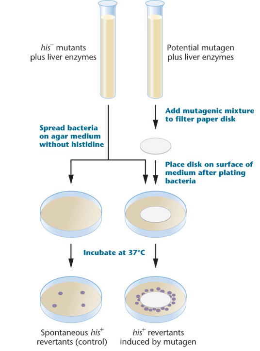
Mobile genetic elements in corn plants found using
Ac–Ds system
Dissociation (Ds) and Activator (Ac)
Two mutations of transposable elements in corn
Mobile controlling elements (transposable elements)
• Movement of Ds gene is dependent on Ac gene
• Barbara McClintock
[figure this out]
[figure out what retrotransposons are]
Eukaryote differences from prokaryotes - Transcription Regulation
Eukaryotic genes on chromosomes w/ distinct location
Chromosome territory: In interphase, each chromosome occupies a domain and stays separated from others
Interchromosomal domains: Channels between chromosomes that contain little or no DNA
*Chromosome structure is continuously rearranged; transcriptionally active genes are cycled to edges of chromosome territories
Eukaryotic DNA combined with histones and nonhistone proteins to form chromatin
Compact chromatin structure inhibits transcription, replication, and DNA repair
Nucleosome modifications
*posttranscriptional modifications:
to histones:
type of gene regulation; involves changes to either nucleosome or DNA
– Histones contain normal histone H2A
– Variant histones (H2A.Z) affect nucleosome mobility and positioning on DNA
– Nucleosome position may repress or activate transcription via gene promoter
DNA methylation:
– Associated with decreased gene expression
– Methylation occurs most often on cytosine of CG doublets in DNA (CpG Islands)
– Methylation can repress transcription by binding to transcription factors of DNA
– Regulated by methylases and demethylases (5’-mCpG-3’ 3’-GpCm-5)
cis-acting-sequences
non‑coding DNA element that regulates the transcription of a gene
promoters (transcription initiation): Nucleotide sequences as recognition sites for transcription machinery
Transcription start site, position +1
double helix is denatured/unwound to make template strand accessible
Located directly next to the regulatory genes (control the expression of other genes)
Core promoter: Determines accurate initiation of transcription
Proximal-promoter elements: Modulate efficiency of basal levels of transcription
Enhancers (increase rate of transcription)
Located on either side, some distance from, or within gene
Important in reaching maximum level of transcription
Insulators: Found between an enhancer and a promoter for a non-target gene
allow some enhancer–promoter interactions and block others
Silencers (block transcription): Repress the level of transcription initiation
Transcription factors pt 2
*EUKARYOTES
Transcription factors: regulatory proteins that bind to enhancers and promoter elements and fine-tune the level
Target cis-acting sites of genes regulating expression
Activators increase transcription initiation
Repressors decrease transcription initiation
2 functional domains (clusters of amino acids with a specific function):
DNA-binding domain
Binds to specific DNA sequences in the cis-acting regulatory site
Trans-activating domain
Activates or represses transcription by binding to other transcription factors or RNA polymerase
Characteristic domains of DNA-binding proteins
Helix-turn-helix (HTH): eukaryotic and prokaryotic transcription factors - DNA binding domain
Zinc-finger: found in many transcription - enables binding
Basic leucine zipper (bZIP): Allows for protein–protein dimerization
TFIIA | Stabilizes transcription complex. |
|---|---|
TFIIB | Positions RNAP II at promoter. |
TFIID | Contains TBP; first factor to bind promoter. |
TFIIE | Recruits and regulates TFIIH. |
TFIIF | Escorts RNAP II to promoter. |
TFIIH | Unwinds DNA and phosphorylates RNAP II. |
TBP | TATA-binding protein; binds TATA box. |
DABEFH (TFIID → TFIIA → TFIIB → TFIIF → TFIIE → TFIIH)
Formation of RNA Pol II initiation complex
General transcription factors (GTF): Proteins
Required at promoter to initiate basal (find where else basal is used and fix it) or enhanced levels of transcription
Assembly of proteins in specific order forms pre-initiation complex (PIC)
PIC provides platform for RNAP II to recognize transcription start sites
—
Transcription activators and repressors change RNA Pol II transcription
DNA looping delivers activators, repressors, and general transcription factors to promoter
Recruitment model: enhancers and silencer elements act as donors; affect regulatory proteins at gene promoters
– Chromatin alteration model: looping results in alteration of the chromatin via remodeling complexes
– Nuclear relocation model: looping relocates region to a favorable nuclear environment
GAL gene system
yeast
Composed of four structural genes and three regulatory genes
Products of structural genes transport galactose into cell for metabolism
Positive control: Activator protein must be present to turn on gene transcription
are inducible
Transcription regulated by presence/absence of substrate (galactose)
Absence of galactose: GAL structural genes not transcribed
Galactose added to medium: Transcription begins immediately
ENCODE
Encyclopedia of DNA Elements Project (ENCODE): Identify all functional DNA sequences and determine how elements regulate expression
findings:
more than 80 percent of human genome contains regulatory elements (used to be considered junk DNA)
Enhancer and promoter elements
Non-coding regulatory RNA
—
genome-wide association study (GWAS):
Identifies genetic variations that are significantly increased in the genomes of patients with diseases
Many GWASs evaluate single base-pair changes/single nucleotide polymorphisms (SNPs)
Do not directly mean disease susceptibility
located near the causative issue- the two are inherited together
90%+ of disease-associated variations in the human genome are located within regulatory regions
Alternative Splicing
*EUKARYOTES
different forms of mRNA from identical pre-mRNA (spliceforms)
expression of one gene = many protiens with similar/diff functions
increases num. of proteins from one gene (isoforms)
Types of splicing:
Cassette Exons: skipping an exon region (joining the before and after region)
most prevalent type of alt. splicing in animals (40%)
Alternative splice site: splicing within an exon splice site (shifting where cutting starts)
Intron retention: including introns - can be used to negatively regulate at post transcriptional level
Mutually exclusive exons: coregulation where only one exon can be included from a cluster
Alternative Promoters: having more than one transcription initiation site (diff 5’ ends)
Alternative polyadenylation: different 3’ poly-A tails created
if an exon that signals the poly-A tail is skipped, another exon with the signal is used
An image can be added!!
Regulation of AS
splicing enhancers/silencers: cis-acting sequences that regulate alternative splicing
SR protiens: bind to enhancers and activate splicing (recruit spliceosome components)
hnRNPS (heterogeneous nuclear ribonucleoprotiens): bind splicing silencers = inhibit splicing
RNA-binding protiens (RBPs): bind to RNA sequences to regulate splicing
*spliceopathies: mutations that affect regulation and cause genetic disorders
mRNA stability
steady-state level of mRNA: amount of mRNA avaible in cell for translation (determined by transcription and mRNA degredation rates)
Half life: (t 1/2): half life of mRNA - at what point it degrades
Degredation of mRNA
1) shortened poly-A tail - called deadenylation-dependent decay
2) decapping enzymes removed cap - makes mRNA unstable
3) endonuclease cleaves (removes) mRNA internally
*regulated bu cisacting sequence elements and trans-acting proteins
adenosine-uridine rich element (ARE): cis-acting sequence that regulates mRNA stability
mRNA surveillance & Nonsense mediated decay
surveillance: degrading mRNAs with premature stop codon that come from a nonsense mutation or RNA pol error
NMD: most studied surveillance pathway - eliminates mRNAs with premature stop codon
RNAi vs longnoncoding RNAs
RNA interference: sequence specific posttranscriptional regulation - short RNA molecules that regulate gene expression (repress and trigger mRNA degredation)
studied in lab and developed as pharmaceutical agent
therapeutic RNAi attacks diseases caused by overexpression
potential use in cancer diagnoses + reduces severity of infections by viruses
in animals:
treatment for biral infection, eye diseases, cancer (potential tool in molecular med)
long noncoding RNAs: act as regulators
Regulation of protiens
phosphorylation: most common type of posttranscriptional modification - induces conformation changes → function changes
kinases catalyze addition of phosphate group to ser, thero, tyro AA side chains
phosphatases: enzymes that remove phosphates
*ubiquitincan mediates protein degregation