ch3 immunity and immunologic
 Call Kai
 Learn
Learn Practice Test
 Spaced Repetition
 Match
Match Flashcards
 Knowt Play
1/41

There's no tags or description
Looks like no tags are added yet.
Name | Mastery | Learn | Test | Matching | Spaced | Call with Kai | Chat |
|---|
No analytics yet
Send a link to your students to track their progress
42 Terms
• Defends the body against injury
• Has memory capability
• Responds quicker than inflammatory response
• Involves complex network of white blood cells
• May result in increased level of tissue damage and disease as it fights
acquired (adaptive) immune response
• Foreign substances (bacteria, viruses) that have these which activates the immune system to defends the body.
• Components of our own cells, our immune system tolerates
• Tumor cells (may develop abnormal ____)
• Cells infected with viruses
• Organ transplant (may have different _____, that cause ur body to reject the organ)
• Tissue graft
• Incompatible blood transfusion
• Cells of own body: Autoimmune diseases
Antigens or Immunogens
• Develop from stem cells in bone marrow
• Mature and reside in lymphoid tissue
• Lymph nodes, tonsils, and other body tissue
• travel to the site of injury when stimulated by antigen
• Two main types:
• Plasma cell: Produces specific antibodies
• B memory cell: Retains the memory of previously encountered antigen and will clone itself in the presence of antigen
B-cell lymphocytes
Round, pinwheel-shaped nucleus with visible cytoplasm
• Produces and releases a protein (antibody) in response to presence of antigen
• Circulating antibodies: Immunoglobulins (Igs
produces specific antibodies
b-cell lymphocytes: plasma cells
• Five different types of immunoglobulins:
• IgA
• IgD
• IgE
• IgG
• IgM
• Antibody titer: Level of a specific antibody
• Immune complex: Antibody + antigen
B-cell lymphocytes: Immunoglobulins
• Develop from bone marrow stem cell
• Travel to the thymus and mature
• Types:
• Memory cells
• T-helper cells: Increase functioning of B cells
• T-suppressor cells: Turn off functioning of B cells
• T-cytotoxic cells: Attack virally infected cells or tumor cells
T-cell lymphocytes

• Destroy foreign cells
• Located within the microcirculation
• Active against viruses and cancer cells
• Activity can be abnormal as in HIV infection
releases substances that cause the target cell to destroy itself
Natural Killer Cells (lymphocytes)
• Accessory cells in immune response that amplify but do NOT have memory
• Located in connective tissue (CT) during inflammation
• Functions:
• Phagocytosis
• Assist B cells and T cells
messenger between inflammatory response and immune response
Activating factor: produce and secrete lysosomal (digestive) enzymes
macrophages

• Type of white blood cell found on skin, mucosa, and in blood
• Antigen-presenting cells (APCs): Process antigenic material and present it to other immune system cells such as T-lymphocytes to activate immune response)
• Messenger between innate immunity and acquired immunity
• Langerhans’ cell: Specialized dendritic cell (NOT isle of langerhans)
Dendritic cells
• Immunomodulating agents: Alter the immune response
• Communication system
• Interleukins (may regulate inflammation/immune activation)
• Macrophage chemotactic factor (attracts macrophages)
• Migration inhibitory factor (keeps macrophages at inflammation site, promotes inflammation)
• Macrophage-activating factor (activates macrophages, makes them better at killing/digesting pathogens)
• Lymphotoxin (contributes to inflammation/cell killing)
• Interferons (important for antiviral defense/immune signaling)
• Tumor necrosis factor (major proinflammatory, actives immune response and contribute to cell death)
Cytokines
1) B cells produce antibodies that bind to antigens
2) Remove from the body, key in extracellular bacteria & toxins.
3) Long term protection through memory B cells & the antibodies they produce
4) Faster response, antibodies produce quickly
humoral immunity
1) Kill cells or activate other immune cells
2) Role in destruction of intracellular viruses & bacteria
3) Long term protection through memory T cells to future infection
4) T cell response can take longer but are highly specific and effective intracellular pathogens.
cell-mediated immunity
Using antibodies created by another person to prevent infectious disease
• Natural: Mother to fetus
• Acquired: Injection
Passive Immunity
Antibodies created by the person himself or herself
• Natural: Microorganism causes the disease
• Acquired: Immunization, vaccination, booster
• Killed-type vaccine
• Live-attenuated vaccine
Active Immunity
• The study of immune reactions involved in disease; the study of diseases caused by the malfunctioning of the immune system
• Hypersensitivity
• Autoimmune diseases
• Immunodeficiency
immunopathology
anaphylactic
anaphylaxis, asthma, allergies
Plasma cells → histamine (inflammation) → edema (dilation) → bronchoconstriction (smooth muscle)
ex. (hay fever, urticaria, asthma, anaphylaxis)
Type I Hypersensitivity
cytotoxic
binding
• Antibody/Antigen bind tissues cells → (RBC) → complement system, IgG and IgM → causes cell tissue destruction w/ antigen (foreign) attached.
ex (blood transfusion incompatibilty, rhesus (Rh) incompatibility
Hypersensitivity Type 2
immune complex
complexes
“serum sickness”
Immune complexes form (antigen attaches to antibody) → deposit in tissues →( inflammation) → causes tissue destruction
ex (autoimmune diseases like SLE)
hypersensitivity type 3
cell mediated
delayed response
Delayed reaction 2-3 days to develop Cell Mediated (T Cells) rather than humoral response (B cells-antibodies) T cells cause tissue damage
ex (TB test, granulomatosis, organ & tissue rejection)
hypersensitivity type 4
• Oral hypersensitive reaction
• Diffuse swelling of the lips (^ permeability of deeper blood vessels)
• Surface skin on affected area appears normal
• Not itchy usually
• Acute self limited episodes
• Type I hypersensitive reaction
• Acetylsalicylic Acid, NSAIDS (can cause vascular permeability)
• this condition involving the of larynx and pharynx can be fatal
• Dx: clinical appearance of lesion
• Tx: Antihistaminic drugs (Benadryl) or Epinephrine
Angioedema

• Hypersensitive reaction on oral mucosa
• Direct contact on the skin w/ an allergen.
• T cells w/ in a cell mediated response
• Type IV hypersensitive reaction
• Erythematous an edematous, burning, itching, smooth shiny appearance where the agent contacted the skin
• Small vesicles may appear in area
Contact Mucositis
• Skin hypersensitive reaction
• Multiple areas of well demarcated and erythema on the skin, itchy (pruritus)
• Cause can be difficult to pinpoint: infection, trauma, emotional stress, certain systemic diseases, ingested allergens
• Dx: clinical appearance of the lesion
• Tx: Antihistaminic Drugs (Benedryl)
Urticaria “hives”

• Skin hypersensitive reaction
• Erythematous, swelling and vesicles
• Later: crusted, scaly, white
• Common causes of allergic reaction:
LA preservatives, topical medications, acrylics, metal-based alloys, epoxy resins, flavoring agents, chewing gum, dentifrices, mouthwashes, cinnamon oil, latex
Contact Dermatitis

• Lesion that appears at the same site each time a drug is introduced.
• Single or multiple sl. Raised, reddish patches or clusters of macules on skin or oral mucosa (rare in oral)
• Appear after latent period of several days
• Subside when drug is discontinued
• Type III sensitivity reaction (immune complex type)
• Barbituates, chlorhexidine, lidocaine, penicillamine, sulfonamides, tetracycline all assoc. w. fixed drug eruptions
Fixed Drug Eruption

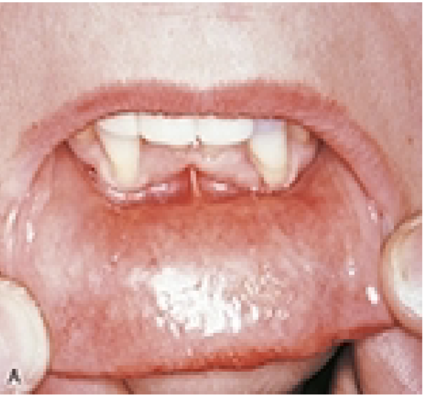
• Most common type
• Discrete, punched out round to oval ulcers, yellowish, fibrin center w/ a halo or erythema.
• up to 1cm in diameter, more common in anterior area of mouth
• small but painful, burning, tingling, soreness, prodromal period
• Single or multiple lesions, heal 7-10 days
• Tx: manage pain – kanka eeze, orabase
Minor Apthous Ulcer “canker sore”
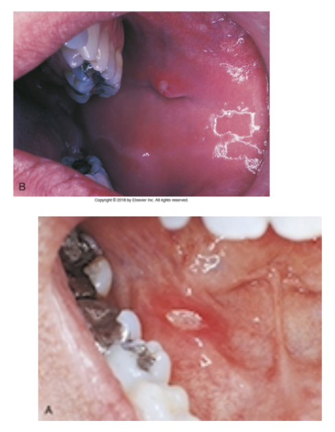
• Smallest 1-2 mm ulcers, least common type
• Herpetiform is misnomer (look like lesions caused by herpes) not caused by herpes
• Do not have a known cause
• Painful and develop anywhere in the oral cavity
Herpetiform Apthous Ulcers

• larger than 1cm in diameter
• deeper and last longer than minor
• more painful, common in posterior area
• several weeks to heal, may result in
scarring
• HIV, Bechet Syndrome, Chron’s Disease, Reactive Arthritis: systemic conditions can typically be reported w/ this
Major Apthous Ulcer (Sutton Disease, Periadentis Muosa Necrotica Recurrens)

• acute self limited disease
• skin & mucous membrane lesions
• Cause not clear (hypersensitive reaction?)
• Young adults under 30, wide range of clinical disease
minor: oral site, some skin sites
• major: 2 or more oral sites, widespread skin involvement
• Explosive onset, prodromal symptoms: fever, malaise, headache, cough, sore throat
• Oral Lesions: diffuse, large, superficial erythematous areas, frequently ulcers on lateral borders of the tongue, crusted bleeding lips
Erythema Multiforme “crusty lips & oral ulcerations”
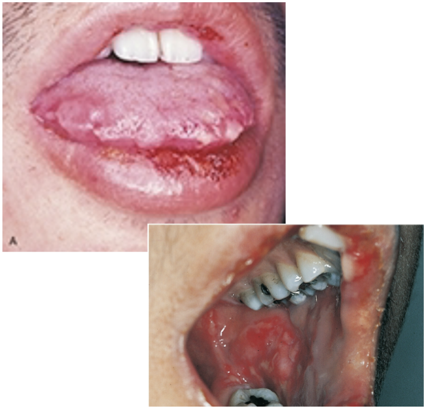
• Skin lesions: target or bullseye lesions, concentric ring of alternating erythema and normal skin color, darkest color at the center
• Triggers: herpes simplex infection, Mycoplasm pneumoniae, drug therapy (antibiotic and analgesics)
• Wide range of skin lesions: macules, plaques, and bullae
• Oral lesions can occur alone or with skin lesions.
• Skins lesion w/out presence of oral lesions.
• Dx: clinical features, exclude diseases, biopsy, micro
• Tx remove cause, topical and systemic corticosteroids, antiviral for reoccurring episodes
Erythema Multiforme “bullseye target lesions”

• Benign chronic condition affecting skin and oral mucosa
• Interconnecting white lines and circles (lace-like) does not wipe off, asymptomatic
• Slender white lines are called Wickham Striae
• small papular, pin-sized, domed or hemispheric glistening white nodule
• Most common site is buccal mucosa but can occur on the tongue, lips, floor of the mouth and gingiva
• 13-78 yrs old, most common middle aged female, can have skin lesions
• Cause unknown, some drugs and chemicals have been implicated
• Dx: Clinical, biopsy, micro, rule out epithelial dysplasia
• Tx: corticosteroids if symptomatic, antifungal if candidiasis results from tx.
Lichen Planus

Types of Lichen Planus
• Reticular Lichen Planus – most common, white lines lacelike,
2-4mm papules
• Erosive Lichen Planus – epithelium separated from ct, results
in erosions, ulcers, bullae, less common
• Bulbous Lichen Planus – same as above, w/ bullae (large
blisters
• Lichen Planus can occur with desquamative gingivitis
• Erosive as been linked to possible development of squamous
cell carcinoma
• Lichenoid mucositis – mixed inflammatory infiltrate rather
than lymphocytes, extends deeper into ct, may be cause by
response to something topical (amalgam, cinnamon), may
develop into malignancy.
• Rec 3-6 mnth intervals for eval and biopsy for premalignant
lesions
Skin Lesions and Desquamative Gingivitis assocaited w/ Lichen Planus

Chronic disease w/ triad of disease
1) Arthritis
2) Urethritis
3) Conjunctivitis
• Develops 1-6 weeks after an sexually transmitted or gastrointestinal infection: Chlamydia, Salmonella, Shigella, Yersinia and HIV, more common in men
• Inflammation of iris, arthritis w/ ankles and knees, TMJ has been reported
• Oral Lesions: anywhere, apthous like lesions erythematous lesions and depilation of the tongue (like erythema migrans)
• Dx: Clinical symptoms, HLA-B27 marker
• Lasts 3 months to a year, aspirin, NSAIDS, antibiotics, corticosteroids, physical therapy
Reactive Arthritis (Reiter Syndrome)

• rare disease
• w/ histiocytes and eosinophils present in lesions
1) Single cell disease (bone, most common site, skin, lymph node, lung)
2) Multisystem disease w/ or w/out organ dysfunction (bone, skin, liver, spleen, bone marrow)
• White children, of northern European ancestry
• Dx: biopsy histopathologic examination
• Young child: have lesions in skull and femur, organ involvement more common
• Over 20 yrs: ribs, shoulder girdle, and mandible
• Punched out radiolucencies , similar to perio, may result in tooth loss
tx: single lesions- radiation
widespread disease – chemotherapy
Eosinophilic Granuloma of bone – localized form, surgical excision needed
Langerhans Cell Histiocytosis (Langerhans Cell Disease)

• chronic systemic, autoimmune disease that affects the salivary and lacrimal glands
• Combo of dry eye and dry mouth (sicca syndrome)
• Cause not known genetic? virus?
• Xerostomia, oral discomfort, erythematous mucosa, mouth feels sticky, dry cracked lips, angular chelitis, loss of papillae on the tongue, difficulty eating, swallowing, taste change (dysgeusia), bilateral parotid enlargement
• High risk of caries, perio disease, oral candidiasis
• Affects major and minor salivary glands
• Dx: 2 of 3 are present dry mouth, dry eye, arthritis or another autoimmune disorder
• Tx: Saliva substitutes, eye drops/meds, NSAIDS, corticosteroid, meticulous homecare, oral lubricants, xylitol, MI paste (Recaldent) fluoride rinses, electric tb, more frequent recall
• Pt should be monitored closely by physician
Sjogrens’ Syndrome “Lack of papillae & bilateral parotid enlargement”

• Severe progressive autoimmune disease affects skin and mucous membranes
• Vesicle formation due breakdown of epithelial cell to epithelial cell adhesion (acantholysis)
• Pt have circulating autoantibodies that react against the epithelial attachment (desmosomal attachment) mechanism
• Most common form “vulgaris” means common
• No gender or age predilection, some genetic and ethic factor reported
• Shallow painful ulcers, vesicles, bullae, size can vary, positive Nikolsky sign
• Tzanck Cells – breakdown of desmosomal cells, found on exam
• Dx: biopsy and micro
• Tx: high does of systemic corticosteroids, disease was once fatal.
Pemphigus Vulgaris

Chronic autoimmune disease that causes blistering and peeling (desquamation) of the gingiva, leaving red, painful, raw areas; may also affect the eyes and other mucous membranes.
Desquamative Gingivitis w/ Mucous Membrane Pemphigoid

• Older than 70 yrs.
• Variant of a single disease? w/ MMP
• Oral lesions are less common than MMP, more extensive and painful
• Dx: circulating auto antibodies detectable, autoantibodies do not always correlate to disease activity.
• Chronic disease w/ periods of remission, not life threating
• Tx: High doses of Systemic corticosteroids, NSAIDS, immunosuppressant medications
Bullous Pemphigoid

• Benign, chronic autoimmune disease affects oral mucosa, conjunctiva, genital mucosa and skin
• 2x more common in women, 50 + yrs of age, not as severe as pemphigus vulgaris
• “cicatricial” means healing w/ scarring (oral scarring is rare)
• Cleavage of the epithelial layer from the underlying ct
• Oral lesions limited to gingiva, marginal and attached gingiva, vesicles, bullae, ulcers are thicker and least longer than pemphigus vulgaris , episodic remission and exacerbation
• Dx: biopsy, microscopic examination
• Tx: chronic disease, topical and systemic corticosteroids, immunosuppressant agents
Mucous Membrane Pemphigoid Benign Mucous Membrane Pemphigoid, Cicatricial Pemphigoid

• Acute and chronic inflammatory autoimmune disease
• Unknown cuase – Genetics ? Environmental influences?
• 8x more frequently affects women, 3x more common in black women than white women
• Chronic and progressive w/ periods of remission and exacerbation (syndrome)
• Wide spectrum of disease activity skin lesion→ organ involvement → Kidney involvement
• Antiuclear antibodies (ANA’s ) present – autoantibodies to the pt DNA
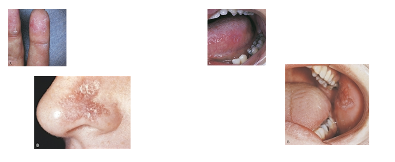
• “Butterfly rash” across the face over the bridge of the nose w/ erythematous lesions on the fingertips
• Oral Lesions: erythematous plaques or erosions involving buccal mucosa, palate, gingiva, may resemble lichen planus but less symmetric, petechiae and gingival bleeding (due to thrombocytopenia)
• Develop other auto immune diseases: Sjogrens’, Rheumatoid Arthritis
• Dx: Circulating ANA, multiorgan involvement
• Tx: once fatal, aspirin, NSAIDS, Hydroxychloroquine (antimalarial), systemic corticosteroids, immunosuppressive agents, topical corticosteroids for oral lesions.
Systemic Lupus Erythematosus
• Toxic epidermal necrolysis (TEN)
• Rare & severe reaction to drugs
• Mucosal lesions, more severe and painful than erythema multiforme
• Lips more crusty & bloody than ER
• Can occur in older and females
• Genital and eye mucosa may be involved
• Tx: removing the drug or agent IV hydration, and corticosteroids
Steven Johnson Syndrome

• Chronic, recurrent multisystem autoimmune disease
• Due to systemic vasculitis
• Possible infectious or environmental antigen
• Oral ulcers (common), genital ulcers, ocular inflammation
• Emerges 30-40 yrs of age
• Oral lesions similar to apthous ulcers, larger red area than aphthous ulcers
• Dx: recurrent oral lesions w/ one or 2 other , pos. pathergy test
• Tx: Systemic, topical corticosteroids, and immunosuppressive drugs
Aphthous- like Ulcer on Tongue in Bechet
Syndrome (Bechet Disease)