cell cycle + infectious diseases + cell structure + microscopy
 Call Kai
 Learn
Learn Practice Test
 Spaced Repetition
 Match
Match Flashcards
 Knowt Play
1/337

There's no tags or description
Looks like no tags are added yet.
Name | Mastery | Learn | Test | Matching | Spaced | Call with Kai | Chat |
|---|
No analytics yet
Send a link to your students to track their progress
338 Terms
what is a disease?
illness/sickness that causes reduced effectiveness of functions
an illness with a set of symptoms that lead to poor physical, mental or social well being
what is an infectious disease?
disease caused by a pathogen that can cause harm to the health of the host
can be passed from one organism to another(communicable/transmissable)
what are pathogens?
disease passing organisms
prokaryote/eukaryotes
eg: bacteria, virus, protoctists, fungi
what is the disease transmission cycle?
pathogens:
1- gain entry to host
2- colonise host tissue(multiply)
3- damage host’s tissues
4- resist host defences
how do we prevent disease from spreading?
break the disease transmission cycle

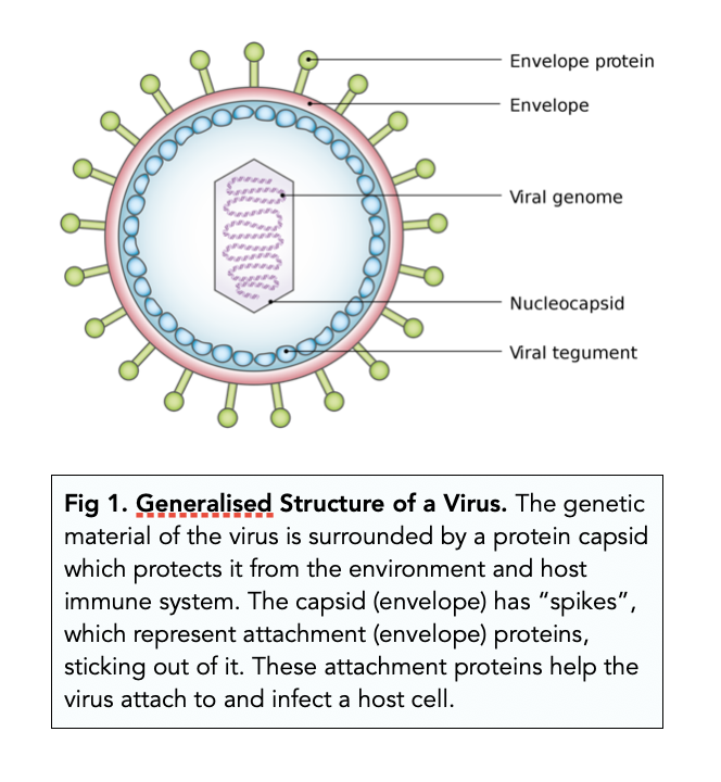
describe the virus structure
non cellular
no plasma membrane, cytoplasm or ribosomes
ONLY DNA or RNA
protein coat=capsid.
it is a protective coat
may have 1 or 2
many viruses also have a lipid envelope
some proteins may be present

describe bacterial structure
all bacteria HAVE:
plasma membrane
cytoplasm
peptidoglycan cell wall made of chains cross linked by amino acids
70s ribosomes
circular DNA
DNA is ‘naked’, not associated with histone proteins
all bacteria do NOT HAVE:
membrane bound organelles
nucleus—DNA lies free in cytoplasm in the nucleoid region


how do viruses reproduce?
all parasitic
can only reproduce by infecting living cells
uses protein synthesising machinery of host cell to replicate

what is the causative agent of cholera?
a bacteria called Vibrio Cholerae
what is the causative agent of malaria?
a protoctist called either:
Plasmodium falciparum
P. malariae
P. vivax
P. ovale
what is the causative agent of HIV/AIDs?
(retro)virus called Human Immunodeficiency Virus
what is the causative agent of tuberculosis?
a bacteria called Mycobacterium tuberculosis/ M. bovis
M. tuberculosis—>TB in humans
M. bovis—> TB in cows + humans
what is the causative agent of smallpox?
a virus called Variola virus
what is an epidemic?
an outbreak of disease that attacks many peoples at about the same time and may spread through one or several countries
what is an endemic?
a disease that exists permanently in a particular region/population(ie malaria)
describe the cholera causative agent
Vibrio cholerae—a bacterium
comma shaped, has flagella, motile
explain how cholera is transmitted
through the ‘fecal-oral’ route:
large numbers of Vibrio cholerae found in faeces of infected people
infected person’s faeces/sewage contaminates water/food
houseflies land on faeces and contaminate food/water
uninfected person eats contaminated food/water
describe the mode of action of cholera in the body
if bacteria is not killed by stomach acid—>reaches small intestine
bacteria secrets choleragen toxin
toxin binds to complementary receptor on intestinal epithelial cell—>enters via endocytosis
disrupts function of intestine epithelium lining
loss of Cl- ions and Na + ions from epithelial cells
W.P decreases, water moves out from blood, down W.P gradient by osmosis thru partially permeable membrane
causes severe diarrhoea + dehydration
what are the symptoms of cholera?
diarrhoea
severe dehydration
loss of water and salts
weakness and fatigue
low BP
weight loss
vomitting
how do we diagnose cholera?
microscopic analysis of faeces
how do we treat cholera?
oral rehydration therapy:
use oral rehydration solution(ORS) that has glucose and salts
ensures fluid intake=fluid loss in urine and faeces
maintains osmotic balance of blood and tissue fluids
what is the global distribution of cholera?
endemic in developing countries
outbreak follows natural disasters/war
due to absence of proper sanitation—>no treatment of faecal waste—>water supply contaminated
poor hygiene + poor living conditions
lack of education of transmission
what other problems can cholera cause?
there are many different strains of cholera—>cannot make a vaccine for it
each strain is more virulent than the last
adults can be reinfected with cholera
what is malaria caused by in humans?
plasmodium falciparum—75%
P. malariae, P. ovale, P. vivax—25%
what is a vector?
an organism that carries disease from a person to another
how is malaria transmitted?
vector—>female Anopheles mosquitoes
only females take blood meals to supply eggs with nutrients
blood transfusions, use of unsterile needles, and can pass across placenta from mother

symptoms of malaria
fever
anaemia
nausea
headaches
muscle pain
shivering
enlarged spleen

diagnosis for malaria
microscopical analysis of blood
dip stick test for malaria antigens in blood
treatment for malaria
anti-malarial drugs:
quinine, artemisin
chloroquine—>inhibits protein synthesis + prevents parasite from spreading in body
proguanil—>inhibits sexual reproduction of plasmodium in the mos
combination therapy—>multiple drugs are used at the same time to prevent drug resistance
how do we prevent malaria?
use preventative drugs
reduce number of mos
prevention of bites
global distribution of malaria
endemic in tropical + subtropical areas
this is because:
the vector survives and breeds in hot + humid areas
needs still/stagnant water to reproduce
plasmodium reproduces within mos >20 degrees C
what other problems does malaria cause?
drug resistant plasmodium
insecticide resistant mos
global warming resulted in more area for mos to breed
what does HIV have?
it is an RNA virus/retrovirus
contains single stranded RNA as genetic material
has a protein coat/capsid made of capsomeres
has outer viral envelope made of a lipid bilayer + proteins mostly derived from host
has viral glycoproteins on the outer envelope
2 enzymes:
reverse transcriptase: uses RNA as template to produce DNA in host cell
protease: cleave/process new viral proteins
life cycle of HIV
the viral RNA + reverse transcriptase(RT) enters T helper lymphocytes
RT converts RNA into DNA
the viral DNA is incorporated into the host DNA
the cell’s machinery is used to express viral proteins(through transcription + translation)
viral proteins are assembled into many new viruses
describe the transmission of HIV
direct exchange of bodily fluids
virus is unable to survive out of the human body
semen, vaginal fluids during sexual intercourse
blood transmission via infected blood transfusion/contaminated syringes
mother to baby transmission across placenta/breast milk
anal intercourse increases risk—>less natural lubrication in the rectum—>rectal lining is easily damaged + virus can pass from semen into blood
symptoms and effects of HIV
slow infection—virus can stay dormant for years
changes surface proteins to hide from immune system
HIV infects T helper cells—>destroyed T helper cells—>numbers decrease—>body unable to defend itself against infection
flu like symptoms and then symptomless
opportunistic infections can occur as a result of a compromised immune system
collection of opportunistic diseases associated with immunodeficiency caused by HIV=AIDs
symptoms and effects of AIDs
opportunistic infections:
oral thrush
pneumonia
cancers
neurogenerative diseases like dementia
TB
malaria
malnutrition+ weight loss + diarrhoea + fever + sweating
how is HIV/AIDs treated?
no cure for AIDs
drug therapy for HIV:
Zidovudine: similar to Thymine—>binds to viral enzyme reverse transcriptase and inhibits it
drugs can target viral protease and inhibit it
expensive + many side effects
combination therapy:
strict pattern + timing of medication
if not followed properly, patients may develop strains of HIV resistant to drugs
how do we prevent HIV?
protected sex
HIV testing
contact tracing= where the person with HIV traces people he/she has put risk of infection and that person to provided an HIV test
no needle sharing
donated blood is screened for HIV + heat-treated to kill viruses
control mother-to-child transmission using drugs
HIV+ women of high income countries should use formula
what other problems does HIV/AIDs cause?
difficult to reach people for widespread testing in rural area
many symtomless carriers
drugs are expensive + cause side effects
pandemic
how is TB transmitted?
mycobacterium tuberculosis:
by aerosol infection
pathogen is in airborne droplets
infected person coughs/sneezes—>uninfected person breathes it in
mycobacterium bovis:
from infected cows/cattle
eat undercooked + contaminated meat
drink unpasteurised milk containing bacteria
effects of tuberculosis
site of primary infection: lungs
secondary infection: lymph nodes + bones + gut
slow infection—many infections are controlled by the immune system
bacteria may be activated after many years when immune system is weakened by other infections(ie HIV)
incubation period is a few weeks-years

symptoms of TB
central:
appetite loss
fatigue
lungs:
chest pain
coughing up blood
prolonged cough
skin:
night sweats
pallor

how do we diagnose TB?
x ray
microscopical examination of sputum(mucus + pus) for bacteria
how is TB treated?
long term because bacteria is slow growing + not responsive to drugs
combination therapy:
use multiple antibiotics to prevent drug resistance
DOTS( direct observation treatment, short course):
makes sure patients take medicine regularly and reduce spread of drug resistance
how do we prevent TB?
BCG(Bacillus Calmette-Guerin) vaccine derived from M. bovis
isolate patients during infectious stages
contact tracing
cattle tested for TB
pasteurise milk
other problems that emerge from TB
multiple drug resistant TB
drugs like isoniazid no longer effective
not completing course of treatment increases risk of drug resistant TB
describe the global distribution of TB
endemic all over the world
high rates in cities due to high migration rate
overcrowded area
areas where HIV/AIDs is prevalent—>TB is an opportunistic infection—>HIV/AIDs causes weakened immune system—>increases risk of TB development + dormant TB more likely to become active in HIV+ person
what are antibiotics?
drugs used that are derived from microorganisms
kill/inhibit growth of bacteria without harming infected organism
bacteriocidal antibiotics—kill bacteria
bacteriostatic antibiotics—inhibit bacterial growth
prevent spread of bacteria in body
have no effect on human cells AND viruses

how do antibiotics work?
inhibit bacterial cell wall synthesis
inhibit activity of specific membrane protein/glycoprotein—>block binding to cells
block specific enzyme action
inhibit protein synthesis + nucleic acid synthesis—>target enzymes that bacteria have but humans don’t

what happens to bacteria in the absence of penicillin?
bacterial wall is made of peptidoglycan
when bacterial cells grow—>secretes autolysins
autolysins make tiny holes to allow the cell wall to stretch
new peptidoglycan formed
peptidase enzyme forms cross links between peptidoglycan chains—>form cell wall
what happens to bacteria in the presence of penicillin?
penicillin inhibits peptidase enzyme
stops formation of cross links between peptidoglycan polymers in the cell wall
autolysins make tiny holes but new peptidoglycans that are formed cannot link up
cell wall is weaker
cell walls unable to withstand turgor pressure when water moves by osmosis—>bacteria lyses and dies
penicillin is only effective when bacteria is growing
what is the spread plate method and what is it used for?
antibiotic discs are placed in a petri dish with growing bacteria
the diameter of the zone of inhibition shows how effective the antibiotic is
method:
sample is pipetted onto surface of agar plate
sample is spread evenly over surface of agar using sterile glass spreader
sample is incubated
final results: the one with the bigger inhibition zone=most effective
why are antibiotics not effective against viruses?
viruses do not have a peptidoglycan cell wall, have a protein coat
viruses do not have their own metabolism, rely on host cells
viruses have no cell structure/very few organelles—>very few sites for antibiotics to act on
viruses live inside host cells—>out of reach for antibiotics
antivirals exist—>target viral glycoproteins on viral envelope—>prevent binding of virus to host cells + inhibit specific viral enzymes
what is antibiotic resistance?
when antibiotics are no longer effective against bacteria
can spread from bacteria to bacteria
many bacteria have penicillinase enzymes which break down penicillin—>resistant to penicillin
what is antibiotic resistance caused by?
spontaneous mutation in bacteria
mutation causes change in protein/production of new protein that cannot be targeted by antibiotics
natural selection enables resistance genes to spread
antibiotic is the selection pressure—>antibiotics only kill non resistant bacteria
resistant bacteria survive + reproduce—>antibiotic gene is passed to next gen + bacteria

what causes antibiotic resistance?
patients not completing their course for antibiotics—>some susceptible bacteria survives—>bacteria replicates + have increased chances of mutation/becoming resistant
what are the consequences of antibiotic resistance?
bacteria can carry several antibiotic resistant genes—>develop multiple resistance
become ‘super bug’
wounds do not heal + continuously getting infected with bacteria
bacteria cannot be killed by common antibiotics—>need to be controlled by stronger antibiotics—>but we are running out of antibiotics
what is the solution to antibiotic resistance?
discover more antibiotics by slightly modifying preexisting ones but discovery takes time
prevention:
only use antibiotics when prescribed
always finish course
never use left over antibiotics
do not share antibiotics
good hygiene
What are the two types of microscopes?
light and electron
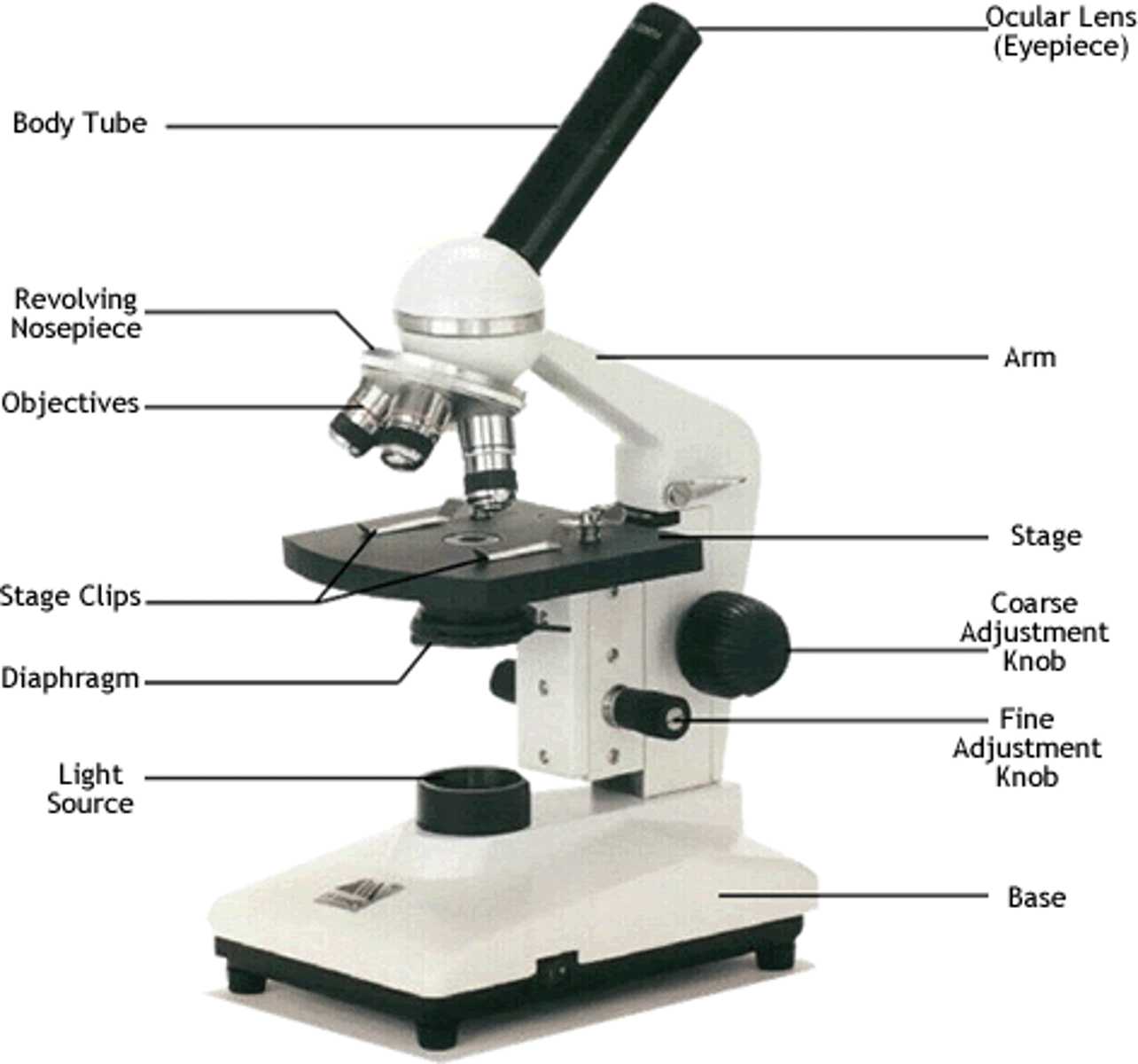
light microscope
An optical instrument with lenses that refract (bend) visible light to magnify images of specimens.
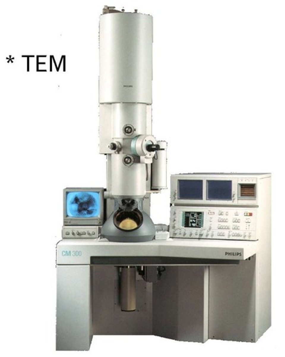
electron microscope
a microscope that focuses a beam of electrons to magnify objects
what do light microscopes use as their source of light radiation?
visible light
What is the typical magnification of a light microscope?
10x
How many lenses are typically below the eyepiece in a light microscope?
three to four lenses, known as an objective lens
what is the function of an objective lens?
helps to magnify the object of interest
How do you work out magnification using the magnification of the eye piece and the objective lens?
eye piece M x objective lens M
ie: 10x 10= 100
What can light microscopes be used for?
- Light microscopes & stains can be used to visualise cells
- The light from the microscope shines through the specimen, allowing it to be visualised
- Watch cell division take place
what do electron microscopes depend on?
Rely on the specimens to be stained with heavy metal elements and placed in a vacuum, hence, only non-living organisms can be viewed with it
What type of specimens do electron microscopes show?
-biological specimens
-inorganic specimens
what biological specimens does the electron microscope show?
-microorganisms
-cells and tissues
what is magnification?
The ratio of an object's image size to its real size
What is resolution?
The ability to clearly distinguish the individual parts of an object, the higher the resolution, the closer the two objects are to each other, meaning
What is resolution determined by?
The wavelength of light used and the numerical aperture of the lens
What is aperture?
The size of the opening of the lens
what is the general rule of resolution to visible light?
The longer the wavelength, the lower the resolution
what is the wavelength of visible light?
400-700nm
What is the resolution of visible light?
200nm, meaning objects closer together than 200nm cannot be seen as two distinct object points
electron microscope vs light microscope
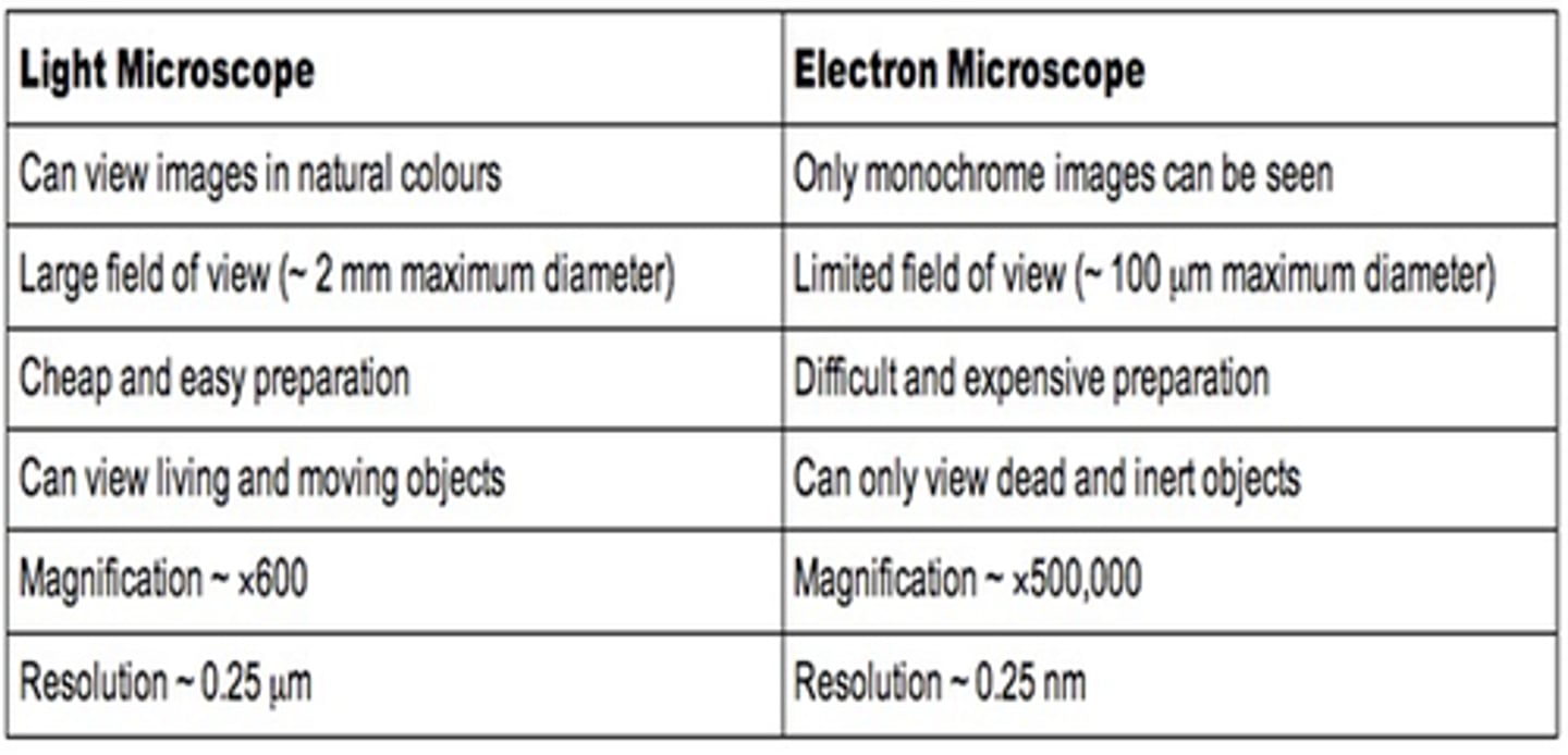
How do you prepare a microscope slide using a liquid specimen?
- Add a few drops of the liquid sample to a clean slide using a pipette
- Lower a coverslip over the specimen and gently press down to remove air bubbles
How do you prepare a microscope slide using a solid specimen?
- Use scissors or a scalpel to cut a small sample of the tissue, and peel away or cut very thin layers of cells
- Place the sample onto a slide and add a drop of water
- Apply iodine stain
- Lower a coverslip over the specimen and press down to remove air bubbles
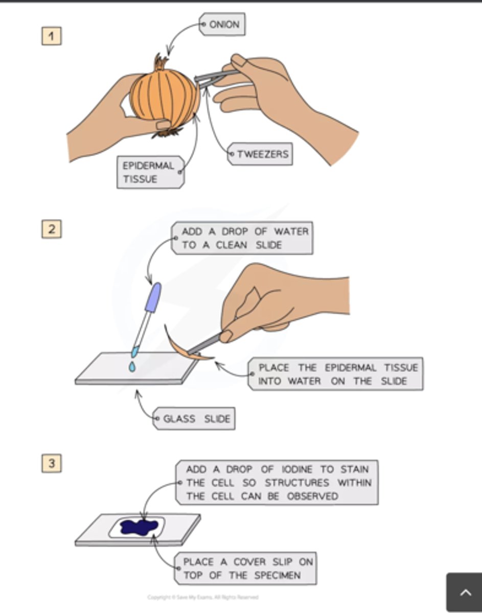
Why do we prepare small samples in a solid slide prep?
to allow light to pass through
how do you prepare a slide using human cells?
1- Brush teeth thoroughly to remove any bacteria, so as not to obstruct the view of the cheek cells
2- Take a sterile cotton swab and gently scrape the inside cheek surface of the mouth for -10 seconds
3- smear the cotton swab on the centre of the microscope
4- Add a drop of methylene drop solution
5- This causes the nucleus and mitochondria to appear darker than their surroundings
6- Place a coverslip on top
7- Lay the coverslip down at one edge from a 45-degree angle and then gently lower the other edge until it is flat to remove gas bubbles
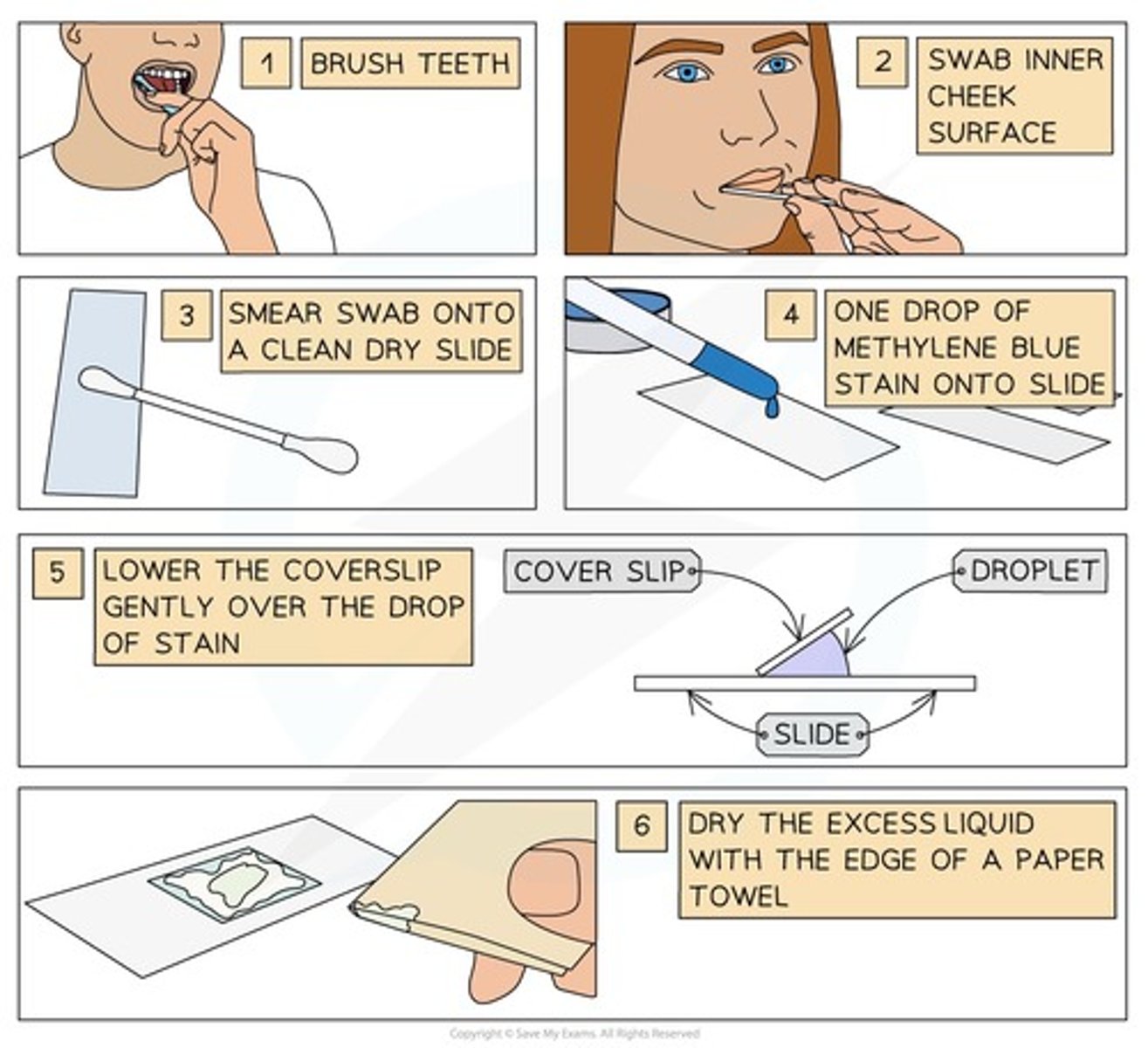
why do we use methylene blue drop solution when preparing a slide using human cells?
Methylene blue stains charged molecules in the cell, including DNA and RNA
why do we use coverslips?
to protect the microscope lens from liquids and help prevent drying out
what are permanent slides?
microscope slides that have a specimen preserved and mounted in a solid mounting medium, making them suitable for long-term storage and observation
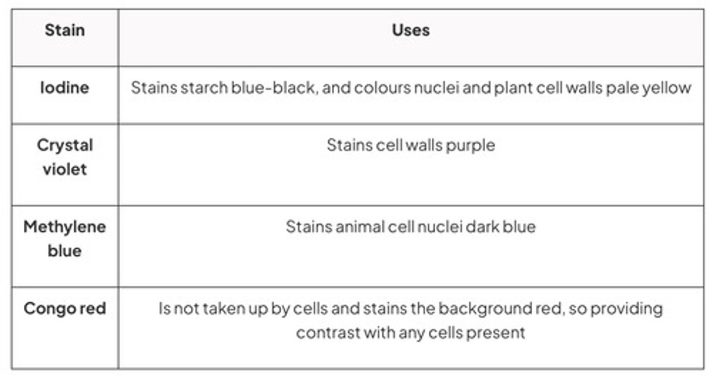
What is staining?
colouring microorganisms with a dye that emphasises certain structures
common microscope stains and uses
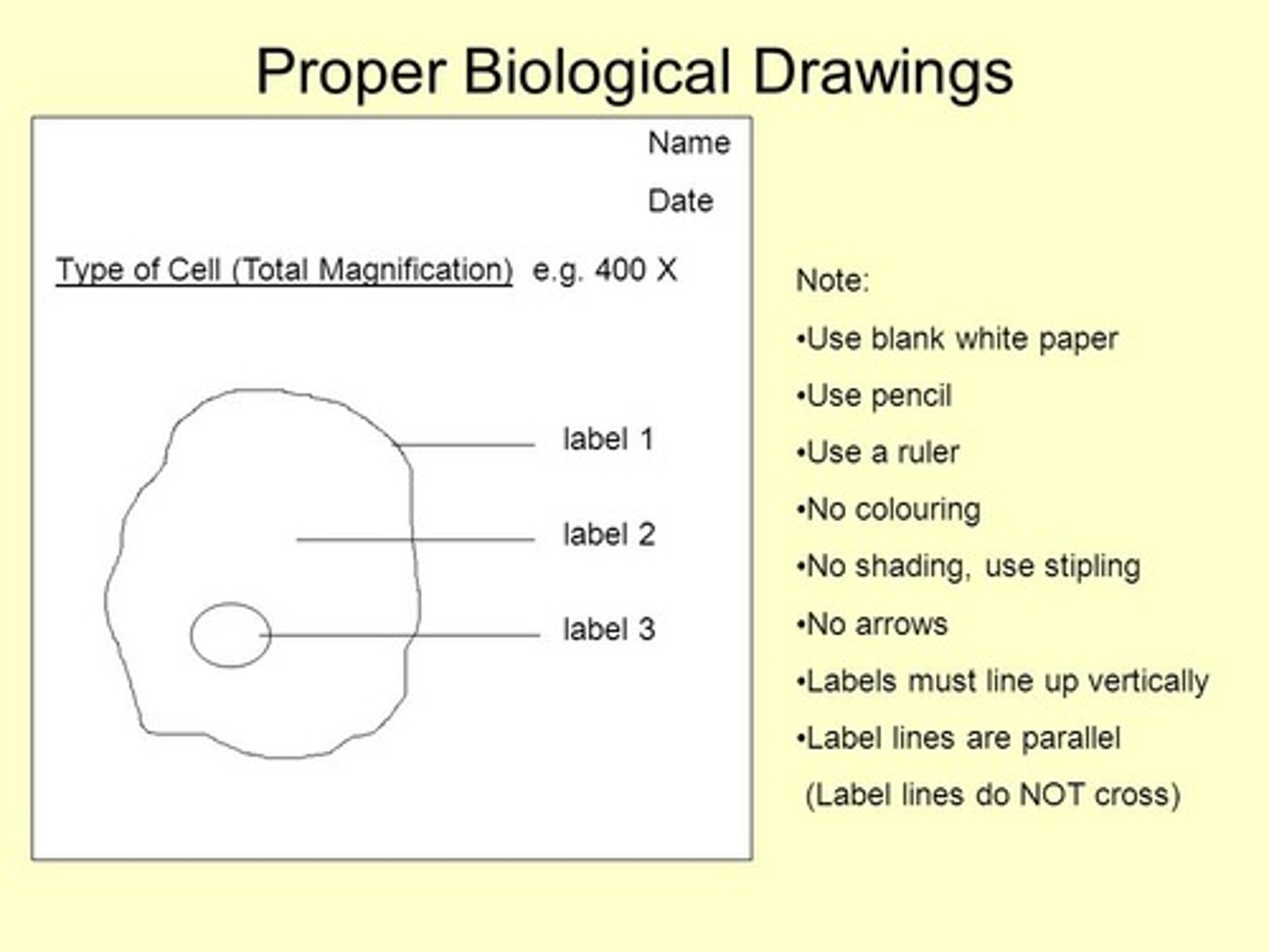
what is the function of a biological drawing?
to record the observations seen under a microscope
what are biological drawings?
Biological drawings are line pictures which show specific features that have been observed when the specimen was viewed.
What are the rules when drawing a biological drawing?
1- title
2- The magnifications under which the observations shown by the drawings are made and recorded
3- no shading or no a lack of clear lines
4- Well-defined structures should be shown
5- Drawing takes up most of the page and is made with proper proportions
6- clearly labelled without arrows and connected directly to the labelled part
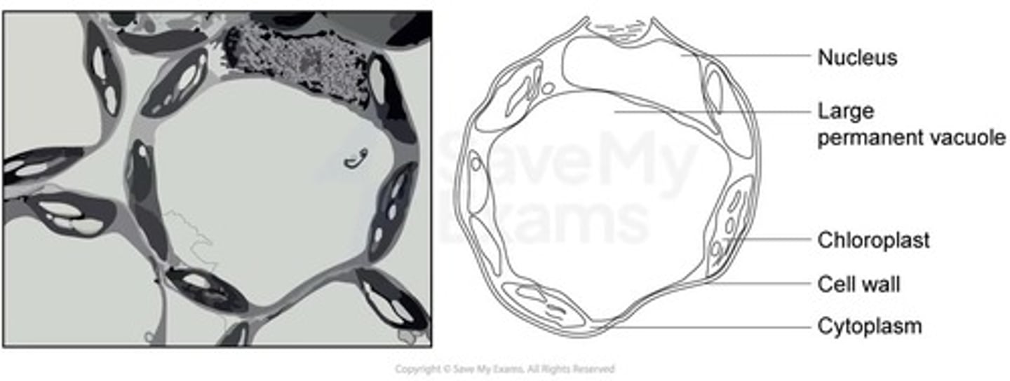
when are drawings with higher magnification made?
when visualising cells at a higher magnification power
when are drawings with lowermagnification made?
tissues viewed under lower magnifications
plant cell biological drawing
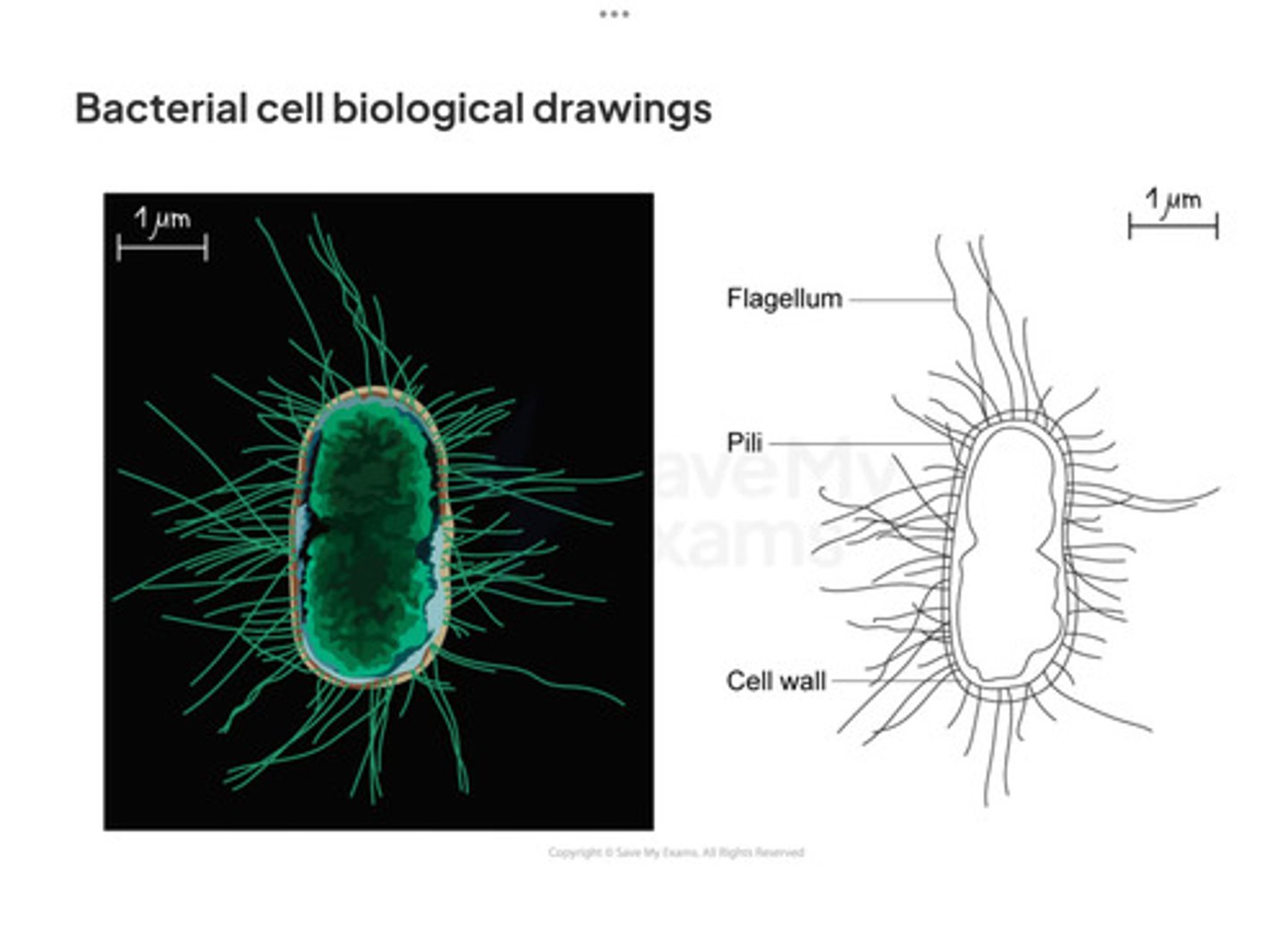
bacterial cell biological drawing general
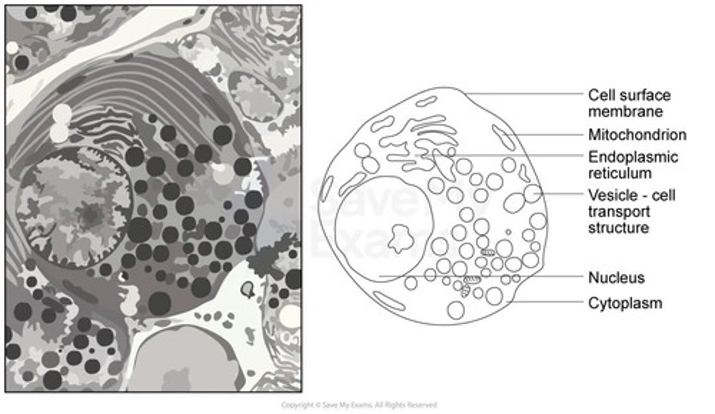
animal cell drawing
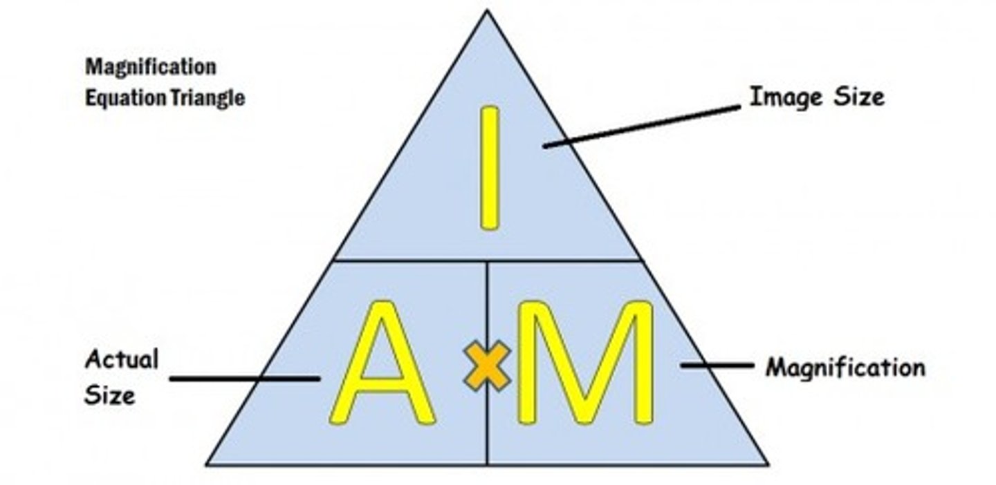
how do you calculate magnification?
magnification = image size/actual size
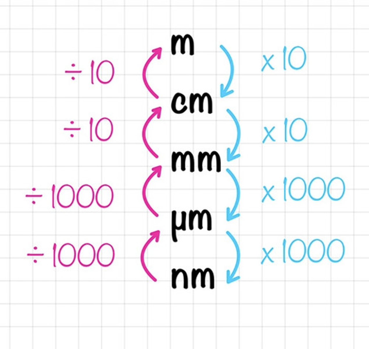
How many micrometres are in a millimetre?
1000 µm = 1 mm
How many nanometers are in a micrometer?
1000 nanometers
converting units