3 BIO
 Call Kai
 Learn
 Practice Test
 Spaced Repetition
 Match
 Flashcards
 Knowt Play
1/45

There's no tags or description
Looks like no tags are added yet.
46 Terms
What is BIO used to assess?
evaluates ocular fundus from the posterior pole out to the ora serrata
What type of view does BIO give?
→ stereoscopic, wide-angled, high resolution view of the entire retina and overlying vitreous
gives 1.5x - 3.5x mag
What type of image is formed between the examiner and condensing lens?
Real, magnified, inverted (upside down), reversed (backwards)

List 8 indications for conducting BIO.
Comprehensive exam
Dilated fundus exam (DFE)
Follow up exam
Symptomatic patients: (+) flashes/floaters, ‘curtain’ in vision
Hx peripheral vision loss
Blunt ocular trauma
Pre- and post- ocular surgery exam (cataract, refractive)
Intraocular inflammation pt
Systemic disease affecting retina (ie. DM, HTN, sickle cell) pt
What are the contraindications for BIO?
Occludable angles
Pregnancy
List the pros of BIO.
Stereoscopic views
↑ FOV
Bright light
Minimal peripheral distortion
When coupled with scleral indentation, you get a view of very far periphery
Variety of filters
List the cons of BIO.
Requires pupil dilation
Moderate glare discomfort for pt
Low mag (20 D lens)
Initial adjustment for examiner to inverted and reversed images
Takes time to get proficient in this skill
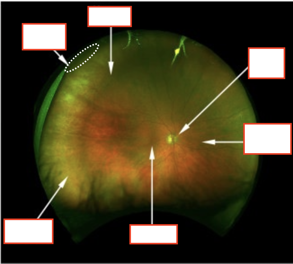
Label.

Compare the optical properties, working distances, and primary indications for +20D, +15D, and +28D condensing lenses used in BIO.
Rule: Bi-convex aspheric lens held with the silver ring (more concave side) facing the pt
.
+15D Lens
↑ mag (4.11x) = ↓ FOV
WD = ~7.5 cm
(+) Best for detailed evaluation of the posterior pole
.
+20D (Standard Lens)
~2.5x mag = 8 disc FOV
WD = ~5 cm from cornea
(+) Best general-purpose lens for fully dilated pupils
.
+28D Lens
↓ mag (~2.2x) = ↑ FOV
WD = ~3 cm
(+) Best for poorly dilated or small pupils (e.g., diabetic pt)
What is a main reason you’d want to do reclined BIO?
strong suspicion of a retinal tear/detachment
List 3 reasons to perform Seated BIO.
Frail, elderly pt with neck or back problems
Breathing or cardiac issues and can’t be reclined
Vestibular issues/suffering with vertigo
How should the pt’s chair be when evaluating superior vs inferior retina?
Elevated = superior retina
Lowered = inferior retina
What direction would the pt need to into if you see a lesion?
Look in the direction of retina you would like to examine
What do you do if a pt refuses dilation, but you still must get the best view of the fundus?
undilated 90D
What effects could dilation have on the patient’s vision?
Reading (especially if a younger pt or latent hyperope)
Ability to drive (cataracts can make this worse)
Light sensitivity
Troubleshooting - If you’re getting an image, but it’s blurred what do you do?
be sure you are not too close to the pt (your arms are fully extended, but not locked)
you’re fully corrected
Troubleshooting - If only seeing a red reflex or the lens is only partially filled with fundus details. What do you do?
Trombone the lens, and make sure the pt’s pupil, condensing lens and oculars are all lined up
make sure BIO light is completely in the lens and not the pt’s face
Troubleshooting - What do you do to get far peripheral views?
try tilting your head slightly and re-directing the light
Troubleshooting - What do you do if the pt is having difficulty maintaining eyes open/fixation?
↓ illumination
Move light out of patient’s eye more frequently
Yellow filter
Troubleshooting - What does it mean if you’re seeing vertical diplopia?
Head or instrument is tilted
examiner decompensated vertical phoria
Troubleshooting - What does it mean if you’re seeing horizontal diplopia?
BIO PDs are off
you’re too close to the patient
examiner decompensated horizontal phoria
What would you document if everything was normal?
Retina: flat, no holes, tears, RD or abnormalities 360o OU
What would you document if everything was abnormal?
What are you seeing (do you have a diagnosis?)
dot and blot hemorrhages
choroidal nevus
chorioretinal scar
Location (at the equator, ~ 1:00 vs. ‘superior’)
Size/ extent – you can use disc diameters (DD)
Shape and color (if don’t have an actual diagnosis for your finding)
Elevation? (if a growth/pigmentation)
Diagnose this lesion.

CHRPE (~1.5DD, with flat, distinct margins, located at inferior arcades,~3DD inferior temporal to macula)
Diagnose this lesion.

OS: Atrophic hole @3:00 approaching ora, ~½ DD in size, surrounded by pigment
No tears or RD 360o
What are the 3 regions used to segment the intraocular views, and what are their boundaries/structures?
Posterior Pole
macula, ONH
vascular arcades
.
Mid-periphery
anterior edges of vascular arcades to the vortex veins
.
Periphery
anterior to vortex veins

What are the retinal landmarks?
Equator – not really a landmark, but an imaginary line connecting the ampullae of the vortex veins
Vortex veins – approx. 1/quadrant but 70% of eyes can have more than 4 vortex veins
Long ciliary nerves – 1 at 3:00 + 1 at 9:00, extend mid-periphery to ora, sensory innervation the eyeball
Short ciliary nerves – ~6:00 + 12:00, numbers vary

What are these?

Vortex veins (look like a spider web)
What are these?
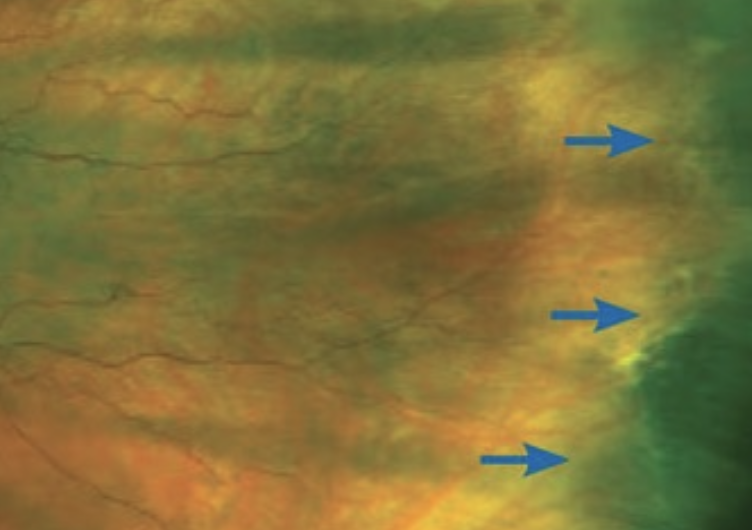
Ora serrata
What is this white arrow pointing to?

Long posterior ciliary nerve
What is this?

Ora serrata pearls
What kind of fundus is this?

Patient with albinism (⭐ choroidal vessels easily visible due to lack of pigment)
What kind of fundus is this?

Tessellated/Tigroid fundus (high myope)
⭐staphyloma, tilted optic nerve
What is White Without Pressure (WWP)? Mention who it’s commonly found in and how it’s managed?
→ Distinctive white appearance of the peripheral retina visible without any depression or indentation
Whitish-gray, distinct borders
bilateral, located temporally
occurs b/w the ora and equator (may extend posterior to equator)
hard to see on fundus examination
associated with abnormal vitreo-retinal surface changes but may just be a shift in the reflectivity of the PR outer layers (more reflective than adjacent retina on OCT)
common in even healthy patients (e.g., myopes, pale fundus pt)
Management: No treatment, Annual DFE


What is Dark Without Pressure (DWP)? Who is it common in? How is it managed?
→ Flat, darker area on the retina, surrounded by normal appearing retina
involves a temporary change in how light reflects off the EZ → can shift location, change in size
common in darker pigment retina
visible without any depression or indentation
located in mid-periphery
benign (can be assoc with sickle cell disease)
exact cause is unclear, but OCT shows it
Treatment: None

What is Lattice Degeneration? Who is it common in? How is it managed?
→ Oval or elliptical area of retinal thinning
vitreoretinal degeneration affecting inner retinal layers and vitreo-retinal surface
located b/w the equator and posterior border of the vitreous base
occurs PARALLEL to the ora
unilateral or bilateral
common in moderate myopes (also in some hyperopes)
may have small round holes within the lattice
may develop tears at the lattice border
may see pigmentation in/around areas of lattice
may see sclerosed vessels within (⭐ why its called lattice)
Management: Depends on whether lattice has holes, any pigment, lattice location, previous h/o retinal tears


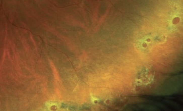
What are Atrophic Retinal Holes?
→ Full thickness breaks in the retina due to retinal thinning (not caused by V-R traction)
might see pigment around it b/c the retina is trying to heal it with pigment
can also see fluid near it
can pinpoint to 2DD in size
single hole or multiple
longstanding, can be surrounded
Management: Depends on size of hole, if any fluid around hole, if any pigment around the hole

What do Retinal Tears, Retinal Detachments (RD) occur due to?
due to vitreous pulling on the retina
What is this an image of?

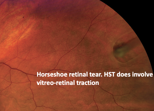
What is this an image of?


What is this an image of?

White Without Pressure
What is this an image of?

Dark Without Pressure (DWP)
What is this an image of?

Lattice Degeneration
What is this an image of?

Lattice Degeneration
What is this an image of?

Atrophic Retinal Holes
What is this an image of?

Atrophic Retinal Holes