Cross-Sectional Abdominal Anatomy and Ultrasound
 Call Kai
 Learn
 Practice Test
 Spaced Repetition
 Match
 Flashcards
 Knowt Play
1/59

There's no tags or description
Looks like no tags are added yet.
60 Terms
largest organ within the body that is inferior to the diaphragm and covered by the ribs
liver
potential space that surrounds the liver and goes anterior to the stomach; where fluid may accumulate
greater sac
area where the greater and lesser sacs meet; where fluid can spill out into the greater area
foramen of Winslow (epiploric foramen)
part of the pancreas that is the most inferior; sits behind duodenum and infront of IVC
head
part of the pancreas that sits more midline and is anterior to the portal confluence
neck
part of the pancreas that sits directly behind the stomach
body
most superior part of the pancreas that sits towards the splenic hilum
tail
what two organs are in front of the pancreas
stomach and liver
organ posterior to the right lobe of the liver that courses anterior
gallbladder
kidney should be ________ or slightly more ________ than the liver
isoechoic; hypoechoic
where does the gallbladder sit in relation to the right lobe of the liver?
posterior
in TRV what should you see in the most superior part of the liver?
hepatic veins as tubes dumping into the IVC
in TRV what should you see in the mid part of the liver?
part of the portal system and its branches; caudate lobe sitting anterior to IVC and ligamentum venosum
in TRV what should you see in the inferior part of the liver?
main portal vein as a tube and gallbladder starting to come in
what regions does the liver occupy?
Rt lumber, Rt hypochondriac, part of the epigastric and the Lft hypochondriac
appers more hypoechoic than pancreas, more hyperechoic than kidney
liver
organ that lies posterior and lateral to the left lobe of the liver
stomach

part of an organ that lies inferior to the left lobe of the liver
body of the pancreas
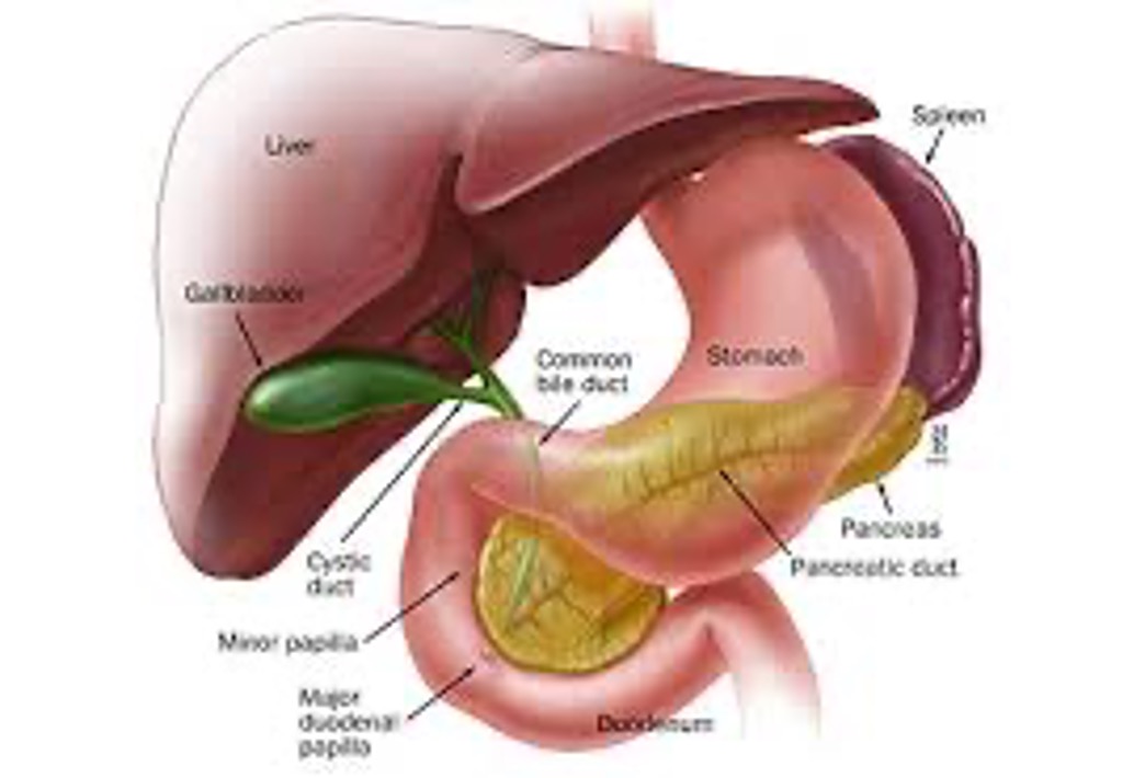
sits on the anterior surface of the liver; secures the liver to the diaphragm and the anterior abdominal wall
falciform ligament
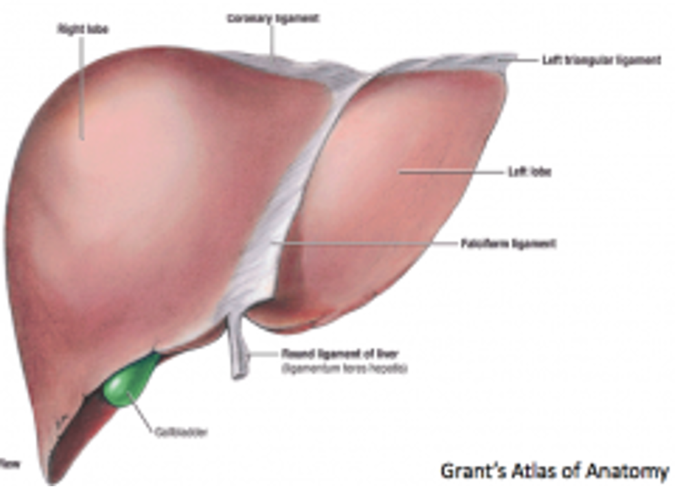
a large area that rests on the diaphragm which is not covered by peritoneum; an area where fluid would NOT accumulate
bare area

space located inferior to the Rt lobe
Rt subheptic space

area in the the Rt subheptic space that lies between the posterior Rt lobe and the Rt kidney; first place where fluid WOULD accumulate
Morison’s pouch

lies in the epigastric and Lft hypochondriac regions
left lobe of the liver
has two segments (medial and lateral)
left lobe of the liver
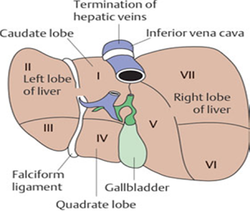
segment I of the liver
caudate lobe

segment II of the liver
superior lateral left lobe
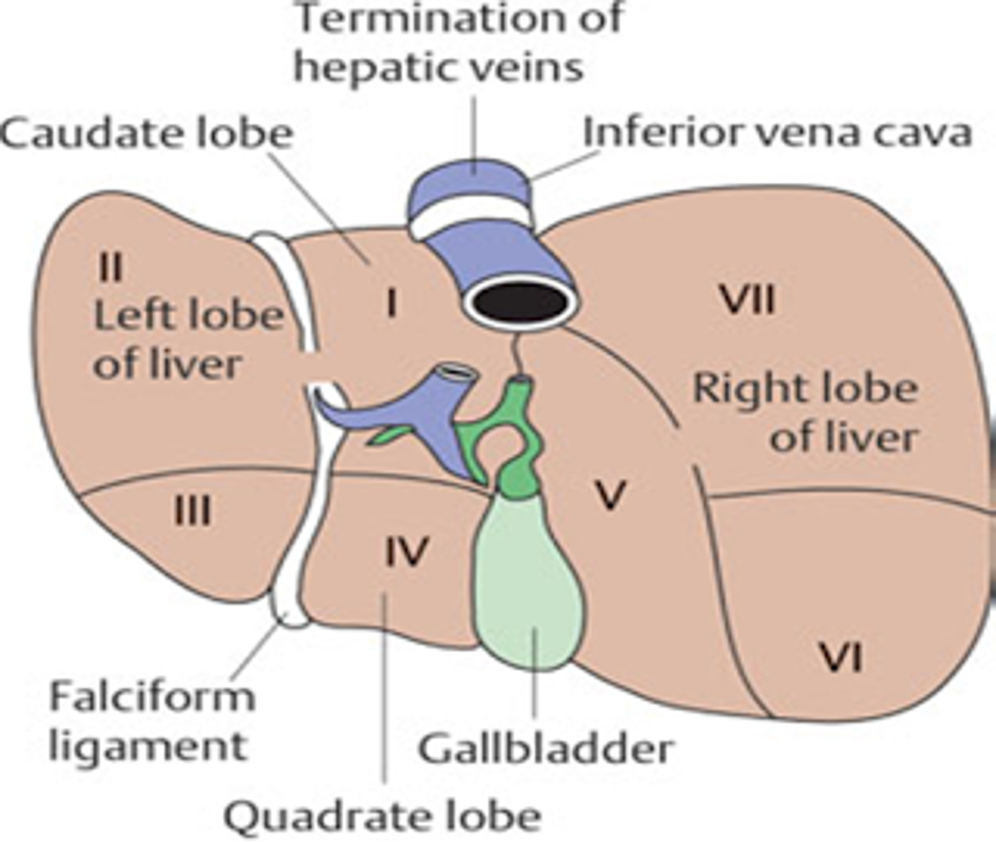
segment III of the liver
inferior lateral left lobe

segment IV of the liver
quadrate lobe; inferior medial segment

what structures border segement IV?
porta hepatis is superior and GB fossa is lateral on the right side

What anatomical structure separates Segment III from Segment IV?
falcifrom ligament

small lobe on the posterior superior surface of the liver; segment 1
caudate lobe
what is the right side of the caudate lobe bounded by?
IVC fossa
what is the left side of the caudate lobe bounded by?
ductus venosum
largest lobe of the liver
right lobe
part of the liver that occupies the Rt hypochondrium
right lobe
part of the liver that contains 3 fossa
right lobe
what are the 3 fossa of the right lobe?
porta hepatis, GB fossa, and the IVC fossa
normal anatomical variant, extension of rt lobe of liver down into abdomen or pelvis; past right kidney
Reidel’s Lobe

appear echogenic or hyperechoic due to the composition of collagen and fat
ligaments and fissures
connective tissue that completely surrounds the liver
Glisson’s capsule
separates Rt and Lft lobes of the liver
main lobar fissure (median fissure)

hyperechoic line going from the portal vein to the neck of the gallbladder; used to find gallbladder in LNG
main lobar fissure (median fissure)

extends from the umbilicus to the diaphragm in the sagittal plane and contains the liagmentum teres
Falciform Ligament

rounded termination of the falciform ligament that divides the medial and lateral segments of the left lobe of the liver
Ligamentum Teres (round ligament)
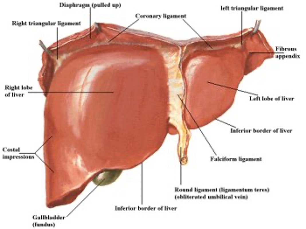
separates the left lobe from the caudate lobe
Ligamentum Venosum
seen just inferior to the dome of the liver as a horizontal line just anterior to the caudate lobe of the liver and the IVC
ligamentum venosum

pear-shaped sac in the anterior aspect of the right upper quadrant closely related to the liver surface
gallbladder

rounded fundus projects below the inferior margin of the liver
gallbladder

occupies epigastric and Left hypochondriac regions
pancreas
appears isoechoic or more echogenic than adjacent left lobe of the liver
pancreas
extends in a horizontal oblique position from the 2nd portion of the duodenum to the splenic hilum
pancreas
located in the retroperitoneum posterior to the stomach, duodenum and proximal jejunum of the small bowel
pancreas
divided into head, neck, body and tail
pancreas
lies in the posterior left hypochondrium along the shaft of the 10th rib. is an intraperitoneal organ covered in peritoneum
spleen

organ that lies between the fundus of the stomach and the diaphragm
spleen

inferior medial surface is in contact with the stomach, left kidney, pancreas and splenic flexure
spleen

attaches the spleen to the kidney; lies in contact with the posterior peritoneal wall
splenorenal ligament

what other ligaments also support the spleen?
the splenophrenic ligament and the gastrosplenic ligament

important structure that seperates the greater and lesser sacs
Gastrosplenic Ligament
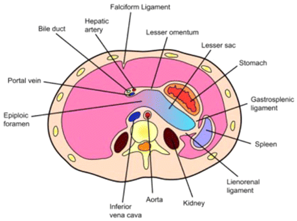
“spaces” within the abdominal cavity where fluid may accumulate
the greater and lesser sacs
