Dermatomes mine
 Call Kai
 Learn
Learn Practice Test
 Spaced Repetition
 Match
Match Flashcards
 Knowt Play
1/77

There's no tags or description
Looks like no tags are added yet.
Name | Mastery | Learn | Test | Matching | Spaced | Call with Kai | Chat |
|---|
No analytics yet
Send a link to your students to track their progress
78 Terms
an area of skin supplied primarily by sensory fibers from a SINGLE spinal nerve root
dermatome
muscle or group of muscles supplied by a specific spinal nerve
myotome
what are the four options for documentation of dermatomes
normal, diminished, absent, altered
to assess myotomes, the joint is placed in ___ ___ to optimize muscle function and the patient holds position against 5 seconds of sustained resistance
mid range
myotomes use ___ ____ contractions
resisted isometric
normal strength for myotome
5
movement against some resistance for myotome
4
movement against gravity for myotome
3
minimal to no movement for myotome
2

what is the representative movement for C5
shoulder aduction

what is the representative movement for C6
wrist extension

what is the representative movement for C7
elbow extension

what is the representative movement for C8
finger flexion, thumb abduction

what is the representative movement for T1
finger abduction

what is the representative movement for L2
hip flexion
what is the representative movement for L3
knee extension

what is the representative movement for L4
ankle dorsiflexion

what is the representative movement for L5
great toe extension
what is the representative movement for S1
ankle plantarflexion

what is the representative muscle for C5
deltoid, supraspinatus

what is the representative muscle for C6
extensor carpi radialis longus, brevis
what is the representative muscle for C7
triceps brachii

what is the representative muscle for C8
flexor pollicis longus, flexor digitorum profundus

what is the representative muscle for T1
dorsal interossei

what is the representative muscle for L2
iliopsoas
what is the representative muscle for L3
quadriceps

what is the representative muscle for L4
tibialis anterior

what is the representative muscle for L5
extensor hallucis longus

what is the representative muscle for S1
gastrocnemius, soleus

what is C5-C6 representative of
elbow flexion
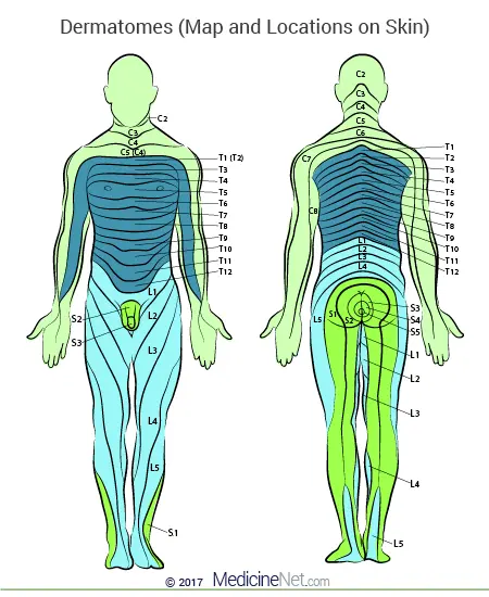
what is the C4 dermatome
AC joint

what is the C5 dermatome
lateral elbow
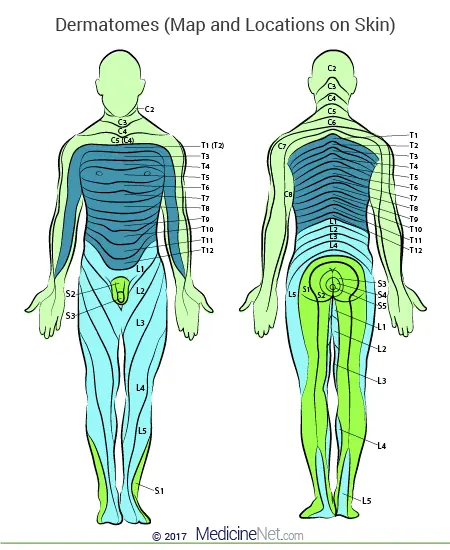
what is the C6 dermatome
dorsal surface thumb

what is the C7 dermatome
dorsal surface index

what is the T1 dermatome
medial elbow
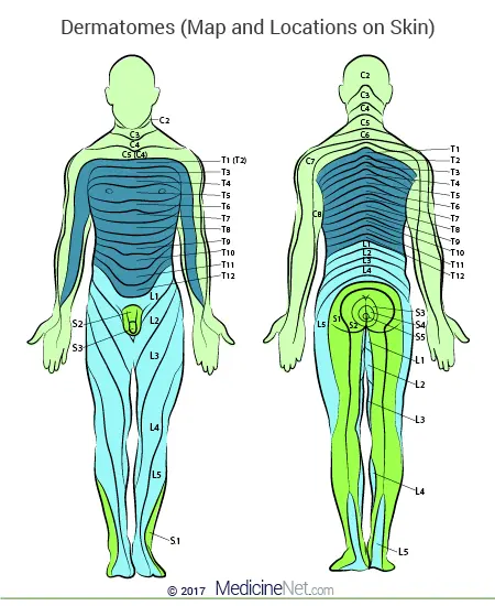
what is the T2 dermatome
apex axilla

what is the C1-2 myotome action
cervical flexion

what is the C3 myotome action
cervical lateral flexion

what is the S2 myotome action
knee flexion

what is the L1 dermatome
upper anterior thigh
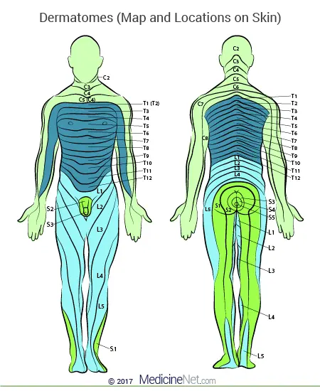
what is the L2 dermatome
mid anterior thigh

what is the L3 dermatome
medial femoral condyle

what is the L4 dermatome
medial malleolus

what is the L5 dermatome
dorsum of great toe

what is the S1 dermatome
lateral malleolus
what is the S2 dermatome
popliteal fossa
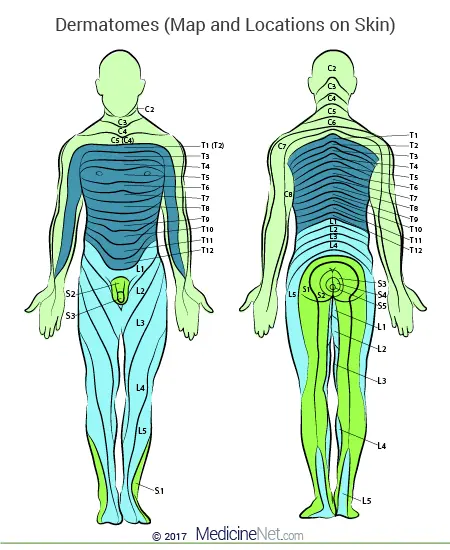
Dermatome
A strip of skin innervated by a single spinal nerve
(these nerve fibres may travel through dif. peripheral nerves)
Dermatome derive from somite cells
Myotome
Group of muscles innervated by a single spinal nerve root
Derive from somite cells
(myotome is part of somite that develops into muscle)
Dermatomes are of great clinical importance. Why?
There is some overlap, but sensory abnormalities in the dermatomes can allow you to work out if the spinal cord is damaged, and which spinal nerve is damaged
Origin of dermatomes and myotomes
Embryogenesis
Day 20 = trilaminar disc, middle layer is mesoderm, paraxial mesoderm is next to neural tube
After day 20 = paraxial mesoderm differentiates into 44 pairs called "somites", 13 break down, left with 31 pairs of somites
Correspond to 31 pairs of spinal nerves
Ventral somite portion - scelerotome
(ribs + vertebral column)
Dorsal somite - dermomyotome
Myotome proliferates
Dermatome disperses into dermis
Limb growth, dermatome associate with growing limb is stretched and moved down limb, creating segmental innervation
(Keegan and Garrett dermatome map of 1948)
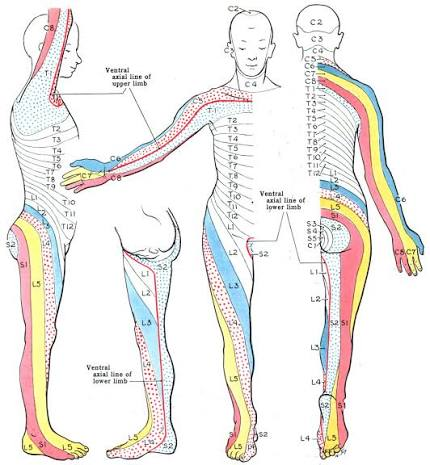
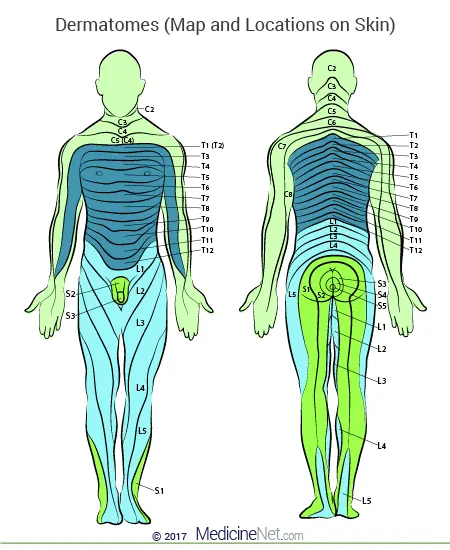
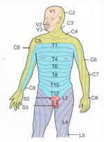
Dermatome maps
Depict dermatome distribution
Show progression of limb growth around an axial line
The general pattern is similar in all people, but significant variations exist in dermatome maps from person to person.

Axial line
The line between two adjacent dermatomes that are not represented by immediately adjacent spinal levels. Although dermatomes are shown to be discrete segments on dermatomal maps, they are in fact not; adjacent dermatomes overlap with one another
Once leaving the spinal cord, nerves divide and branch, and the branches combine with branches from other roots to form ...
A new nerve
Eg. median nerve at elbow contains nerve fibres from different spinal roots
EG of virus infection and link with dermatomes?
Herpes Zoster
"shingles"
Viral infection of spinal nerve
Dormant in dorsal root ganglion, activates and spreads along sensory nerve to affect only area of skin (dermatome) supplied by that nerve
*Viruses that infect spinal nerves can outline dermatomes well

The 31 spine segments give rise to ...
(..cervical, ..thoracic, ..lumbar, ..sacral, ..coccygeal)
Dermatomes exist for each spinal nerve, except ....
Sensory info. from specific dermatome is transmitted by ... to spinal nerve of specific...
... nerve roots emerge ... respective vertebrae
... nerve root emerges between ...
Remaining nerve roots emerge ... respective vertebrae
Along the thorax and abdomen, the dermatomes are ..., each supplied by ...
Dermatomes along the ... differ from pattern of trunk dermatomes - because ...
31 spinal nerves on each side (31 pairs)
8, 12, 5, 5, 1
first cervical spinal nerve
sensory nerve fibers
specific segment of the spinal cord
C1-C7 above
C8 between C7 and T1 vertebrae
below
evenly spaced segments stacked up on top of each other
by a different spinal nerve.
arms and legs
dermatomes run longitudinally along limbs
Myotomes:
Most muscles of the upper and lower limb are made up of a number of myotomes. Why?
Muscles of upper/lower limb tend to be innervated by more than one spinal nerve root
EG: biceps brachii muscle.
Innervated by the musculocutaneous nerve and able to perform flexion at the elbow.
Musculocutaneous nerve is derived from C5, C6, C7 nerve roots.
All three of these spinal nerve roots can be said to be associated with elbow flexion.

Myotome function can be tested clinically and graded (0 = complete paralysis, 5 = normal movement)
to see if spinal cord damage, and where the damage is located.
Movements MOST STRONGLY associated with each myotome:
C5 - Elbow flexion
C6 - Wrist extension
C7 - Elbow extension
C8 - Finger flexion
T1 - Finger abduction
L2 - Hip flexion
L3 - Knee extension
L4 - Ankle dorsiflexion
L5 - Great toe extension
S1 - Ankle plantarflexion

C2 Dermatome/Myotome
D: Temple, forehead, occiput
M: Neck Flexion

C3 Dermatome/Myotome
D: Entire neck, posterior cheek, temporal area, prolongation forward under mandible
M: Neck Side Flexion

C4 Dermatome/Myotome
D: Shoulder area, clavicular area, upper scapular area
M: Shoulder Elevation

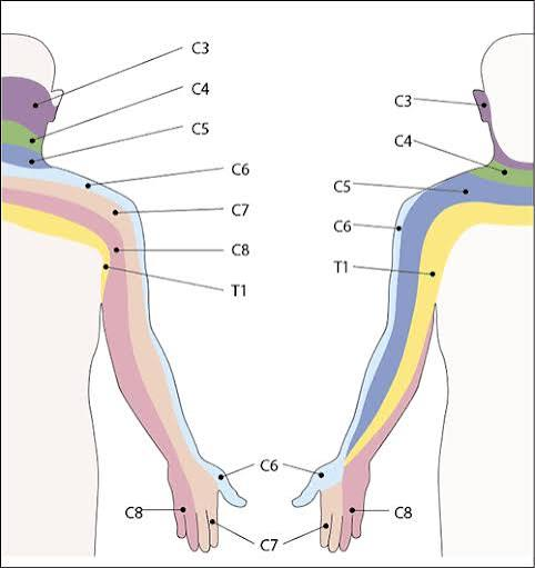
C5 Dermatome/Myotome
D: Deltoid area, anterior aspect of entire arm to base of thumb
M: Shoulder Abduction

C6 Dermatome/Myotome
D: Anterior arm, radial side of hand to thumb and index finger
M: Elbow Flexion & Wrist Extension

C7 Dermatome/Myotome
D: Lateral arm and forearm to index, long, and ring fingers
M:
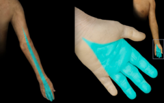
C8 Dermatome/Myotome
D: Medial arm and forearm to long, ring, and little fingers
M:

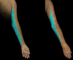
T1 Dermatome/Myotome
D: Medial side of forearm to base of little finger
M:
T2 Dermatome/Myotome
D: Medial side of upper arm to medial elbow, pectoral and mid scapular areas
M:

T3-T12 Dermatomes/Myotomes
D: T3-T6: upper thorax
T5-T7: costal margin
T8-T12: abdomen and lumbar region
M:

L1 Dermatome/Myotome
D: Back, over trochanter and groin
M:

L2 Dermatome/Myotome
D: Back, front of thigh to knee
M:

L3 Dermatome/Myotome
D: Back, upper buttock, anterior thigh and knee, medial lower leg
M:

L4 Dermatome/Myotome
D: Medial buttock, lateral thigh, medial leg, dorsum of foot, big toe
M:

L5 Dermatome/Myotome
D: Buttock, posterior and lateral thigh, lateral aspect of leg, dorsum of foot, medial half of sole, first second and third toes
M:

S1 Dermatome/Myotome
D: Buttock, thigh and posterior leg
M:

S2 Dermatome/Myotome
D: Buttock, thigh and posterior leg
M:

S3 Dermatome/Myotome
D: Groin, medial thigh to knee
M:
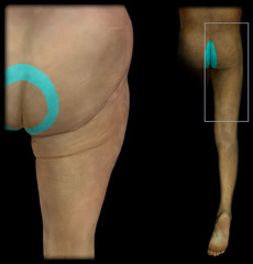
S4 Dermatome/Myotome
D: Perineum, genitals, lower sacrum
M:

S5 Dermatome/Myotome
D: Perianal area
M:
