Physiology 209
 Call Kai
 Learn
 Practice Test
 Spaced Repetition
 Match
 Flashcards
 Knowt Play
1/150

There's no tags or description
Looks like no tags are added yet.
151 Terms
Physiology
The study of the normal functional activities in the healthy living organism

Milieu Intérieur
the internal environment surrounding individual cell
Basic Cell functions

Claude Bernard Belief
Environment surrounding individual cells is vastly different from external environment
Internal environment remains relatively constant under conditions of health
Homeostasis
a state of dynamic constancy
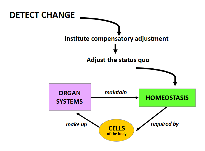
Fundamental Principle
“At all levels of organization, the functional activities are directed at maintaining optimal and relatively constant INTERNAL CONDITIONS”
What are the factors that must be homeostatically controlled?
Concentration of Nutrient Molecules
Concentration of O2 and CO2
Concentration of Waste Products
Concentration of Electrolytes
pH
Temperature
Volume and Pressure
Failure to maintain homeostasis causes. .
disease
Body water is the medium in which…
Solutes are dissolved
Metabolic reactions take place
What accounts for the large variation in water content in different individuals?
Fat (adipose tissue) varies in % in each individual based on factors unrelated to water
fat contains only 10% water, unlike the 70-80% of other body tissues
What is a physiological reference individual? What should one adjust for when the subject varies from the reference individual?
21 yr old white 70 kg male
age, gender, weight
babies are 70% water
mid-life women 50%, men 60%
elderly women 45%, men 50%
How do you calculate body water volume? How can this be important?
(weight * %body water)/100
important when administering water-soluble medication (helps calculate accurate concentration)
need to know how to convert between mili, micro, nano, and pico (look at attached photo PLS)

What are the intakes necessary to maintain dynamic water balance in the body?
Oral fluid (drank fluids, they have water)
Oral intake as food (stuff you eat has water)
Oxidative water from metabolism (metabolic reactions produce water as a byproduct)
What are the main types of output necessary to maintain dynamic water balance in the body?
2 ways to categorize, insensible vs sensible and obligatory vs facultative, one can be insensible and facultative at the same time or any other combo between the different ways of categorization
Insensible (automatic processes you cannot control, you can’t sense it, hard to measure)
Lungs (water molecules lost through breathing by accident, out of your control)
Skin (passive evaporation from pores, NOT SWEAT)
Sensible (necessary processes that you do, like removing waste, you can sense it, you can measure it)
kidneys (urine)
stool (feces)
sweat (NOT OBLIGATORY OR FACULTATIVE, JUST SENSIBLE)
Obligatory (a set, unchanging amount of water that must be lost each day to get rid of waste and maintain bodily function regardless of consumption)
lungs and skin (insensible)
urine and stool (sensible)
Facultative ( a variable amount of water that is lost based on the given intake of that day to maintain homeostasis in the body)
urine (kidney is the main homeostatic organ for water balance)
What is the difference between insensible precipitation and sweat?
Insensible precipitation
pure water
passive evaporation (no energy, not on purpose, just affected by ambient (surrounding) temperature and relative humidity
takes place on the entire skin surface, so it can occur in people that don’t have sweat glands
Continuous and classified as obligatory
Sweat
electrolyte solution (not pure)
active secretion ( on purpose, requires energy and signals in the body)
requires sweat glands
activated by heavy work or high temperatures
What is the difference in body water turnover in24 hours as an adult vs a baby?
Adults are much lower, 3-4% of body weight
Baby’s much higher, 10% of body weight
What can cause a negative water balance? (Water Loss > Water Intake)
reduced intake
excessive loss from gut (vomiting)
excessive sweating
excessive loss in expired air (high altitude has dry air, increasing amount of water lost from skin pores)
excessive loss in urine
What can cause water intoxication? (water intake > water loss)
excessive water intake (difficult)
renal system failure
Body water volume is remarkably constant under conditions of health (water intake = water loss) This helps maintain…
normal solute concentrations
normal blood volume and pressure
both of these together allow for adequate supply of O2 to tissues
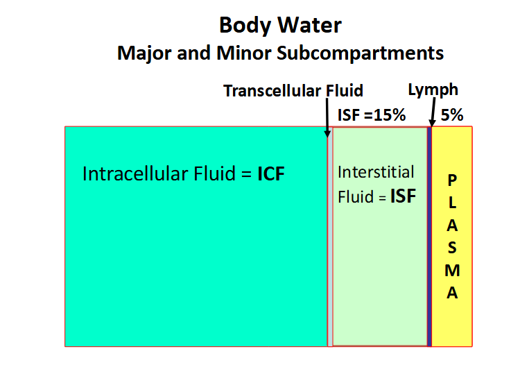
What are the two major body water compartments?
Intercellular Fluid (ICF)
Aggregate of fluid bound by internal surfaces of all cell membranes
true “Milieu Interieur”
The fluid which percolates between individual cells
2/3rds of all body water
Extracellular Fluid (ECF)
1/3rd of all body water
divided into 2 Major ( Plasma and Interstitial Fluid) and 2 Minor (Lymph and Transcellular Fluid)
Hematocrit (Ht)
The percentage of Blood Volume that is occupied by Red Blood Cells (erythrocytes)
calculated by: (height of erythrocyte column/whole blood column)

Plasma
The fluid medium in which blood cells are suspended
Explain the Lymphatic system from the smallest piece to largest
A network of blind-ended terminal tubules come together to form larger lymphatic vessels
these converge to form large lymphatic ducts
which in turn drain into the large veins in the chest
Transcellular fluid
Aggregate of small fluid volumes secreted by specific epithelial cells that line some body cavities
these small fluids have specialized function
does not contribute significantly to overall water exchanges; local changes do not affect body fluid balance
still plays an important role in function locally
What are the 3 important factors that are constant for bodily fluids?
Total Volume in a compartment remains CONSTANT
Relative Distribution between compartments remains CONSTANT
Compartments are in a state of DYNAMIC equilibrium

What are the two types of methods to determine compartment volumes?
direct
just straight up measure
ex: indicator dilution method
indirect
via calculations
How does the Indicator Dilution Method Work?
Introduce known quantity (Q) of indicator into vein
Allow time to equilibrate
Remove known volume of blood, and centrifuge to obtain plasma
Measure concentration c in unit volume of plasma
Calculate
V = Q/c
Need to know:
The total quantity of test substance introduced
The concentration of the substance/unit volume of fluid, after dispersion
What do you need to consider when choosing an Indicator for an Indicator Dilution Method?
Indicator must be:
Non-toxic
Diffuse readily, distribute
evenly throughout
compartment(s) to be
measured (must choose one that only diffuse as far as needed, not further, or shorter, see diagram for designated indicators for each compartment)
Induce no changes in
distribution of water
between compartments
Easy to measure its
concentration

In a clinical situation, what would you have to account for in an Indicator Dilution Method that we do not in class?
any amount of bodily fluid which has been removed from body by metabolism or excretion during the time allowed for mixing.
What is the difference in ion and protein concentrations between the ICF and ECF?
ICF
High in K+ and Mg++
low in Na* and Cl-
high protein anions
ECF
High in Cl- and Na+
low protein anions found only in plasma NOT ISF

What are the Barriers to transport between ICF and External Environment
Cell (plasma)
membrane barrier between ICF and ECF
Capillary wall
barrier between:
ECF and plasma
plasma and the external environment
Explain the simplified directional movement of materials between compartments

What does the viability of the cell depend on and require?
depends on the relative constancy of the “Internal Environment” (the “Milieu Intérieur”)
requires EXCHANGES with the internal and (ultimately) the external environment
Describe what the Cell membrane in permeable, impermeable, and partially permeable to?
Highly Permeable to:
H2O
Lipid-soluble substances
Dissolved Gases (O2, CO2)
Small uncharged molecules
Less Permeable to:
Larger molecules
Charged particles
Impermeable to:
Very Large molecules
Cholesterol
inserted into the phospholipid bilayer
functions as a buffer (prevents extremes, keeps state near constant)
prevents lower temperatures from decreasing cell membrane fluidity and higher temperatures from increasing fluidity too much
involved in the formation of vesicles ( these pinch off the plasma membrane and in lipid rafts)
Slightly amphipathic
Cell Membrane
Bimolecular Phospholipid Layer ( Phosholipid Bilayer)
Amphipathic
Polar head (outside) and Nonpolar tails (inside)
comprised of 40-50% phospholipids by weight
Proteins
Most diverse macromolecules
25-75% of cell membrane by weight
Two types:
Integral
closely associated with phospholipids (found at least partly inside the layer), mostly cross the membrane (Trans-membrane, partly in membrane partly out of membrane)
Peripheral
more loosely associated (attached on the outside), can be found on both sides of the plasma membrane
Glycocalyx
a gel-like, sugar-rich coating
surrounds the cell membrane
formed of:
glycans
glycoproteins
glycolipids
contributes to cell-cell:
recognition
communication
adhesion
protection
How does the Fluid Mosaic Model of the Cell Membrane describe it?
describes the cell membrane as a dynamic, fluid structure where a phospholipid bilayer acts as a "solvent" (like a moving river) for embedded and attached proteins, creating a "mosaic" of molecules that can move laterally (horizontally)
What are the different Cell Membrane Transport Mechanism?
Passive (energy independent, doesn’t need it)
simple diffusion
facilitated diffusion
osmosis
Active (energy dependent, needs energy)
Active Transport
Primary Transport
Secondary Transport
Endo and Exocytosis
Diffusion
the movement molecules from one location to another as a result of random thermal motion

Flux
Amount of a solute crossing a surface area per unit time
Two types:
One-way flux:
Unidirectional rate of movement of a solute
Net flux:
Overall rate of movement of a solute
At equilibrium, net flux is 0 and either direction one-way fluxes are equal

Explain equilibrium for intracellular concentration (inside a cell) vs extracellular concentration (outside of a cell)
While there is equivalent exchange between the two fluids, the volume outside of the cell is so much larger than inside the cell that this exchange of solute between them does not alter the extracellular concentration. It remains constant.
Intracellular fluid has a significantly lower volume than extracellular fluid, so as the exchange occurs, it’s concentration is greatly affected. Since the concentrations need to be equivalent for equilibrium, and extracellular concentration is unaffected by the exchange, intracellular concentration rises or falls to match the extracellular concentration

Explain Fick’s law of Diffusion, include the equation and the meaning of the variables
Equation: J = PA (C0 – Ci)
C0 – Ci: Concentration gradient of the diffusing molecule across the membrane
J: Flux, Amount (moles) of solute crossing a surface area of membrane per unit time - rate of diffusion
P: Permeability (or diffusion) coefficient, a constant based on the ease with which a molecule moves through a membrane
A: Surface area of the membrane
Ion Channels
transmembrane proteins that show ion selectivity
The movement of an ion through it is determined by opposing electrochemical forces
1st by the electrical potential difference across the cell membrane (must balance charges on both sides, positive ions go toward negatively charged side, and vise versa)
2nd by the concentration gradient of that ion across the cell membrane (normal diffusion, goes toward lower concentration
When both are balanced, its called an electrochemical equilibrium
note: electrical potential difference balances the concentration gradient
Channel Gating
The ability of an ion channel to open and close
3 types:
Ligand Gating
channel changes conformation, pinches inside together to close
Voltage Gating
opens and closes based on the electrical potential difference across the membrane
ex: Na+, K+ and Cl- channels
Mechanical Gating
a separate attached gate plugs the pore of the ion channel
Mediated Transport
the movement of ions and other molecules (glucose, amino acids) by integral membrane proteins called transporters, carriers or exchangers
NOT diffusion
slower than ion channels
Can be passive or active, ex:
Facilitated Diffusion (Passive)
Primary Active Transport
Secondary Active transport
What are the characteristics of Mediated Transport?
Specificity
transporter usually transports one particular type of molecule
Competition
occurs when structurally similar substances compete for the same binding site on a membrane carrier
Saturation
rate of transport reaches a maximum when all binding sites on all transporters are occupied
Thus, a limit – the transport maximum (Tm) – exists for a given substance across a given membrane
Facilitated Diffusion
involves the presence of a “transporter” or “carrier” molecule
enables a solute to penetrate more readily than it would be expected to by simple diffusion
How it works:
Solute binds transporter
Transporter changes configuration
Solute is delivered to other side of membrane
Transporter resumes original configuration
transporter (carrier) mediated
passive (no-energy)
net flux from high to low concentration
Hormones may increase the number and/or affinity of transporters in some membranes
Glut-4 transports glucose in muscle that is increased by insulin

Active Transport
transporter mediated
requires supply of chemical energy (usually derived form enzymatic hydrolysis of ATP)
Susceptible to metabolic inhibitors
Can transport solute against its concentration gradient (i.e., “uphill transport”)
Primary Active Transport
involves the hydrolysis of ATP by a transporter (carrier)
Phosphorylation of the transporter changes the conformation of the transporter and its solute binding affinity
Changes in the binding site affinity for a transported solute are produced by phosphorylation and dephosphorylation of the Na+/K+-ATPase
How it works (using Na+/K+-ATPase as an example):
1. Na+ binds to a transporter outside the cell (where the Na+ concentration is high) allowing glucose or amino acid to bind to the same carrier (atp is used, bound to transporter)
2. Through a change in configuration, the transporter “delivers” both Na+ and glucose or amino acid into the cell (configuration changes when atp is phosphorylized, used)
3. The transporter then reverts to its original configuration, and the Na+ is extruded from the cell by the Na+/K+ -ATPase (when the attached phospho group leaves the shape reverts to original)
note: He writes it in this order where Na+ leaves somehow at the end after the transporter reverts back to its original position, but both his diagram and previous classes (biol 112, gr 12) say that Na+ stays in the cell after it is delivered stays in the cell and detaches with glucose or amino acid
other examples of transporters:
Ca2+-ATPase
Maintain low intracellular Ca2+ levels
H+-ATPase, Vacuolar-type H+-ATPase (V-ATPase)
Maintain low lysosomal pH
H+/ K+- ATPase
Acidification of the stomach
Secondary Active Transport
the movement of Na+ down its concentration gradient is coupled to the transport of another solute molecule (ion, glucose, amino acid)
uphill against its concentration gradient
uses the energy stored in the electrochemical gradient to move both the Na+ and the transported solute
The creation and maintenance of the electrochemical gradient depends on primary active transport
Symport/Cotransport
when solute X is transported in the same direction as Na+ in Secondary Active Transport
ex:
Na+/HCO3− cotransporter
(NBC) Na+/amino acid cotransporter (NAcT)
Na+/glucose cotransporter (SGLT)

Antipor/Countertransport/Exchange
when solute X is transported in the opposite direction to Na+.
ex:
Na+/H+ exchanger (NHE)
Na+/Ca2+ exchanger (NCX)
Endocytosis
Active transport mechanism (energy-dependent) involving participation of the cell membrane itself
the cell membrane invaginates (opens a pore to let outside solute in) and pinches off to form a vesicle
3 types:
Pinocytosis
also known as fluid endocytosis
Phagocytosis
the process by which cells bind and internalize particulate matter (>0.75 μm, solids, particles) such as small-sized dust particles, cell debris and microorganisms
Receptor-mediated Endocytosis
molecules in the extracellular fluid (ligands) bind with high affinity to specific protein receptors
on the plasma membrane

Exocytosis
Active transport mechanism (energy-dependent) involving participation of the cell membrane itself
the process of moving material from the inside to the outside of the cell
2 Types
Constitutive Exocytosis
Non-regulated
replaces plasma membrane
delivers proteins to the cell membrane
secretes collagen and other substances from the cell.
Regulated Exocytosis
triggered by extracellular signals and the increase ofcytosolic Ca2+
secretes of hormones, digestive enzymes, and neurotransmitters
Pinocytosis
also known as fluid endocytosis
an endocytotic vesicle engulfs the extracellular fluid including whatever solutes are in it
nonspecific and constitutive (always active)
vesicles travel into the cytoplasm and fuse with other vesicles such as endosomes or lysosomes

Phagocytosis
the process by which cells bind and internalize particulate matter (>0.75 μm, solids, particles) such as small-sized dust particles, cell debris and microorganisms
specific and triggered
Extensions of the cell membrane called pseudopodia fold around the particle and fully engulf it
The pseudopodia fuse to form large vesicles, called phagosomes, that pinch off the membrane
Phagosomes migrate to and fuse with lysosomes where the contents of the phagosome are degraded
Defends against infection and scavenge senescent and dead cells (macrophages, neutrophils and dendritic cells)

Receptor-Mediated Endocytosis
molecules in the extracellular fluid (ligands) bind with high affinity to specific protein receptors
on the plasma membrane
2 Types:
Clathrin-dependent
When the ligand binds the receptor undergoes conformational change and clathrin is recruited to the plasma membrane
Potocytosis
the process by which small molecules are sequestered and transported by tiny vesicles called caveolae
clathrin-independent

How does the LDL receptor work with cholesterol?
Cholesterol is transported in the blood in low-density lipoprotein (LDL) particles (vesicles).
The lipoprotein particles bind to PM LDL receptors
Endocytosis occurs right after
This a clathrin-dependent receptor-mediated endocytosis
The endocytized vesicles fuses with early endosomes where LDL dissociates from the receptor protein
The LDL continues to late endosomes and then lysosomes where cholesterol is released

Clathrin Dependent Receptor-Mediated Endocytosis
When the ligand binds the receptor undergoes conformational change and clathrin is recruited to the plasma membrane
Adaptor proteins link the ligand-receptor to the clathrin
The complex forms a cagelike structure that leads to the aggregation of ligand bound receptors
A clathrin coated pit is formed which then invaginates and forms a “clathrin-coated vesicle”
Once the vesicle pinches off it sheds the clathrin coat and vesicles can fuse with the membrane of cellular organelles such as endosomes and lysosomes
Or, they can sometimes fuse with the membrane on another side of the cell (transcytosis)
Receptors and clathrin protein are recycled back to the cell membrane
ex: LDL receptor

Potocytosis/ Clauveola-Dependent Receptor-Mediated Endocytosis
the process by which small molecules are sequestered and transported by tiny vesicles called caveolae
clathrin-independent
The caveolae concentrate molecules within their lumen by releasing receptor-bound species, using enzymes to convert or detach extracellular substances from soluble carriers
Caveolae can deliver their contents directly into the cell cytoplasm, to the endoplasmic reticulum (ER) and other organelles or to the plasma membrane on the opposite side of the cell (transcytosis).
suggested they’re used in consumption of low molecular weight molecules such as vitamins

Osmosis
the net diffusion of H2O across a semipermeable membrane (i.e., permeable to solvent, but not to all solute)
Note: Water diffuses though cell membranes via water permeable channels called aquaporins
Osmotic Pressure
The pressure required to prevent the movement of water across a semi-permeable membrane
equal to the difference in the hydrostatic pressures of the two solutions across the membrane
proportional to osmolarity (Osm)
Calculated by using the Van’t Hoff Equation (π = MRT)
M (Osmolarity)
R (Gas constant) 0.0821 L ! atm/mol * K
T (Temperature) Absolute temperature in Kelvin
Osmolarity (Osm)
the total solute concentration of a solution
1 mol of solute particles = 1 osmol
Ex:
1 mol glucose = 1 osmol of solute
1 mol NaCl = 1 mol Na+ + 1 mol Cl- = 2 mol = 2 osmol
1 mol glucose/L = 1 osmol/L = 1 Osm solution
proportional to osmotic pressure
Calculate by:
First find the Molarity (concentration)
Determine Osmolarity (account for each ion that would show in aqueous solution)
ex
0.15 M NaCl solution = 0.15 moles/L Na+ + 0.15 moles/L Cl-
= 0.15 osmol/L + 0.15 osmol/L = 0.30 osmol
= 0.30 osmol/L H2O = 0.30 Osm = 300 mOsm (miliosmol)
Isosomotic
Solutions that have the same osmolarity (concentration
of osmotically active particles) as normal extracellular or
intracellular solution
must be 300 mOsm or 0.30 Osm
Hypoosmotic
Solutions which have an osmolarity lower than 300 mOsm
Lower osmolarity than normal extracellular or intracellular solution
Hyperosmotic
Solutions which have an osmolarity greater than 300 mOsm
Higher osmolarity than normal extracellular or intracellular solution
Extracellular Na+ behaves as a nonpenetrating solute because . . .
the Na+ that moves into the cell is pumped out by the Na-K ATPase
What kind of particles are effective in exerting sustained osmotic pressure?
nonpenetrating molecules that cannot cross the membrane
Isotonic
=300 mOsm
extracellular solution has a concentration of 300 mOsm of nonpenetrating solute particles
no net shift of water
Hypotonic
<300 mOsm
extracellular solution has a concentration of nonpenetrating solute particles less than 300 mOsm,
water enters the cell
cell swells
Hypertonic
>300 mOsm
extracellular solution has a concentration of nonpenetrating solute particles greater than 300 mOsm
water leaves the cell
the cell shrinks
Capillaries
An adult has ~40 km
contains ~5% of total circulating blood
~1mm long
inner diameter ~8 μm
Capillary Wall
A single layer of flattened endothelial cells and a supporting basement membrane
Means of transportation:
1, 2- (labeled for attached diagram) DIFFUSION
most important means of transport
also occurs through water filled channels (intercellular clefts and fused vesical channels)
3. Transcytosis
Endocytosis on the luminal side followed migration of the vesicle across the cell and then exocytosis on the interstitial side
Simplifies: Endocytosis on one end of the cell membrane, then exocytosis on the opposing end
4. BULK FLOW
distributes the extracellular fluid volume between the plasma and ISF
Magnitude of bulk flow is proportional to the hydrostatic pressure difference between the plasma and the ISF
acts as a filter that permits protein free plasma to move from capillaries to the ISF (through BULK FLOW)
Blood
a highly dynamic tissue, part of the cardiovascular system
supporter of life – “lifeblood”
associated with emotions – “bad blood”
reflective of relationship – “blood brothers”
Ancient Chinese Medicine –blood flow linked to energy flow
Medieval Western Medicine – blood inhabited by good and evil spirits
Modern Time – blood as a carrier of disease
Ancient Greece – bleeding as treatment for disease
Functions:
Transport
Nutrients
Respiratory Gasses
Wastes
Hormones
Temperature Regulation
Acid-Base Balance
Normal pH range 7.35 - 7.45
Protection
White Blood Cells (WBCs)
Plasma Proteins
Contains:
After centrifuge: 55% plasma (ECF) 45% red blood cells (ICF, also called erythrocytes) and very thin BUFFY LAYER (white blood cells + platelets)
accounts for ~7-8% of Body Mass
NORMOVOLEMIA
normal blood volume, “norm”
HYPOVOLEMIA
lower than normal blood volume, “hypo”
HYPERVOLEMIA
Higher than normal blood volume, “hyper”
Composition of Plasma
Similar to ISF but with proteins
>90% water
Ions: Na+, K+, (Ca++, Mg++), Cl-, HCO3-, (PO4--)
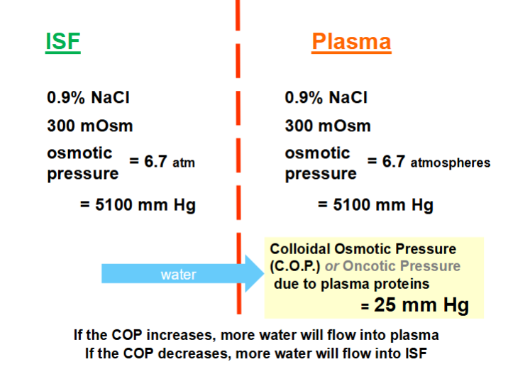
May be approximated by physiological saline 0.9 % NaCl (= 300 mOsm, 6.7 atmospheres or ~ 5100 mm Hg) since most of it is Na+ and Cl-)
Nutrients, Respiratory Gasses, Wastes
Glucose, Amino Acids, Lipids, O2, CO2, Urea, Lactic Acid
Proteins (colloids) = 7 %
Albumins
Globulins
Fibrinogen
* Proteins are ONLY FOUND IN PLASMA, NOT in ISF
Electrophoresis
a method of separating plasma proteins
fractionation method based on movement of charged particles along a voltage gradient
The rate of migration is influenced by the number and distribution of charges and by the molecular weight, MW, of each protein
Each protein migrates at its own characteristic rate

What is the origin of plasma proteins
Liver
Albumin
α1, α2, β Globulins
Fibrinogen
Lymphoid Tissue - Gamma (Y) Globulin
Since most in liver, if liver is diseased plasma protein levels decrease
Plasma Protein Properties + Importance
Albumin is most abundant (highest []) and the smallest
oval
Fibrinogen is the largest and least abundant (lowest [])
elongated thin oval
Globulin has the most variety in shape and size
They play a major role in determining the distribution of fluid between the plasma and the ISF compartments by controlling transcapillary dynamics

How is the different bodily fluids distributed in the body (fractions) and what membranes separate them?
ICF - 2/3
ECF - 1/3
Of that 1/3:
ISF - 3/4
Plasma - 1/4
ICF and ECF separated by cell membrane (relatively impermeable to ions)
ISF and Plasma separated by capillary wall (freely permeable to H2O and ions but impermeable to proteins)

What can or cannot contribute to the effective Osmotic Pressure of a solution?
Diffusible solutes do NOT contribute, because they become equally distributed on the 2 sides of the membrane
Plasma Proteins are Non-Diffusible therefore, they can exert an osmotic effect
This effect is known as the Colloidal Osmotic (Oncotic) Pressure (C.O.P.) of Plasma = 25 mm Hg
For a net flow of water between compartments (ISF and Plasma) there must be a difference in osmotic pressure
Hence:
If the COP increases, more water will flow into plasma
If the COP decreases, more water will flow into ISF
PLS LOOK AT DIAGRAM IT HELPS
Transport across the Capillary Wall
two major forms of fluid transport across – osmotic flow and filtration
1. The C.O.P. of plasma (osmotic flow)
determines how much water will flow into or out of capillaries
plasma proteins, tend to “pull in” or retain fluid inside the capillaries
2. Filtration / Bulk Flow / Filtration
BULK FLOW – flow of molecules subjected to a pressure difference
Magnitude of bulk flow α hydrostatic pressure difference
FILTRATION = bulk flow across a porous membrane which acts as a “sieve” (strainer) withholding some particle
because fluid in the blood vessel is under pressure, it tends to “push out” fluid from the capillaries into ISF
1 AND 2 TOGETHER ARE CALLED THE STARLING FORCES
The balance between Colloidal Osmotic Pressure and Hydrostatic Pressure will determine how water moves between these two compartment
Diffusion VS Starling Forces
DIFFUSION is responsible for the exchange of nutrients, gases, and wastes across the capillary wall
STARLING FORCES determine the distribution of ECF volume, the movement of water between the Plasma and ISF
Capillary Exchanges
Nutrients, wastes, O2, CO2 move by simple diffusion
Starling’s Transcapillary Dynamics
Exchanges filtration and absorption take place along the whole length of the capillary bed not just at the 2 ends
determines the distribution of ECF volume between Plasma and ISF
Filtration – tends to “push fluid out” from inside the capillaries
Osmotic Flow – tends to “pull fluid into” or retain fluid in the capillaries
Only ~90% of the fluid filtered out of the capillaries is reabsorbed into the capillaries ~ 10% is drained from the tissues by the lymphatic vessel

Lymphatic System + Vessels
System:
A network of blind-ended terminal tubules
which coalesce to form larger lymphatic vessels
which converge to form large lymphatic ducts,
which drain into the large veins in the chest
Vessels
The walls are made up of a single layer of endothelial cells
highly permeable to all ISF constituents (including proteins which may have leaked into the ISF from the plasma)

Which proteins contribute the most to C.O.P.?
The osmotic pressure of a solution depends on the NUMBER of osmotically active particles/unit volume
NOT their configuration, size, or charge
Each protein fraction exerts an osmotic pressure which is
directly related to its [] in the plasma
inversely related to the Molecular Weight of that protein
Hence need a light and abundant protein - ALBUMIN
Diagram gives metaphor to help

Daily Lymphatic & Blood Flow Volumes
Total Blood Flow 6,000L
Volume filtered into ISF 20L
Volume returned by absorption 17L
Volume returned by lymph drainage 3L

Edema
accumulation of excess fluid in the interstitial spaces
Caused by decreased venous fluid return (less fluid returned to veins from lymphatic drainage and absorption)
Conditions that can lead to it:
Increased Hydrostatic Pressure
Decreased Plasma Protein (lower C.O.P.)
In extreme cases:
due to failure to synthesize plasma proteins e.g., Liver disease (since they are made there)
or severe protein malnutrition
can lead to swelling of the abdomen (Kwashiorkor)
Increased Capillary Permeability
Normally, there is very little protein in ISF.
If the capillary wall becomes more permeable, some plasma proteins escape into the ISF where they can exert oncotic pressure (colloid osmotic pressure), this pulls water out of the capillaries
Obstruction of Lymphatic Drainage
Extreme case: Elephantiasis. blockage of lymphatic drainage resulting from parasite infestation (Filaria nematode)
Typical Symptoms: swollen ankles and feet
Roles of Plasma Proteins
Determine the distribution of fluid between the plasma and the ISF compartments
the Starling Forces control transcapillary dynamics
Contribute to the viscosity of plasma
Viscosity contributes to blood pressure
Contribute to the buffering power of plasma
Normal pH range ~7.4
Fibrinogen and some globulins are essential to clotting
Gamma (γ) - globulins (Immunoglobulins/Antibodies) provide specific resistance to infection
Albumin and some globulins act as carriers for lipids, minerals, hormones
What are the different cells in the blood?
Red Blood Cells (Erythrocytes)
Diameter: 7.2 μm
Lifespan: 120 days
Number: 5 x 10⁶/μL
Platelets (Thrombocytes)
Diameter: 2-3 μm
Lifespan: 7-8 days
Number: 250,000-400,000/μL
White Blood Cells (Leukocytes)
Diameter: 10-18 μm
Lifespan: hours-years
Number: 8,000-10,000/μL
Note: Slide shows them coming from the Blood Progenitors, this is just their “ancestor” its a “blood ancestor” not a specific term you need to know

Hematopoiesis
The process through which all blood cells are derived from a common, multipotential (pluripotential) hematopoietic stem cell in the bone marrow
is a general pattern
Examples for different cells:
Erythropoiesis is the production of Red Blood Cells (Erythrocytes)
Thrombopoiesis production of Platelets (Thrombocytes)
Leukopoiesis production of White Blood Cells (Leukocytes)
How it works (according to diagram)
An inducer stimulates a Pluripotential Multipotential Stem Cell to self replicate (Division)
The self-replicating stem cell becomes a committed stem cell (committed to becoming a specific type of cell, from the options above, think about it like committing to a career path) (Differentiation)
The committed stem cell interacts with a stimulant and goes through the specific process (from options listed above) of production to become the specific cell type they have committed to

Cytokines
substances (proteins or peptides) which are released by one cell and affect the growth, development, and activity of another cell
those that influence the proliferation and differentiation of blood cell precursors are known as Hematopoietic Growth Factors (HGFs)
Sites of Hematopoiesis
Pre-natal:
Yolk Sac til 3 months
Liver & Spleen from 1 month to 9 month
Post-Natal (mainly, starts off in prenatal at ~4 months): Axial Skeleton
Flat bones of skull
Shoulder blades
Sternum
Vertebrae
Ribs
Pelvis
Proximal epiphyses of long bones: over time (as shown in diagram) sites move to the epiphyses (long end, cap area) of long bones (femur, those in the arms, etc, not ribs!)
ex: moves to the top joint bone area of femur

Red Blood Cells (RBC)
FUNCTION: facilitate transport of respiratory gases
“biconcave” disk shape due to the presence of “spectrin” (a fibrous protein, forms a flexible network linked to cell membrane)
Shape is advantageous
Maximal surface area and minimal diffusion distance
Increases the efficiency of O2 and CO2 diffusion
High degree of flexibility
Allows them to squeeze through narrow capillaries
Cell Size
Normocytic (normal size)
Microcytic (smaller than normal)
Macrocytic (larger than normal)
Cell Shape
Sickle Cell (looks like a sickle, half moon when its not supposed to
Spherocyte (completely circular no ovality or dip, also an illness, not normal)
Rate of production = Rate of destruction ~2 x 10⁶/second
Numbers (MvsF)
determined by the O2 requirements by the body and the O2 availability
Increases with decreased O2 (higher altitudes)
Males: 5.1 – 5.5 x 10⁶/μL (more than women)
Females: 4.5 - 4.8 x 10⁶/μL
Components:
contain no subcellular organelles (No Nucleus, No Mitochondria)
Glycolytic Enzymes to generate energy anaerobically
Carbonic Anhydrase for CO2 Transport
Converts CO2 to bicarbonate which is easier to transport
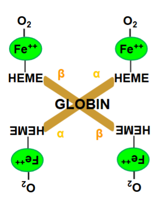
33% deoxyhemoglobin (Hb), called hemoglobin when O2 is actively being carried (HbO2)
Each molecule of Hb can bind a maximum of 4 O2 molecules


Hemoglobin
200-300 x 106 molecules/RBC
In lungs, becomes saturated with O2
blood appears bright red
In tissues, O2 dissociates (no longer has O2)
blood appears dark red
LOOK AT BOTH ATTACHED DIAGRAMS FOR DETAILS IN STRUCTURE + COMPONENTS
Functions
Transport of O2 (Hb + O2 reversibly reacts to produce HbO2)
Transport of CO2
Acts as a buffer
Despite O2 solubility being super low in plasma, due to Hb in RBC O2 solubility in overall Blood is very high ( 0.3 ml O2/100 ml plasma vs 20 ml O2/100 ml blood)
Values in Blood (MvsF)
Males: 16 g/100 ml blood
Females: 14 g/100 ml blood
When Hb is fully saturated with O2 each gram of Hb holds 1.34 ml O2
Therefore, the O2 carrying capacity of blood is: 20 ml O2/100 ml blood (15 grams x 1.34 ml = 20)
Why have Hb inside the red blood cell rather than dissolved in plasma?
Increases Plasma Viscosity (hard for the heart to pump, tougher to diffuse in)
Plasma C.O.P.
adding yet another non-diffusing substance would increase osmotic pressure higher than normal, too much flow of water into veins
Would lose them via the kidney