Genetics (Ch.10-#)
 Call Kai
 Learn
 Practice Test
 Spaced Repetition
 Match
 Flashcards
 Knowt Play
1/47

There's no tags or description
Looks like no tags are added yet.
48 Terms
Molecules responsible for heritability must:
Be able to STORE complex information
REPLICATE faithfully/copy with fidelity (from parent to offspring)
ENCODE phenotype (through effects on RNA and Protein)
Must allow for VARIABILITY between individuals (due to sexual reproduction)
Levene (guy)
He proposed that DNA consisted of repeating units containing all four bases in equal proportions, theory incorrect because fixed repeating arrangement would not allow enough variation to encode diverse phenotypes of different organisms
DNA (polymer) made of nucleotides (monomer)
Each nucleotide is composed of a phosphate, base (purine, pyrimidine), and sugar (deoxyribose, ribose)
Adenine, Thymine, Guanine, Cytosine
Tetranucleotide Theory (WRONG)—> Doesn’t allow for variation, cannot encode different phenotypes
Chargaffs Rules:
Distribution of the bases varies from species to species
A=T
G=C
Chargaff’s Findings
Compared DNA from organisms (E.coli, Yeast, Sea Urchins, Rats, humans)
Comparing multiple species matters because a single sample such as E.Coli would not establish how base composition varies across organisms
observed that base composition varies among species, A approx=T, G approx=C
Ratio >1 numerator more abundant A/T , G/C
Purines=A,G Pyrimidines=C,T
Griffith
Bacterial Transformation
studied bacterial transformation
living bacteria can take up material released from dead bacteria and acquire new traits
When bacterial cells break open (dead, weak membrane), DNA fragments and plasmids can enter the environment
Fragment integration (one of the ways bacteria can acquire variation)???
Avery, MacLeod, and McCarty
Identifying the transforming substance
they investigated the chemical nature of Griffith’s transforming substance
Prepared material from heat-killed virulent bacteria and treated separate samples with:
RNase, degrades RNA
DNase, degrades DNA
Protease, degrades protein
Substance that doesn’t KILL lacks the transforming substance
Tested whether the treated material could transform nonvirulent bacteria
Transformation continued after RNA or Protein degrades, but not after DNA degrades (it is transforming substance)
NOTE: transforming substance also absorbed UV light like DNA and was not degraded by protease and rnase
Hershey and Chase
Testing whether DNA or protein enters bacterial cells
Used bacteriophages (viruses that infect bacteria)
T2 phage contained DNA and a protein coat
Used radioactive isotopes to track the two components (35s, 32p)
After phages infected E. Coli, the samples were blended (to shear protein coats) and centrifuged (to separate protein from cells)
Results supported the conclusion that DNA, not protein, entered bacterial cells
Wilkins and Franklin
Used X-ray crystallography to investigate DNA structure
X-ray passes through crystallized DNA to produce a diffraction pattern, pattern provides evidence about the structure. Diffraction pattern is analyzed
Franklin and her student Gosling produced image photo 51, provided evidence of DNA helical structure
Reflection
Light bounces back
Refraction
Light bends as it passes between media
Watson and Crick
1953, proposed a model for DNA structure based on:
Levene
Chargraff’s Rules
Franklin and Wilkins’ DNA structure X-ray data
DNA
A polymer made of nucleotides held together via phosphodiester linkages (bond between the phosphate group of one nucleotide and the 3’ hydroxyl group of another nucleotide’s sugar)
This forms the backbone
Deoxyribose is more stable due to missing Oxygen
Phosphodiester Linkage
The bond between the phosphate group of one nucleotide and the 3’ hydroxyl group of another nucleotide’s sugar
Holds nucleotides together
Forms the backbone of nucleic acids (DNA, RNA)
Primary Structure of DNA
The linear sequence of nucleotides, joined together by phosphodiester linkages.
Nucleotide Structure
Contains:
Phosphate
Ribose sugar (Ribose or Deoxyribose)
Nitrogenous base (Purine/Pyrimidine bases; ATGCU)
Nitrogenous Base binds to the 1’ Carbon of the sugar (purine corner N of house)(pyrimidine bottom N/N1)
Negative charge of Phosphates (binds to 5’ carbon of sugar) makes DNA Acidic
Purine Nitrogenous Bases
Adenine (A6), Guanine (O6A2)
Pyrimidine Nitrogenous Bases
Cytosine (O2,A4), Thymine (U+5Methyl), Uracil (O2,4)
DNA Secondary Structure
Double helix formed by complementary base pairing
Antiparallel strands (double stranded)
Bases are H-bonded to each other
Sugars & Phosphates are on the outside (Backbone; deoxyribose sugars linked by phosphate))
DNA tertiary and quaternary structure
The 3D folding and coiling of double helix into complex structures (e.g. chromosome)
B Form DNA (watson and crick form)
(aqueous cellular environment)
10 base pairs per turn
Bases spaced 36 degrees apart
Complete turn 3.4 nm
Helix has major and minor grooves
RIGHT HANDED Helix
A-DNA
Exists when water is limited
shorter and wider than B form
DNA-Protein complexes (found in)
RIGHT HANDED Helix
Z Form DNA
LEFT HANDED Helix
Rich in CG repeats
Sites of Active Transcription
RNA Secondary Structure
Single stranded
Base pairing can exist between the bases on SAME strand
Hairpin Structure (consists of paired stems and loops)
Stems
May contain numerous hairpins allowing them to fold up into complex structure
Special Structure (can form in DNA & RNA): H-DNA
Three-stranded (triplex)
long sequence of only purines or only pyrimidines
Common in mammalian genomes, more prone to breakage/breaks more often
Methyl Groups (DNA Methylation)
Some enzymes add Methyl groups to DNA
Bacterial DNA is frequently methylated (allows for the cell to detect foreign DNA (A and C))
Eukaryotic cells use methylation to regulate gene expression (5-methylcytosine) think Epi genetis
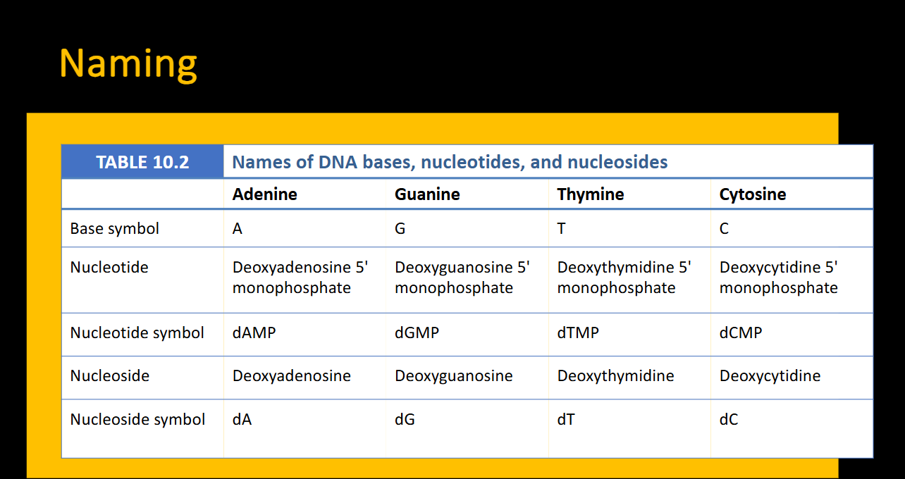
Naming system for bases
Nucleotide:
dAMP (deoxyadenosine 5’ monophosphate)
Nucleoside:
dA (deoxyadenosine)
Eukaryotes
Have a nucleus
Linear Chromosomes
Multicellular
Membrane-bound organelles (ex: mitochondria)
EX: Fungi, plants, animals, diatoms, malaria
Prokaryotes
No nucleus
lack membrane-bound organelles
DNA is long and CIRCULAR supercoiled chromosome in nucleoid region
single celluar
EX: Archaea and Bacteria
Endosymbiotic Theory
Ancestral prokaryote with nucleoid—> Infolding of plasma membrane forming protonucleus—>Engulfs aerobic bacterium—>Engulfs cyanobacterium—>Ancestral Eukaryote (with mitochondria and chloroplasts)
Supercoiling
Makes DNA structure more compact and is stabilized using proteins
Occurs in both prokaryotic and eukaryotic cells
Topoisomerases
(+) Over wound
(-) Under Wound
Topoisomerases
Enzymes that add or remove rotations of DNA (to manage its supercoiling)
(+) Supercoil (Overwound)
Closes down DNA, makes it tighter and more compact
(-) Supercoil (underwound)
Allows 2 strands to move or twist off of one another
(During processes such as transcription)
Type IA (Topoisomers)
(+) Supercoiling
5’ ssDNA (single-stranded) cleavage
Cleaves One side
Does NOT require ATP
EX: Reverse gyrase with helicase
Only enzyme that introduces (+) supercoil in DNA
also found in Archaea
Type IB Type IC (Topoisomerases)
(+) Supercoiling
3’ ssDNA (Single-stranded) Cleavage
Cleaves one side
does NOT require ATP
Controlled rotation
Chromatin
Consists of DNA and packing proteins (histones)
Eukaryotic DNA remains packed during interphase
Epigenetic changes effect chromatin structure
Euchromatin
Less condensed structure of DNA where transcription occurs
Located on Chromosome arms
Unique Sequences
Many genes
Replicated throughout S Phase
Often transcribed
common in crossing over
Heterochromatin
More condensed (“true” chromatin) structure
Located at centromeres, telomeres
Repeated sequences
Few genes
Replicated in Late S phase
uncommon in crossing over
Types:
Constitutive
Facultative
Constitutive Heterochromatin
Remains condensed (ALWAYS heterochromatin)
like at centromeres
Facultative
Is condensed (heterochromatic) only at particular developmental stages
How does the cell package long amounts of DNA into the nucleus?
Through the tertiary structure of DNA (chromatin) wrap around Histone proteins and Nucleosomes
Nucleosome (NCP)
The basic repeating unit of chromatin
147 base pairs of DNA
Protein octamer of 4 histone proteins (2 of each; H2A, H2B, H3, H4)
Separated by Linker DNA (a stretch of DNA whose length varies in different cell types)
Histone Core Particle (HCP)
Octamer
High in Lys & Arg (positively charged amino acids)
The positively charged amino acids attract DNA’s negatively charged sugar phosphate backbone (167 base pairs)
Excludes H1
Histone tails
11-37 Amino acids long
can be modified, affecting transcription and replication
H1
Not apart of HCP octamer
Binds to 20 base pairs of DNA (and linker dna?) to lock the nucleosome components in place.
Epigenetic effect on chromatin
Compaction prevents transcription of DNA into RNA
even euchromatin (that is reactive/actice) has nucleosomes which block transcription of DNA
Chromatin remodeling is required for DNA transcription (to unblock it)
Telomeres
The caps/tips of LINEAR chromosomes
usually consists of repeating A and T followed by several G
There is a G rich 3’ overhang of 50-500 bases
The overhang can form a T-loop
Helps protect chromosome ends from degradation
Shelterin protects the ends from repair
multiprotein complex
binds to telomeres and protects end
Telomere shortening
DNA polymerase cannot replicate the ends of chromosomes, so telomeres get Shorter with every Cell Division
Germ cells and stem cells have telomerase enzyme to lengthen telomeres and prevent shortening
Cancer cells sometimes have increased telomerase activity
Too Short Telomeres…
Leads to Senescence
Senescence
Process of deterioration with age