UF BSC2011 Exam 2 (Animal Unit 2024)
 Call Kai
 Learn
 Practice Test
 Spaced Repetition
 Match
 Flashcards
 Knowt Play
1/99

There's no tags or description
Looks like no tags are added yet.
100 Terms
What are the four general characteristics of animals, and what is one type of animal that does not have all of these characteristics?
Four General Characteristics of Animals:
- Multicellular
- Heterotrophic Metabolism
- Internal Digestion
- Movement and nervous system
One animals Lacking all these characteristics are sponges (no nervous system or movement)
The common ancestor to all animals is likely similar to what type of extant organism?
Most likely a colonial flagellated protist, similar to existing choanoflagellates and sponges
- Came together as colonies because more efficient and capturing prey than single cells
- Cells in these colonies began to specialize for different functions - ex movement, nutrition
- Developed into animals
What is an important difference between spiral cleavage and radial cleavage, and what are some examples of types of organisms that have each type?
Cleavage: Early cell divisions of an embryo. Patterns of cleavage are used to characterize major animal groups
Radial Cleavage: Yolk is evenly distributed zygote and descendant cells divide completely and evenly
- central axis of division (typically a symmetrical ball)
- Deuterostomes (most complex organisms like frogs)
Spiral Cleavage: Complex permutation of radial cleavage (new cells at angles to existing ones)
- each layer of cells is angled to the cell beneath it making a spiral
- Protostomes (mollusks and arthropods)
- ex. spiral of snail shells is due to spiral cleavage

What is the difference between diploblastic and triploblastic organisms? Where do these layers come from (how are they formed)?
Monoblastic: one cell layer - sponges only
Diploblastic Organisms: Animals with two cell layers
- Ectoderm (outer layer) - skin, neural crest
- Endoderm (internal layer) - Digestive tube, Respiratory tube
Triploblastic Organisms: Animals with three cell layers
- Ectoderm, Endoderm, and Mesoderm (middle layer)
- Mesoderm ex. red blood cells
- more complicated and advanced animals that form a clade
Layers form during Gastrulation: Cell migration inward of embryo
- during cleavage, form blastula (hollow ball of cells)
- cells migrate inward (invagination) and forms distinct layers and a pocket for the gut

What is the main difference between a protostome and a deuterostome organism? How does the blastopore form?
Gastrulation - Embryo is a hollow ball of cells that indents to from a cavity, the blastopore
Both are Bilaterians: Bilateral symmetry during embryonic development
- Protostome: blastopore develops into the mouth; anus forms later
- Deuterostome: blastopore develops into the anus; mouth forms later
Each group is a distinct clade
radial vs bilateral symmetry
Radial: body parts arranged around a central axis
ex. starfish
Bilateral - can be divided into mirror image (halves on only one plane
ex. frog or human - left and right
- Bilateral Symmetry is correlated with Cephalization: concentration of sensory organs (eyes ears) and nerve tissues at the anterior end or head
- Has been evolutionarily favored; the anterior end encounters the environment first
Asymmetrical means no plane of symmetry ex. sponges

What features differentiate a true coelomate from an acoelomate or pseudocoelomate?
Coelom: a fluid-filled body cavity that forms within the mesoderm layer
Acoelomate: no fluid-filled body cavity
- space between gut and body wall is filled with cells called mesenchyme; move by beating cilia
Pseudocoelomate: body cavity is a pseudocoel
- a fluid-filled space in which internal organs are suspended; no mesoderm surrounding internal organs only not inside wall
Coelomate: body cavity is a coelom that develops within mesoderm; mesoderm surrounds both organs and inside wall

What broad categories of animals would be expected to have nerve nets instead of a central nervous system?
Nerve Nets: Diffuse nervous systems in animals such as ctenophores and cnidarians
Bilaterians: Have well-coordinated central nervous systems
- Muscle action is coordinated to allow movement of appendages and body parts
- Sensory information is gathered and processed
Compare and contrast some of the defining features of sponges, cnidarians, ctenophores, and placozoans.
- Sponges, ctenophores, placozoans, and cnidarians are not bilaterians
- Many believe that sponges and ctenophores were the first lineages to split from the remaining animals but disagree on which split first
- Common ancestor was probably very different from any living animals - large multicellular animals appear to have evolved several times in different lineages
Sponges:
- no distinct embryonic cell layers and no true organs
- monoblastic
- have hard skeletal elements called spicules
cnetophotes (comb jellies):
- radial summetry
- diploblastic
- Move by beating cilia arranged on eight comb-like plates called ctenes
Placozoans:
- simple; only 4 cell types
- diploblastic
- Two life stages: Pelagic swimming stage and stage that adheres to surfaces
Cnidarians (jellyfish, corals):
- Gastrovascular cavity: function in digestion gas circulation
- diploblastic
- Nematocysts: harpoon like structures with toxins
What are heterotrophs and how do they get their energy and building blocks for cells and tissues?
Heterotrophs: Organisms that require preformed organic molecules as a source of energy and chemical building blocks (ex. those who eat plants since they cannot perform photosynthesis to make their own food)
- Obtain energy by breaking the chemical bonds of organic compounds from other organisms
- Build their tissues from the matter within these preexisting organic compounds as well
- Animals required to do this to grow/replace cells their whole life
In terms of the second law of thermodynamics, explain why animals need to continuously obtain energy.
Animals need to continuously obtain energy so they can do work to maintain their organization
ex. Cells maintain their charge across the cell membrane
ex. Tissues have layers of organized cells (neurons and axons must be in correct positions)
Second Law of Thermodynamics:
- Organization doesn't just happen
- Any organized system, left to itself, tends to lose organization and become more random (entropy)
- It takes energy to create and maintain organization
What are essential amino acids and essential fatty acids? In terms of these essential nutrients, what would be the significance of eating tortilla chips with bean dip?
- some molecules can be interconverted (ex. lipids can be converted to carbohydrates) but some molecules must be obtained, fully formed, from their food
- Essential nutrients: Require but cannot be synthesized by animals
- Standard Amino Acids: 20 amino acids animals use to build proteins; animals can synthesis some of these (nonessential)
- Essential Amino Acids: Amino acids that cannot be synthesized and must come from food
- Essential Fatty Acids: includes omega-3 and omega-6 fatty acids
MEMORIZE THESE 8 ESSENTIAL AMINO ACIDS: (PVT Tim HLL)
- phenylalanine
- valine
- tryptophan
- threonine
- isoleucine
- methionine
- Histidine
- leucine
- lysine
Beans and Grains contain all of these amino acids when combined

What are two examples of essential vitamins, two examples of essential minerals for humans, and what are they used for in the body?
Essential Vitamins: Vitamin A (vision), Vitamin K (blood clotting)
Essential Minerals: Chemical elements that animals require in addition to C, O, H, and N
ex. Calcium and phosphorus are needed for bone, iodine is required for synthesizing thyroid hormones, iron is required to synthesize hemoglobin
How is one calorie defined? What are "calories" on food labels?
Energy content of food can be measured by the amount of heat the food produces when completely burned in the presence of O2 producing CO2 and H2O
Calorie: amount of heat needed to raise 1 gram of H2O 1 degree Celsius
- Food labels, that show "calories," are actually kilocalories (1000 calories)
How is metabolic rate defined, and how is it typically measured?
Metabolic Rate: amount of chemical bond energy consumed and converted to heat per day
- Measured by determining rate of O2 consumption
- When organic matter is oxidized during aerobic metabolism, O2 is used and heat is produced
Animals can have different metabolic rates:
- Animals with high rates need to find and eat a lot of food per unit of time to replace the energy being consumed
- Animals with lower metabolic rates have lower food needs
Physical activity increases metabolic rate:
- for humans, it increases linearly as running speed increases
- for fish, it increases exponentially because resistance posed by water increases exponentially
- for birds, flight at low speed has a high metabolic rate just to stay airborne
What are the three basic types of nutrient molecules?
Thee types of food molecules:
- Lipids (fats and oils)
- Carbohydrates (sugars starch)
- Proteins (amino acids)
When an animal eats foods containing more chemical-bond energy than it needs, some can be stored, usually as energy dense lipids (fat on body)
Which would need more energy per unit body weight- a mouse or a horse? Why?
Scaling Relationships: animals characteristics as a function of size
- Basal metabolic Rate (BMR): measured when animal is in a comfortable thermal environment and has not eaten recently
- BMR per gram of weight decreases as animal size increases
- Small mammals need more food per from of body weight than large mammals do
So, a mouse would need more energy per unit body weight as they are smaller than horses and have a higher BMR per gram of weight
Which graph represents a temperature conformer? Which represents a regulator?
(One with a constant line an one with a positive slope)
Cells in an animals body are bathed with body fluids called tissue fluids (interstitial fluids)
- Tissue fluids are the animal's internal environment
- The outside world is the external environment
Cells also have intracellular fluids: fluids inside cells
- The rest of the fluid in the body is the extracellular fluid; includes blood plasma and interstitial fluid
Regulation: occurs when the internal environment stays constant despite changes in the eternal environment (constant line)
- animals that maintain constant internal conditions such as temperature are regulators
Conformer: when the internal environment varies so that it always matches the external environment (sloped line)

What are some advantages and disadvantages of thermoregulation verses thermoconforming?
Thermoregulation: provides stability but is energetically expensive; tends to increase metabolic rates (Hot Blooded)
- Small Mouse in the winter
Thermoconforming: energetically cheap but the animal's cells must be able to cope with changes in their environment (Cold Blooded)
- Alligator in Gainesville in January
In terms of digestion, what are some important functions of epithelial tissue?
Epithelium: sheet of epithelial cells that covers a body surface or organ or lines a body cavity
Simple Epithelium: Single layer of cells on a nonliving basement membrane; lines all blood vessels, intestines, and other tubules
Functions: PROTECTION ABSORPTION SECRETION
- pumps ions between fluids on either side of the epithelium
- Secretes substances such a hormones, mucus, digestive, enzymes, milk, sweat
- Absorbs nutrients from the gut
- Serves sensory functions, including smell and taste

What is the midgut, and what is its importance in terms of digestion and absorption?
Midgut: the small intestine - part of a multi-organ system responsible for food processing
Digestion: breakdown of food molecules by enzymes into smaller chemical components, such as glucose
Absorption: uptake of digestion products into the blood
The Epithelia lining of the midgut provides a huge surface area for absorption and enzyme secretion

Explain the concept of homeostasis, and explain the difference between a homeotherm and poikilotherm.
Homeostasis: Stability of the internal environment and the mechanisms that maintain it
- Similar to regulation in that it has pros in cons (is energetically expensive)
- Animals exhibit different degrees of homeostasis
Homeotherm: Animals that maintain a constant internal body temperature (thermoregulation)
- ex. mammals and birds
- Thermoneutral zone (TNZ): Range of external temperatures in which metabolic rate is minimal and does not change with external temperature - outside the TNZ, the metabolic rate rises
Poikilotherm or Ectotherms: Body temperatures are variable and are determined by the external temperature (conformers)
- ex. cold-blooded reptile
- Vary widely in their body temperature limits
- Can control body temperatures through behavior ex. alligator can go into the sunlight when cold or into the water when hot
- If too cold, body functions decrease ex. can't digest food
Explain the advantages and disadvantages of homeothermy vs. poikilothermy.
Homeothermy:
- Maintains a stable internal environment for optimal enzyme function and can travel through range of external temperatures
- Energetically expensive and more prone to suffering extreme temperatures
Poikilothermy:
- Energy efficient and survive on less food
- Activity is tied to environmental conditions
- Restricted to specific habitats
Explain the ways that homeotherms heat and cool their bodies to maintain homeostasis.
Homeotherm mechanisms increase metabolic rate in cold environments:
- Shivering - skeletal muscles contract and energy is converted from ATP to heat
- Non-shivering thermogenesis - some mammals have brown adipose tissue in which oxidative phosphorylation is uncoupled from ATP production; energy goes to heat production instead
Insulation is also important for maintaining body temperature; for example, fur and feathers
- specialized blood flow patterns can also conserve heat
- In hot environments, homeotherms can increase heat loss by evaporative cooling (sweating or panting): water absorbs a lot of heat energy to change from liquid to gas state; sweating and panting allow water to evaporate from body
Be able to draw a negative feedback loop for homeothermic thermoregulation, showing the controlled variable, sensors, control mechanism, and effectors. (look at PP 3)
Controlled variable: Characteristic being controlled, such as body temperature
Sensors: detect current level or state such as temperature receptor in the skin and brain
Control mechanism: uses information from sensors to determine which effectors to activate. Nerve cells of the hypothalamus in the brain are the temperature control mechanism (compares TNZ)
Effectors: Tissues or organs that can change the level of the controlled variable ex. shivering muscles

Explain (in terms of homeostasis) how a positive feedback system differs from a negative feedback system.
Negative Feedback System: control mechanism that activates effectors to reduce or negate differences that exists between the controlled variable and the set point
- a stabilizing influence that is often involved in
maintaining homeostasis
Positive Feedback: deviations of a controlled variable from its existing level are increased or amplified (rarer than negative feedback)
- Destabilizes a system moving away from homeostasis, but can be useful if it is ultimately brought under control
ex. child birth when infant is pushed out pressure is on cervix which signals to release oxytocin

Explain some differences in how (and reasons why) small and large mammals might deal with thermoregulation differently.
Smaller bodied animals often deal with the stress of an extreme environment by using behavior
- larger surface area to volume ratio
- Higher metabolic rate = more energetically expensive to maintain body temperature
- can access cooler/hotter microenvironments
Larger bodied animals often have fewer behavioral options; they depend on more physiological defenses to survive
ex. reindeer exhibit phenotypic plasticity between summer and winter in the relationship between metabolic rate and environmental temperature
- lower rate of metabolism compared to humans
- add insulation in winter and regional hypothermia - allows tissues in appendages to be cooler than core tissues
Explain the concepts of phenotypic plasticity and regional hypothermia in terms of thermoregulation.
Reindeer exhibit phenotypic plasticity between summer and winter in the relationship between metabolic rate and environmental temperature
- lower rate of metabolism in winter compared to humans
- add insulation in winter
- regional hypothermia
Regional hypothermia: tissue in appendages doesn't freeze, even when environmental temperatures are very low
- ex. arctic fox: heat is metered to foot pads so that tissues cool to near the freezing point but never colder than freezing. Feet are kept dry because they are too cold to melt snow or ice
Explain how countercurrent heat exchange can be used to maintain parts of the body within certain temperature ranges.
Countercurrent Exchange System: Between arteries and veins in limbs conserves heat
- arteries and veins are contact and in parallel so heat is transferred between the two
- prevent loss to the environment
- keeps most of the heat in the body core but enough prevents the paws from freezing
- energetically/metabolically efficient
Explain some ways (and examples) that behavioral thermoregulation and/or physiological adaptations can be used to maintain physiological function while reducing energy and water demands.
Hot deserts lack water and are hot - high temperatures
Small mammals are mostly nocturnal and stay in burrows during the day avoiding the high temperatures
- ex. kangaroo rats
- Lizards may use shade and stay close to ground
- Ants live underground but forage during day and eat recently dead insects with juices to get water and nutrients
Large animals such as Grant's gazelle can tolerate a rise in body core body temperature about 46 degrees Celsius
-This reduces water need because they don't sweat and have a countercurrent system to keep brain cool
Cold Environments are dangerously cold:
Hibernation: state of low body temperature and thermal conformity that persists for a long period of time in winter
-Animals allows its core body temp to match external temperature
- Animal lives on body fat or stored food collected the previous summer
- uses Metabolic Depression: biochemically induced reduction of metabolic rate
Some insects stabilize in a supercooled state so they can't freeze and some just freeze solid
- Antifreeze blood
Some Fish adapt completely to environment temperatures and cannot live anywhere else (warmed 5 degrees = death)
Explain O2 diffusion in terms of partial pressure gradients.
O2 diffuses passively (no energy required) down the partial pressure gradient (higher in lungs ~100mmHg, lower in blood ~40mmHg)
- Partial pressure refers to portion of pressure a gas contributes to a system
- Gases move from higher partial pressure to lower partial pressure
Internal Respiration: In mitochondria, Oxygen is used as the final electron acceptor in aerobic respiration
External Respiration: Animals obtain Oxygen from the environment which then follows a pathway to the mitochondria
CO2, the waste product of aerobic respiration, must be removed as it can acidify body fluids
Explain the difference between diffusion and bulk flow in terms of O2 movement.
O2 and CO2 are transported by diffusion and bulk flow (no active transport)
In Figure: 1 and 3 are bulk flow and 1 and 2 are diffusion
Diffusion of O2: Diffuses across the alveolar epithelium and capillary epithelium going down the partial pressure gradient (higher in lungs)
- Individual gas molecules moving across epithelial tissue
- O2 binds to hemoglobin on red blood cells
- Then diffuses from from blood to body cells
Bulk Flow of O2: Passive flow of matter (material contained within a medium) from one place to another
- Large quantities of gas moving in a medium catching a train through blood and diffusing where needed ("going with the flow")
- O2 is carried in the bulk flow of air into the lungs and alveoli
- also carried by the bulk flow of blood from capillaries to interstitial fluid
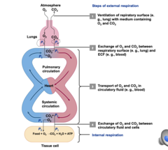
Overview of Human Respiratory system
1. Air fills the lungs
2. Oxygen diffuses across the alveolar epithelium that layers the lungs and the capillary epithelium of the pulmonary veins
3. O2 diffuses down the partial pressure gradient from 100mmHg in the lungs to 40mmHg in the blood
4. O2 binds to hemoglobin on red blood cells and transfers it back to heart where it is sent throughout body
5. O2 diffuses from blood into body cells down the PP gradient from 100 to 40 again

Explain what occurs at each of the 4 main steps of external respiration. Refer to the figures on slides 10 and 11 of the PowerPoint slides.
Breathing organs have a large surface area of gas exchange membranes, achieved by folding and branching (lungs are folded inward to body and gills are folded outward into environment)
4 Steps of External Respiration:
Ventilation: Inhale air into lungs; exhale air out.
Gas Exchange in Alveoli: O₂ diffuses into blood; CO₂ diffuses into alveoli.
Transport in Blood: O₂ binds to hemoglobin; CO₂ transported as bicarbonate, dissolved, or bound to hemoglobin.
Gas Exchange at Tissues: O₂ diffuses into tissues; CO₂ diffuses into blood.
Explain the difference between tidal and flow-through ventilation. Which would typically result in greater partial pressure differences for gas exchange?
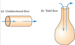
Ventilation: Bulk flow of air or water between the gas exchange membrane and the outside world
- Tidal Ventilation Air moves in and then out the same passageways (not so efficient)
- Unidirectional/Flow-Through Ventilation: In animals with gills, water moves in a one way stream across the gills
Flow Through ventilation is typically more efficient as the partial pressure difference for gas exchange is greater as there is no mixing of fresh air and stale air
How does perfusion affect gas exchange across membranes?
Perfusion: Blood flow through capillaries or other small blood vessels of a tissue
- Perfusion of gas exchange membranes must be fast enough for O2 to be efficiently picked up from breathing
- Blood must the circulate fast enough to efficiently deliver O2 to body cells
TLDR: the faster blood can move oxygen, the more it can pick up and more gas can diffuse across membrane
Why is respiration more difficult (less efficient) in water than in air? How would sea turtle eggs be affected if the nest was flooded with water?
Diffusion in Water vs Air:
- Diffusion is much faster through air than water
- O2 Solubility in water is low and decreases with increasing temperature resulting in lower PO2 in water compared to air
Sea Turtle Result:
- Sea turtles lay eggs in sand - eggs get plenty of O2 if the sand is moist but no wet
- If sand is flooded, PO2 is much lower and the eggs die due to lack of oxygen
What makes gas exchange in fish gills so efficient? Explain countercurrent exchange as it relates to gas exchange in fish gills.
Fish gills have and enormous surface area for gas exchange
Each gill arch has two rows of gill filaments with numerous secondary lamellae (epithelial cells) on its upper and lower surfaces
The fish pumps water unidirectionally: through mouth cavity and gill slits, over gills, and out from under the opercula flaps, Blood flows through each lamella in the opposite direction , a concurrent system (water goes one way and blood goes other)
Countercurrent Gas Exchange: The two fluid streams flow in opposite directions
- Fresh water is always in close proximity to fresh (deoxygenated) blood
- Maintains a high PO2 gradient across the entire surface
Co-Current Gas Exchange: The two fluid streams flow in the same direction (not as good as gradient deminishes
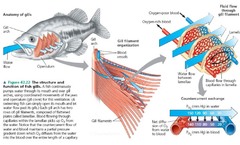
What makes gas exchange in fish gills so efficient? Explain countercurrent exchange as it relates to gas exchange in fish gills.
Concurrent gas exchange: the two fluid streams flow in the same direction.
Countercurrent gas exchange: the two fluid streams flow in opposite directions.
- Fresh water always in close proximity to fresh [deoxygenated] blood
- Maintains high PO2 gradient across the entire surface
Explain the differences between ventilation in birds vs. mammals in terms of air sacs and crosscurrent exchange. Which system is more efficient for gas exchange?
Mammals:
Most terrestrial vertebrates have tidal ventilation
- Air passages in the lungs end in closed dead-end sacs of gas exchange membrane
- Inner surfaces of vertebrate lungs are coated with lung surfactant that reduces surface tension and prevents gas exchange tissue from sticking together
Birds:
- Use unidirectional flow air flow to supply O2 to tissues at high rates
- Air sacs expand and contract pushing air past gas exchange surfaces in the lungs (Air sacs themselves don't participate in gas exchange)
- Crosscurrent exchange: occurs almost counter-currently between air and blood capillaries which makes gas exchange very efficient

What are tidal volume, respiratory minute volume and residual volume? How do these change with exercise?
Tidal Volume: Amount of air that moves in and out of lungs per breath
Respiratory Minute Volume: total amount of air inhaled and exhaled per minute
- exercises results in and increase in tidal volume, and thus, an increase in respiratory minute volume
Residual volume is stale air that is left in the lungs to prevent them from completely closing and collapsing
What parts of the brain control breathing rhythm in mammals?
Breathing in Mammals is controlled by the medulla oblongata in the brain and the pons.
- negative feedback system; If CO2 increases and PH increases it increases breathing; if O2 falls chemoreceptors iin aorta send nerve impulse to get more oxygen
Mammalian lungs are suspended in a thoracic cavity with the diaphragm (sheet of muscle) at the bottom
- Inhalation: Contraction of the diaphragm expands the thoracic cavity, which pulls on the lungs and expands then; air is sucked in
- Exhalation: Diaphragm and intercostal muscles relax and lungs recoil in thoracic cavity, pushing air out
If partial pressure of CO2 in the blood increases, how does this affect blood pH and breathing rate? Where are the detectors for blood pH changes?
If PCO2 increases, Carbonic acid is made which H+ increases and pH falls (acidic blood)
- detected in the medulla oblongata and breathing is stimulated to increase respiratory minute volume
If partial pressure of O2 in the blood decreases, how does this affect breathing rate? Where are the detectors for blood O2 levels?
If PO2 falls, chemoreceptors in the aorta and carotid arteries send nerve impulses to the medulla oblongata to stimulate increased breathing
Explain the important components of a closed circulatory system.
- HEART
A circulatory system consists of:
Muscular pump - Heart
- Myocardium: The muscle tissue of a heart
- Stroke Volume: Volume of blood pumped per heartbeat
- Cardiac Output: Volume of blood pumped per minute
During exercise both stroke volume and heart rate increase
2 atrium and 2 ventricles:- O2 depleted blood from systematic tissue enter right atrium from vena cava and flows into right ventricle
- Right ventricle contracts pushing blood through pulmonary arteries to lungs
- O2 rich blood from lungs enters left atrium and flows into left ventricle
- Left ventricle contracts pushing the O2-rcih blood through the aorta to the systematic tissues
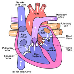
Explain the important components of a closed circulatory system.
- BLOOD
Fluid - Blood
Bulk Flow of blood transports O2 rapidly throughout the body
Also transport of nutrient molecules, hormones, heat, and metabolic waste
term-44
Blood consists of plasma and cells:
- Blood Plasma: water and solutes such as glucose, ions, wastes, hormones, clotting proteins, and O2
Blood cell Types:
- Red (erythrocytes) - contain hemoglobin; to transport O2
- White (leukocytes) - participate in immune responses
Platelets (cell fragments) are involved in blood clotting

Explain the important components of a closed circulatory system.
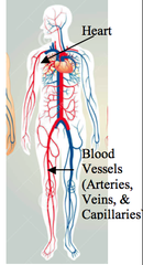
- BLOOD VESSELS
Series of Conduits - Blood Vessels
Vertebrates have a closed circulatory system: Blood always remains in the blood vessels
- Arteries: places where heart pumps blood which branch out into smaller and smaller vessels, eventually into capillaries (microscopic vessels)
- As blood leaves tissues and organs, if flows into larger and larger vessels, the veins, back to the heart
- Arterioles, capillaries, and venules (small veins) comprise the microcirculation
Capillary walls consists of only vascular endothelium, thin layer of tissue less than one micron thick
- Small Blood cells must pass through one at a time (squeeze through)
- capillaries are found near every tissue cell and are primary sites where O2, CO2 and other materials are exchanged
- In many tissues there are pores between epithelial cells
What mechanism in the arterioles controls the rate of blood flow through the capillaries?
Arteriole walls have a layer of smooth muscle under the control of the autonomic nervous system
- Vasomotor control: the smooth muscle adjusts arteriole diameter
- contraction causes vasoconstriction
- relaxation causes vasodilation
- Arteriole diameter determines rate of blood flow through capillaries
Explain the basic characteristics of an open circulatory system.
Open Circulatory system; Blood exits vessels as it flows through the body (arthropods and most mollusks)
- No distinction between blood and interstitial fluid - biologists use the term hemolymph instead of blood due to mixture - no need for veins
- Tissue cells are bathed by the blood
- They readily take up O2 and other materials from the blood and release waste

Where is hemoglobin in the body and what does it do?
Respiratory pigments greatly increase the amount of O2 that can be carried
- Hemoglobin in humans combines reversibly with O2
- Oxygenation: when a pigment combines with O2 in the lungs (picks it up)
- Deoxygenation: when a pigments releases O2 in the tissues
(Oxygen equilibrium curve can show loading and offloading of O2) (on pp)
How does partial pressure of O2 in the blood affect the percent of hemoglobin that is bound to O2?
As partial pressure of O2 in blood decreases, so does the percent of hemoglobin bound to O2
As partial pressure of blood increases so does the percent of hemoglobin bound to O2
- Exercise lowers percent as partial pressure of O2 is lower
How does exercise affect heart performance (stroke volume, heart rate) and hemoglobin-O2 interactions?
The circulatory system increases O2 delivery during exercise in two ways:
- Blood circulated more rapidly by increasing heart rate and stroke volume
- Hemoglobin release more than twice as much O2 at the exercising muscles
- Lower overall partial pressure of O2
- During exercise both stroke volume and heart rate increase
How many heart chambers do mammals, birds, amphibians, and fish typically have?
4 Chambers: humans, mammals, and birds
2 Chambers: Fish
3 Chambers: Amphibians
In a mammal, starting with the vena cava, explain the blood flow through all chambers of the heart, through pulmonary blood flow, and then out of the heart to the systemic tissues. Explain when/where the blood is oxygen-rich or oxygen-depleted.
- O2 depleted blood from systematic tissue enter right atrium from vena cava and flows into right ventricle
- Right ventricle contracts pushing blood through pulmonary arteries to lungs
- O2 rich blood from lungs enters left atrium and flows into left ventricle
- Left ventricle contracts pushing the O2-rcih blood through the aorta to the systematic tissues
What are systole and diastole, and how do they relate to blood pressure readings?
Systole: when ventricles contract (high pressure)
Diastole: when ventricles relax (low pressure)
Left ventricle is more muscular than the right so:
- systolic blood pressure is the higher number and diastolic is the lower number in blood pressure
What are the functions of the SA node, the AV node, and the conducting system in the electrical activity of the heart?
SA node: Sinoatrial node contains pacemaker cells in the right atrial wall that initiate heartbeat
- electrical depolarization and contraction spread rapidly throughout the muscle
AV node: Atrioventricular node relays depolarization from SA node and delays it until atrium is empty to the conducting system and depolarization spreads rapidly and the ventricles contract

How do epinephrine, sympathetic, and parasympathetic stimulation affect heart rate and stroke volume?
Sympathetic Stimulation: Increases heart rate and stroke volume
- Epinephrine: Secreted by adrenal gland in response to fright or stress (increases heart rate and stroke volume)
Parasympathetic Stimulation: decreases heart rate (calms you down)
In an ECG/EKG readout, what do the P wave, the QRS complex, and the T wave represent?
Electrocardiograms record heart-related voltage differences on the surface of the body over time
P wave: Corresponds to the depolarization of the the atria
QRS Complex: corresponds to the depolarization of the ventricles
T wave: corresponds to the repolarization of the ventricles

What is coronary circulation, and why is it so dangerous if coronary vessels are blocked?
Coronary circulation: provides blood flow and O2 to heart tissues which are necessary for healthy heart function
- Coronary artery disease is when there is a blockage which can starve heart tissue of oxygen, causing a heart attack
Which types of blood vessels function as reservoirs, holding large amounts of blood at very low blood pressure?
Veins serve as a blood volume reservoir - any blood not held elsewhere in the circulatory system is held in veins
- blood pressure in veins is very low
- needs contraction of skeletal muscle to help propel blood through the veins and one way valves prevent backflow
What characteristics make the blood-brain barrier different from other parts of the circulatory system?
Many types of solutes can't cross the capillary walls in the brain and spinal cord because they have no pores
The blood brain barrier helps protect the neurons from exposure to toxins in the circulating blood (has no pores)
- Permits tight control of solute exchange
- Solutes require specific transport mechanisms to cross capillary walls rather than being able to leak across
What is unique about the capillaries in the kidney glomerulus?
Capillaries in the glomerulus have many pores and allow many types of solutes to pass
- blood pressure in the capillaries drives fluid and solutes into the kidney tubules where urine is formed
- blood pressure in the capillaries is higher here than others
In which type of blood vessel is blood pressure the lowest?
Great Veins (vena cava) have the lowest blood pressure in the body
Blood pressure tends to force fluid out of capillaries. What force opposes this blood pressure by pushing fluid back into the capillaries?
Blood osmotic pressure exceeds blood pressure and pushes water back into the blood
What mechanism returns fluid from interstitial tissues back to the circulatory system?
The lymphatic systems returns interstitial fluid to the blood
- Lymphatic capillaries continually take up excess fluid; merging into larger and larger vessels that ultimately empty into veins in the neck
- Lymph nodes filter the fluid before returning it to bloodstream
Explain how a countercurrent system helps to keep swimming muscles warm in tuna.
Countercurrent heat exchange depends on parallels arrays of arteries and veins
Tuna use a countercurrent system to keep swimming muscles warm
- Numerous arteries and veins going to and from the red swimming muscles are closely juxtaposed and heat from red swimming muscle is retained by transferring to cooler veins
Define hyper-, iso-, and hypoosmotic, and explain how ionic composition of fluids ondifferent sides of a membrane affect osmotic pressures.
Osmotic pressure: measure of the total concentration of solutes; determines direction of water movement
Hyperosmotic: solution with higher osmotic pressure (more concentrated)
Hypoosmotic: Solution with lower osmotic pressure (less concentrated)
Isosmotic: Two solutions have the same osmotic pressure (equally concentrated)
1) Explain each of the following and give an example:
a. Hyperosmotic regulator
b. Hyposmotic regulator
c. Marine osmoconformer
Hyperosmotic regulators are animals that must use energy to compensate and gain ions from water
- Ex. Fish in freshwater produce large volumes of dilute urine, and actively transport na+ can Cl- back across teh gills into their bodies
Hypoosmotic regulators are animals that tend to lose water by osmosis and gain ions by diffusion and must expend energy to compensate
- Ex. Fish in saltwater drink seawater to gain water, in the intestine and gills, ions are pumped out, and excess ions are excreted
Marine osmoconformers maintain a similar osmotic pressure as their environment
- cannot survive a variety of environments and restricted to certain salinities
How do seabirds and marine reptiles get rid of excess salts in their bodies?
seabirds and marine reptiles get rid of excess salt by excreting from extrarenal organs called salt glands (around head area) that use energy from ATP
- Ex. sea turtles cry out salt solution
If a fish moved from fresh water to salt water, which hormone would help it to compensate for the saltier environment? How about if a fish moved from salt water to fresh water?
Cortisol is a saltwater hormone: it directs shift to hypoosmotic regulation
- Ex. fish moving to saltier water excrete more cortisol to pump excess salt faster
Prolactin is a freshwater hormone: directs shift to hyperosmotic regulation
- Ex. fish moving to fresh water excrete more prolactin to pump in more salt
In fish, where are chloride cells located, and what do they do in fresh water and salt water?
Chloride cells are located in the gills of fish (either fresh or saltwater) and they secrte salt in saltwater but import salt in freshwater
- these cells use active transport against concentration gradient
Freshwater Kidney's can also help by diluting urine more to expel less salty urine
Saltwater Kidneys produce a small amount of urine with same osmotic pressure as blood plasma
On this graph, which is an osmoconformer and which is an osmoregulator? (view doc for picture)
osmotic conformers include most marine invertebrates:
- no regulation of ions
- osmotic pressure of body fluids is always same as the water they live in
- many cannot survive a very wide range of salinities but some can like mussels
Osmotic regulators include coastal invertebrates:
- maintain a constant internal osmotic pressure despite experiencing a range of external salinities
- Ex. blue crab
What is the primary form of nitrogenous waste that each of the following would be expected to excrete:
a. A freshwater fish
b. A dog
c. An iguana (lizard)
Wastes are produced from breakdown proteins and nucleic acids
- too much nitrogen is toxic to body and the type of nitrogen waste correlates with water availability
Ammonia: most aquatic animals that breathe water (ammonotely)
- diluted to nontoxic concentrations
- ex. used by fish
- squids may hold ammonia to hep be more buoyant in water
Urea: most terrestrial animals (ureotely)
- more expensive metabolically and less toxic than ammonia
- relatively non-toxic so can hold for longer
- ex. used by dogs
Uric Acid: insects reptiles and birds (uricotely)
- most expensive metabolically and highly insoluble
- need to conserve a lot of water
- ex. used by lizards or iguanas
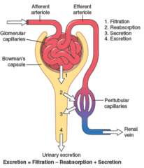
1) Explain the 4 basic processes that take place in the renal tubules:
a. Filtration
b. Secretion
c. Reabsorption
d. Osmoconcentration
- Filtration: Water and small solutes pass through a barrier while cells and large molecules remain behind
- Secretion: Transport epithelia move specific solutes into the tubulelumen for excretion
- Reabsorption: Transport epithelia move specific solutes and water backinto the body from the lumen
- Osmoconcentration: Water is removed from the lumen, leaving solutes behind, producing a more concentrated excretory fluid
Explain the 3 types of renal organs (protonephridia, meso/metanephridia, malpighian tubules) and provide an example of a type of animal with each.
Protonephridia (simple invertebrates):
- Blind end ducts project into body cavity
- Ultrafiltration driven by cilia moving fluid outward
Mesonephridia and Metanephridia:
- Ultrafiltration driven by fluid pressure
- Requires a circulatory system
- Mesonephric tubules in aquatic vertebrates
- Metanephric tubules in terrestrial animals
Malpighian tubules:
- Filtration driven by active secretion of ions
- Arthropods
Identify the key parts of a nephron on a figure and briefly explain the function of each part: Look at PP 6
Vascular Component:
a. Afferent arteriole
- Carries blood to the glomerulus
b. Glomerulus
- A tuft of capillaries that filters a protein-free plasma into the tubular component
c. Efferent arteriole
- carries blood from the glomerulus
d. Peritubular capillaries
- split from efferent arteriole and supply the renal tissue; involved in exchanges with the fluid in the tubular lumen
Tubular Component:
e. Bowman's capsule
- collects the glomerular filtrate
f. Proximal tubule
- uncontrolled reabsorption and secretion of selected substances occur here
g. Loop of Henle
- establishes an osmotic gradient in the renal medulla that is important in the kidney's ability to produce urine of varying concentration
h. Distal tubule and Collecting duct
- variable, controlled reabsorption of Na+
and H2O and secretion of K+ and H+
occur here; fluid leaving the collecting
duct is urine, which enters the renal
pelvis
Combined Vascular Tubular Component:
i. Juxtaglomerular apparatus
- produces substances involved in the control of kidney function

The descending limb of the loop of Henle allows reabsorption of _______ but not ________.
Descending limb of loop of Henle allows reabsorption of water but not Na+
The ascending limb of the loop of Henle allows reabsorption of _______ but not ________.
ascending limb of loop of Henle allows reabsorption of Na+ but not water
What is meant by the "countercurrent multiplier" of the kidney?
Loop of Henle allows kidney to concentrate solution in and around the base of the loop
What causes aquaporin 2 channels to be added to the kidney, where are they added, and what do they accomplish?
AQ2 channels are added due to vasopressin (the antidiuretic hormone) that increases water reabsorption in distal tubule and collecting duct
What do the vasa recta accomplish in the kidney and, very briefly, how do they do this?
The Vasa Tecta help conserve vertical osmotic gradient in the renal medulla (must reach 1200 mosm to concentrate urine)
- Countercurrent exchange occurs when the vasa recta form a hairpin loop

What is meant by "urea recycling" in the kidney, and how does this affect water reabsorption?
Urea Recycling contributes to medullary hypertonicity:
- Top and middle portions of the collecting duct are impermeable to urea
- Urea concentration in the collecting duct increases as water is reabsorbed
- The lowest section of the collecting duct is permeable to urea, so urea diffuses out, increasing the solute concentration of the medulla
- Urea concentrations is
- High levels of urea results in high osmotic pressure
- As substances are reabsorbed, urea becomes more concentrated and makes it favorable to diffuse into outside tissue
What are the stimuli for renin secretion from the juxtaglomerular apparatus, and how does this ultimately result in aldosterone secretion?
Low blood volume or low blood pressure results in the secretion of renin from juxtaglomerular apparatus (JGA)
- Renin catalyzes conversion of angiotensinogen, a protein, into angiotensin I
- Angiotensin I then converted to angiotensin II by angiotensin-converting enzyme (ACE) in teh kidney
- Angiotensin II then stimulated for aldosterone secretion from adrenal cortex
What are the primary functions of aldosterone?
- Increases Na+ reabsorption by distal tubes and collecting ducts
- Stimulated secretion of vasopressin (ADH) which promotes water retention in kidneys
- Potent constrictor of systemic arterioles directly increasing blood pressure
Explain the difference between central nervous system and peripheral nervous system, and explain what a "nerve" is in comparison with a "neuron".
Central Nervous System: brain and spinal cord
Peripheral Nervous System: all other parts of system
All parts of nervous system are made up of neurons which are specialized nerve cells
In the PNS, neurons are organized into nerves which are bundles of hundreds to thousands of neurons
What are afferent and efferent neurons, and which of these terms applies to a sensory neuron and a motor neuron?
Afferent Neurons: Carry signals towards the CNS
Efferent Neurons: Carry signals away from CNS
Sensory neurons carry signals from sense organs to CNS (Afferent Neurons)
Motor Neurons carry signals to muscle cells stimulating contraction (Efferent Neurons)
When discussing neuron function, explain what the terms polarized and depolarized mean.
Neurons are excitable cells that can generate and conduct impulses or action potentials
Cell membrane is polarized when the outside has a greater positive charge than the inside
Cell is depolarized when there is a reversal of polarity across the membrane
- rapid reversal of polarity is an action potential
- Action potential travels along axon of neuron
parts of neuron
Dendrites—carry signals to the cell body
Cell body—contains nucleus and organelles; integrates incoming signals.
Axon—conducts action potentials away from the cell body; can be very long
Presynaptic axon terminals—make synaptic contact with other cells
Synapse: cell-to-cell contact point specialized for signal transmission.
- Presynaptic cell conducts signals into the synapse.
- Postsynaptic cell conducts signals away from the synapse.

What are five important functions of glial cells?
res
- Help orient neurons toward their target cells during embryonic development
- Provide metabolic support for neurons
- Help regulate composition of extracellular fluids and perform immune functions
- Assist signal transmission across synapses
- Assist with signal transmission along and between neurons
Which two cell types produce myelin, and how does myelin help with neuron function?
Myelin is formed from glial membranes and myelinated axons conduct action potentials faster than non-myelinated axons
- myelin coated axons form white matter and areas of cell body are gray matter
Oligodendrocytes insulate axons in CNS
Schwann cells insulate axons in PNS
What are the roles of Na+-K+ATPase, K+ leak channels, and K+ equilibrium potential in establishing the resting potential of a neuron?
Resting Potential (-70mV due to leaking potassium)
Negative resting potential means inside of the cell is negative to outside
Only small numbers of positive charges need to move through the membrane for a rapid change of membrane potential
Sodium-Potassium Pump (Na+-K+ ATPase):
- Uses ATP to move 3 Na+ ions out of cell in exchange for 2 K+ ions inward
- Results in a more positive outside and more negative inside
- Creates ion concentration gradient as result too
K+ Leak Channels:
- Allow some Na+ to diffuse in but not much k+ to diffuse out
- Unequal leaking creates negative inside of cell
- Some K+ diffuses back in because it is attracted to the negative charges
- These movements reach an equilibrium, equal to the equilibrium potential of K+
In and around a neuron at rest, what are the relative distributions (locations) of Na+ and K+?
High concentration of K+ inside and low K+ outside
- Causes relatively positive outside and relatively negative inside
Regarding neurons, explain the terms polarized, depolarized, and hyperpolarized. Explain how ligand-gated and voltage-gated Na+ contribute to graded and action potentials.
When charge differential between inside and outside of cells is similar it is said to be depolarized
Hyperpolarized occurs if the charge on the inside becomes even MORE negative
Voltage Gated Channels: Respond to change in membrane potential
Stretch Gated Channels: Respond to tension applied to cell membrane
Ligand Gated Channels; Open or close when a specific chemical (ligand) binds to channel protein
- Speed of Na+ diffusion depends on strength of signal causing a graded potential
- If neurons reaches threshold potential (-55mV), voltage gated Na+ channels rapidly open and an action potential occurs
Why does an action potential travel in only one direction, and what is the role of myelin in action potential propagation?
Action potential can only propagate in the direction of the axon terminals because Na+ channel cannot open again for a brief time (refractory periods) and cannot depolarize
- Myelination increases speed of action potentials as charges move more laterally and depolarizations jump from node to node (nodes of Ranvier) between insulated areas (saltatory conduction)
What is the role of neurotransmitters?
Neurotransmitters are chemical signaling molecules that diffuse across the chemical synapse (small space between cells) of cells that an action potential cannot access
- They bind to receptors on the postsynaptic cells membrane generated a graded, potential,= or action potential
What are the roles of the somatic and autonomic nervous systems?
Somatic: Controls voluntary skeletal muscle
Autonomic: Controls effectors other than skeletal muscle (smooth, cardiac - involuntary muscle)
What are the roles of the sympathetic and parasympathetic divisions of the nervous system?
Within Autonomic:
Enteric Division: Nerve cells internal to the gut wall
Sympathetic Division: reduces digestion and generally prepares body for emergenceis (fight or flight)
Parasympathetic Division: Slows the heart, lowers blood pressure, increases digestion (opposite of Sympathetic)
Explain the key differences between how the nervous system and endocrine system function, in terms of 1) directness of their targeting, 2) how signals are sent and received, 3) speed of their functions, and 4) types of overall functions that they have.
Endocrine System:
- secretes into cells
- signals through hormones
- signals travel more slowly
Nervous System:
- directly wired to other cells affecting less number of cells
- signals through neurotransmitters (released at synapse)
- more direct targeting
- faster response
Explain the difference between hormones and other types of signaling molecules (neurotransmitters, paracrines, autocrines, pheromones).
- Neurotransmitters: move across synaptic cleft: secretion may be controlled by impulses that originate far from the site of release
- Paracrine: Chemicals secreted by one cell that affects neighboring cells
- Autocrine: Chemicals secreted into intercellular fluids that diffuse to receptors on that very same cell
- Hormones: Carried throughout the body by blood circulation
- Pheromones: released into the environment and affect other individual organisms
What is a neurosecretory, or neuroendocrine cell?
Neurosecretory/Neuroendocrine Cells: Excitable cells that propagate action potentials
- Cell bodies are in the CNS; the axon terminals release hormones into blood often within neurohemal organs
Nonneural Endocrine Cells: (epithelial endocrine cells) Not excitable
- typically stimulated to secrete hormone by other hormones (bandwagon)