Chapter 10 - DNA Structure and Analysis
 Call Kai
 Learn
 Practice Test
 Spaced Repetition
 Match
 Flashcards
 Knowt Play
1/88

There's no tags or description
Looks like no tags are added yet.
89 Terms
Learning goals
Understand and be able to describe the experiments that determined that DNA is the hereditary material:
Griffiths; Avery, MacLeod and McCarty; Hershey-Chase; Watson and Crick.
Understand how DNA structure affects its function:
Be able to draw the sugar-phosphate backbone and each nucleotide; be able to find the 5’ and 3’ ends of the chains
Understand the consequences of the antiparallel nature of the duplex.
Be able to find where the hydrogen bonds are between base pairs; understand the differences in the chemical nature of the ends (i.e., the phosphate group(s) and the 3’ hydroxyl).
For a molecule to serve as the genetic material, it must be able to:
Be replicated (copied) and distributed to daughter cells
Store the molecular information (protect it, repair it)
Express the information
Allow variation by mutation
Until 1944, observations favored protein as the genetic material
Proteins and nucleic acids were the major candidates for the genetic material
For a long time, protein was favored to be the genetic material
It is abundant in cells
It was the subject of the most active areas of genetic research
DNA was thought to be too simple to be the genetic material, with only four types of nucleotides as compared to the 20 different animo acids of proteins
Frederick Griffithc
British physician and bacteriologist during WWI
He assayed the pneumococci samples taken from patients and classified them to study the pneumonia epidemiology
He experimented on mice to understand the pathology
Credited as the first to show that the bacterium can change
Died in an air raid in London
Transformation by Griffith
Showed that avirulent strains of the bacterium Diplococcus pneumoniae could be transformed to virulence
The term for bacterial transformation came from this experiment
He speculated that the transforming principle could be part of the polysaccharide capsule or some compound required for capsule synthesis
Avery, MacLeod, & McCarty
Followed Griffith’s experiment
Demonstrated that the transforming principle was DNA, not protein
This work was recognized by some but ignored by many scientists
It was not recognized by the Nobel committee, which later apologized

Hershey-Chase Experiment
The work on DNA as the molecule of heredity was followed by experiments from the “Phage Church” group
Hershey and his technician Chase performed a critical experiment to demonstrate that DNA is the molecule of heredity
Transfection of viral DNA into bacterial cells proved conclusively that the viral DNA alone contains all the necessary information for production of mature viruses

Evidence for DNA as the genetic molecule
Indirect evidence
DNA is found only where the primary genetic function occurs, whereas protein is found throughout the cell
Correlation of UV light absorption and mutagenesis
UV light is capable of inducing mutations in the genetic material and is most mutagenic at a wavelength of 260 nm.
DNA and RNA absorb UV light most strongly at 260 nm, but protein absorbs most strongly at 280 nm, a wavelength at which no significant mutagenic effects are observed.
The strongest direct evidence for DNA as the genetic material comes from recombinant DNA technology.
Recomb. DNA tech. is the expression of eukaryotic genes in bacteria
e.g., making a copy of the human insulin gene (cloning), expressing it in bacteria, and recovering the produced hormone from the growth medium.
The presence of eukaryotic DNA and protein products produced from it are strong direct evidence that DNA is the material of heredity
DNA Structure, KNOW
nucleic acid chemistry
DNA structure which is essential to understand its functions
Nucleotides
Are the building blocks of nucleic acids
They are assembled into polynucleotides of DNA and RNA
They consist of
A nitrogenous base
A pentose sugar (5 carbon)
A phosphate group
Nitrogenous bases can be purines or pyrimidines; they are heterocyclic rings
Purines and pyrimidines are grouped into those with keto bases (G, T, U) or amino bases (A, C)

A sugar can be
Ribose or deoxyribose

Nucleoside
contains the nitrogenous base and the pentose sugar
Nucleotide
is a nucleoside with a phosphate group added
Nucleotides can have
one, two, or three phosphate groups and are called NMPs, NDP, and NTPs, respectively
RNA contains
ribose as its sugar
DNA contains
deoxyribose as its sugar
The C-5’ position is the location of the phosphate group on a nucleotide
There can be one, two, or three phosphates
Ex. NMP, AMP, ADP, ATP
DNA and RNA both contain A, C, G
Only DNA contains T
Only RNA contains U
Nomenclature
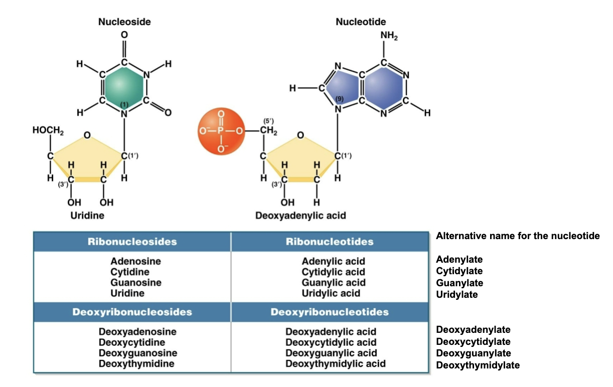
Phosphodiester bonds
Nucleotides are linked by a phosphodiester bond between the phosphate group at the C-5’ position and the OH group on the C-3’ position
An ester is -C-O-C
A phosphoester is -C-O-P-
Phosphodiesters have two adjacent phosphoester bonds; the phosphorus is shared
Chargaff showed
that the amount of A is proportional to T and the amount of C is proportional to G, but the percentage of C+G does not necessarily equal the percentage of A + T
X-ray diffraction of DNA showed
a 3.4 angstrom periodicity, characteristic of a helical structure
This was Rosalind Franklin’s work

Watson and Crick
proposed DNA is a right-handed double helix in which the two strands are antiparallel and the bases are stacked on one another
They built their model based on
Chargaff’s findings
Chemical principles
Bond angles and types
Solvent (water) interactions
Stacking, pi-bond electrons
Franklin’s diffraction data (A and B forms)
B form: 34 angstron periodicity
Modeling: cardboard cut-outs and trial and error
The two strands are connected by A-T and G-C base pairing and there are 10 base pairs per helix turn
B form DNA
The Watson-Crick DNA model is of B-DNA, which is the biologically significant form

DNA base pairs are tightly stacked
Like pancakes
Highly regular spacing
Accounts for 3.4 Angstrom spots
(X-ray diffraction) due to pi-bond interactions
No room for water or ions
Phosphates are far apart
Alternating sugars (gray and white) and phosphates (orange and red)
Sugar-phosphate backbone fully extended
Phosphates are negatively charged - charge repulsion
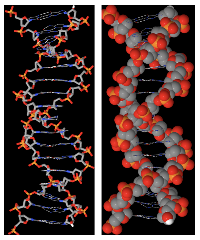
Base complementarily due to hydrogen bonding
Provides complementarily of the two strands
Explains Chargaff’s rule
Provides chemical stability to the helix
Partial positive (amines) and negative charges (keto oxygen)
A-T base pairs form two hydrogen bonds; G-C base pairs form three hydrogen bonds
Base pairs have the same dimension provided a uniform width to the double helix
Base pairs lead to uniform helix width
DNA seen “end on” view
Base pairs (grey and blue) form uniform chentral column
Sugar-phosphate backbone (orange and red) wraps around the outside at a uniform distance ~20 angstroms

Base-pair edges are each unique
Provides a mechanism for regulatory proteins to bind to specific DNA sequences without separating the strands

Other conformations of DNA
Alternative forms of DNA exist
A-DNA is slightly more compact than B-DNA
Double-stranded RNA and RNA-DNA hybrids are in A-form
Z-DNA forms a left-handed double helix

Comparison of A- and B-DNA
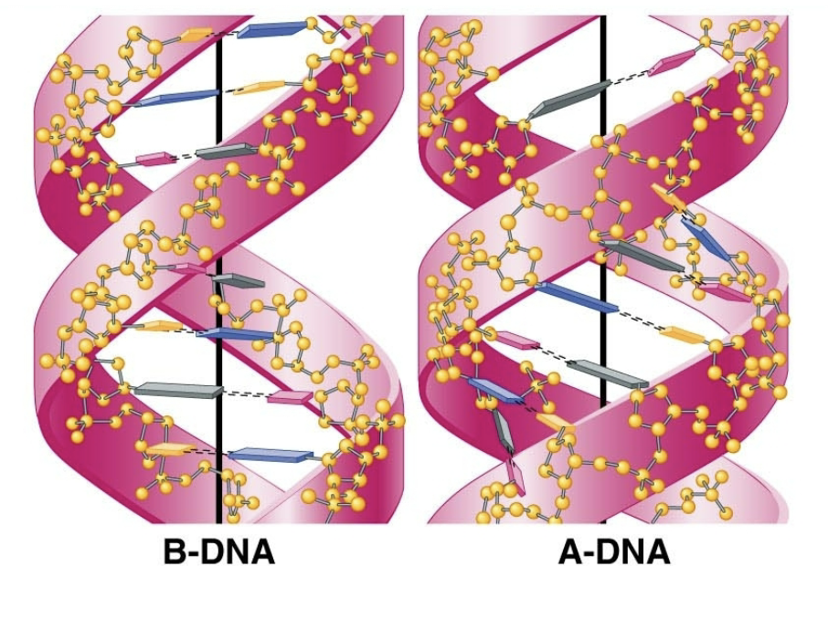
Other DNA structures

RNA
the sugar ribose replaces deoxyribose
and uracil replaces thymine of DNA
Most RNA is thought of as single stranded, but…
RNAs form double-stranded regions as they fold into different secondary structures; these double-stranded regions can be critical for function
Classes of RNA
All RNAs originate as complementary copies of one of the two strands of DNA
DNA serves as the template for their synthesis (transcription is synthesis of RNA from DNA)
There are four classes of cellular RNAs (at least four, depending on how they’re grouped):
messenger RNA (mRNA)
ribosomal RNA (rRNA)
transfer RNA (tRNA)
other RNAs: snRNA, snoRNA, siRNA, miRNA, IncRNA
Structure of RNA
Most RNA is thought of as single stranded, but
RNAs form double-stranded regions as they fold into different secondary structures; these double-stranded regions can be critical for function

Learning Goals
Be able to recognize and distinguish the nucleotides from each other; be able to draw them
Understand that there are different shapes of DNA beyond B-form
Understand the key differences between RNA and DNA
Understand how the chemistry & biochemistry of nucleic acids are used in the analysis of the molecule
Van der Waals forces
Weak, distance-dependent electrostatic attractions that occur between neutral atoms or molecules rather than holding individual atoms together inside a single molecule
The current thinking is that RNA was an early evolving molecule that could self-replicate and do catalysis:
It preceded a world dependent on DNA and proteins
Remnants of this world are seen in the ribosome, ribozymes, splicing machinery, reverse transcriptase

U vs T
Thymine is also known as 5-methyl uracil
In biosynthetic pathways, T is made from U
→ UMP → dUMP → dTMP
These suggest that T is derived, and U was present initially
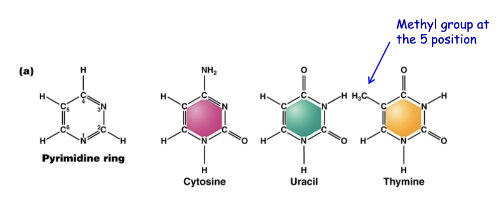
U can form base pairs with…
Watson-Crick A-U base pair
Hoogstein A-U base pair
G-U base pair
U-U base pair
U is the most promiscuous of bases: it forms non-Watson-Crick base pairs
If U pairs with something other than A, this can lead to a mutation (change) in the sequence
The methyl group in T protrudes into the major groove and helps to stabilize A-T base pairs over other pairing options
Watson-Crick A-U base pair
The methyl group in T increases fidelity during DNA replication
C deaminates to U
C spontaneously undergoes deamination (hydrolysis reaction) to become U
If U was in DNA and this deamination occurred, how could cells distinguish a real U from a deaminated C?
Instead, cells use T (methyl-U) to base pair with A
This means that when U occurs in DNA (arising from deamination of C), it can be recognized and removed
Why is there U but not T in RNA
Change to: why have T in DNA instead of U
Increased fidelity during replication and decreased mutation rate (DNA repair will remove U)
RNA chains are more susceptible to cleavage than DNA chains
In alkaline solutions, the 2’ hydroxyl loses a proton; it then acts as a nucleophile to attack the phosphorus atom of the phosphoester linkage, breaking the chain.
DNA lacks the 2’-OH and cannot do this reaction.
Why is there ribose in RNA?
Change to: why is there deoxyribose in DNA?
Increased stability of the DNA chains
Analytical techniques to investigate DNA and RNA
Spectroscopy
Denaturation and renaturation of the double helix
Hybridization of molecules, probes and primers: e.g., melting profiles; in situ hybridization (FISH - fluorescent in situ hybridization and non-fluorescent detections); primers in PCR reactions
Electrophoresis: separation and detection
Cleavage of DNA by restriction enzymes
DNA sequence analysis
DNA/RNA analysis is based on their chemical properties
The chemistry of the nucleic acids allows for analysis
Property 1: conjugated double bonds in the bases
Absorption of UV light at 260 nm; spectroscopy (A260)
Property 2: percent G+C, mass and shape
DNA molecules that differ in GC content have different buoyant densities (sedimentation equilibrium centrifugation)
Molecules with different masses and shapes move differently through gradients (sedimentation velocity centrifugation)
Property 3: hydrogen bond formation & stacking interactions
Denaturation/renaturation (percent G+C; Cot analysis; renaturation kinetics; denaturation in PCR)
Property 4: negative charge of the phosphates
Electrophoresis
Property 5: order of base pairs, i.e., their sequence
Denaturation/renaturation (“probing” in FISH, Southern)
Spectroscopy
Nucleic acids absorb ultraviolet (UV) light most strongly at 260 nm due to interaction between UV light and the conjugated double bonds in the rings of the bases
The absorption at 260 nm can be used to determine the concentration of nucleic acids in a solution
1 A260 = 0.05 mg/ml of DNA
Absorption by itself does not indicate the form of the molecule
single- vs. double-stranded; nucleotide vs. oligonucleotide vs. polynucleotide)
The action spectrum is the wavelength of light that causes mutations.
The energy from UV light causes covalent bonds to form between adjacent pyrimidine bases.
Nucleic acids absorb UV light most strongly at 260 nm
protein absorbs most strongly at 280 nm, due to absorption by tryptophan, tyrosine and phenylalanine

Centrifugation
Nucleic acids can be separated by gradient centrifugation procedures.
Sedimentation equilibrium centrifugation separates by their buoyant density on a salt gradient.
Opposing forces: centrifugal force (sedimentation) vs. diffusion
A sensitive measure of density
Sedimentation velocity centrifugation measures the velocity of sedimentation through a gradient; measurement is in Svedberg (S) coefficient units.
Opposing forces: viscosity (friction) and diffusion
Measure of density, mass and shape (bigger, heavier and more compact molecules sediment faster. E.g, the 80S ribosome or the 16S rRNA molecule.

Sedimentation velocity centrifugatino
Samples can also be monitored in real time for their movement through a density gradient.
Detection occurs via absorption of UV light.
Movement through the gradient is a function of both mass and shape of the molecule.
Measured in Svedberg (S) units.
E.g., the 80S ribosome; the 18S rRNA molecule

Denaturation and renaturation
Denaturation and renaturation can provide information about the composition and complexity of DNA samples.
Denaturation (melting) is the separation of DNA molecules into two strands.
Breaking of non-covalent bonds • dsDNA → ssDNA
Renaturation is the reforming of double stranded molecules from single strands; formation of base pairs.
Renaturation – generally used when the molecules are the same (i.e., both strands of DNA)
Annealing is used with oligonucleotides as probes or primers
Hybridization is synonymous with annealing, and originally came from forming double-stranded hybrid molecules between DNA and RNA
Misconception: denaturation is “broken”
Denature: to change the nature of
In physics – the addition of material to block nuclear fission In chemistry – the addition of a noxious agent (MeOH) to EtOH to make denatured ethanol
In biology – (a) to cause the structure of a protein to unfold so that its biological activity is diminished or eliminated; (b) to cause the paired strands of double-stranded DNA to separate into individual single strands.
In both (a) and (b), non-covalent bonds, such as hydrogen bonds, are broken. Proteins typically cannot refold to restore their activity (too complex) but DNA can re-anneal because of specific base pairing.
Denaturation and renaturation
Denaturation (melting) and renaturation (annealing or hybridization) by the formation of dsDNA.
Critical parameters include
For melting: The number of G-C base pairs, length of the molecules, salt concentration.
G-C content: Higher GC → more stable
Length: Longer → more stable
Salt concentration: Higher → more stable
For renaturation: The number of G-C base pairs; complexity of the sequences (repetitive or unique); length and concentration of the molecules.
Applications (many important applications)
Melting temperature determination
primer binding for PCR
probe binding in hybridization (Southerns, northerns, microarray, FISH, gene expression studies, etc.)
reassociation (Cot) analysis
Melting temperature determination
The temperature of the sample is raised and the absorption at 260 nm is followed.
Hyperchromic shift:
Single- stranded DNA absorbs more UV light than double stranded DNA
Melting temperature (Tm)
is when half of the molecule is denatured
Higher GC → higher Tm
GC rich DNA is more stable
Melting temperature (Tm) is affected by:
G-C content: Higher GC → more stable
Length: Longer → more stable
Salt concentration: Higher → more stable
Renaturation kinetics (Cot) is affected by:
G-C content: Higher GC → faster
Sequence complexity: Simpler → faster
Length: Shorter → faster
Concentration: Higher → faster
Reassociation kinetics
provides information about the genome size and complexity of DNA from an organism
DNA is broken into pieces ~several hundred bp in length
Sample is heated to denature (dissociate into single strands) the DNA
Temperature is lowered
Reassociation into dsDNA is measured

Reassociation kinetics provides information about the size and complexity of genomic DNA

Melting temperature (Tm) is affected by:
G-C content: Higher GC → more stable
Length: longer → more stable
salt concentration: higher → more stable
Renaturation kinetics (Cot) is affected by:
G-C content: higher GC → faster
Sequence complexity → simpler → faster
Length: shorter → faster
Concentration: higher → faster
Applications - many but most important:
PCR primer binding
Hybridization (Southerns, Northerns, microarray, FISH, gene expression studies, etc)
Reassociation (Cot) analysis - DNA complexity
In molecular hybridization,
DNA strands (or DNA and RNA strands) can be annealed to each other

Fluorescent in situ hybridization (FISH) is used to
identify the chromosomal location of a DNA of interest
The yellow color is due to hybridization of a probe that binds to (i.e., recognizes) the centromere; the probe is labeled with a molecule that fluoresces yellow.
The red color is due to a dye (propidium iodide) that binds to DNA non- specifically.
FISH analysis to identify genes associated with disease
Example of hybridization used to see a chromosome inversion found in a patient with Tourette’s syndrome.
The green and red fluorescent probes are lots of small DNA fragments that specifically bind to a larger DNA region
This was used to map a gene, SLITRK1, and associate it with the disorder.

Position of probes on the chromosome - inversion separates the region
Probes are a collection of small DNA molecules that can target specific DNA sequences. Thus they can cover (interrogate) large portions of the genome.

In situ hybridization
is used to analyze gene expression, and uses a labeled complementary DNA, RNA, or modified nucleic acids strand as a probe) to localize a specific mRNA transcript in a section of tissue (in situ)
Probes are “labeled” with a radioactive isotope, a fluorescent molecule, or chemical stain.
The dark blue stain indicates where RNA is localized that is complementary to a probe for a maternal effect gene hunchback.

Polymerase chain reaction
The Polymerase Chain Reaction Makes DNA Copies without Host Cells.
The polymerase chain reaction (PCR) copies a specific DNA sequence through in vitro reactions that can amplify target DNA sequences present in very small quantities.
PCR requires two oligonucleotide primers that provide specificity and a heat stable DNA polymerase.
PCR
PCR requires two oligonucleotide primers, one complementary to the 3' end of one strand of the DNA to be amplified and one complementary to the 3' end of the other strand. The primers anneal to denatured DNA, and the complementary strands are synthesized by a heat-stable DNA polymerase.
The three steps of PCR — denaturation, primer annealing, and extension — are repeated over and over using a thermocycler to amplify the DNA exponentially.

PCR

DNA analysis is based on chemistry
The chemistry of the nucleic acids allows its analysis
Property 1: conjugated double bonds in the bases
Absorption of UV light at 260 nm; spectroscopy (A260)
Property 2: percent G+C, mass and shape
DNA molecules that differ in GC content have different buoyant densities (sedimentation equilibrium centrifugation)
Molecules with different masses and shapes move differently through gradients (sedimentation equilibrium centrifugation)
Property 3: hydrogen bond formation
Denaturation/renaturation (percent G+C; Cot analysis; reassociation kinetics; denaturation in PCR)
Property 4: negative charge (phosphates) at neutral pH
Electrophoresis
Property 5: order of base pairs
Denaturation/renaturation (“probing” in FISH, Southern/Northern analysis, in situ analysis; primer binding in PCR)
Restriction digestion
DNA sequence analysis
Electrophoresis
Nucleic acid electrophoresis separates DNA and RNA fragments by size such that smaller fragments migrate through a gel at a faster rate than large fragments

Restriction enzymes
Restriction Enzymes Cut DNA at Specific Recognition Sequences.
A restriction enzyme is a endonuclease whose activity is restricted to a specific DNA recognition sequence.
DNA fragments are produced by DNA restriction.
Most recognition sequences are palindromic, and restriction enzymes often cleave these sequences in an offset manner

Restriction enxyme cleavage sites are useful for DNA engineering
Some restriction enzymes cleave the sequences in an offset manner to leave bases that are not paired: these are called “sticky ends”

Restriction enzyme cleavage sites are useful for DNA engineering
Some restriction enzymes cleave both strands at the same place: these are called “blunt ends”.
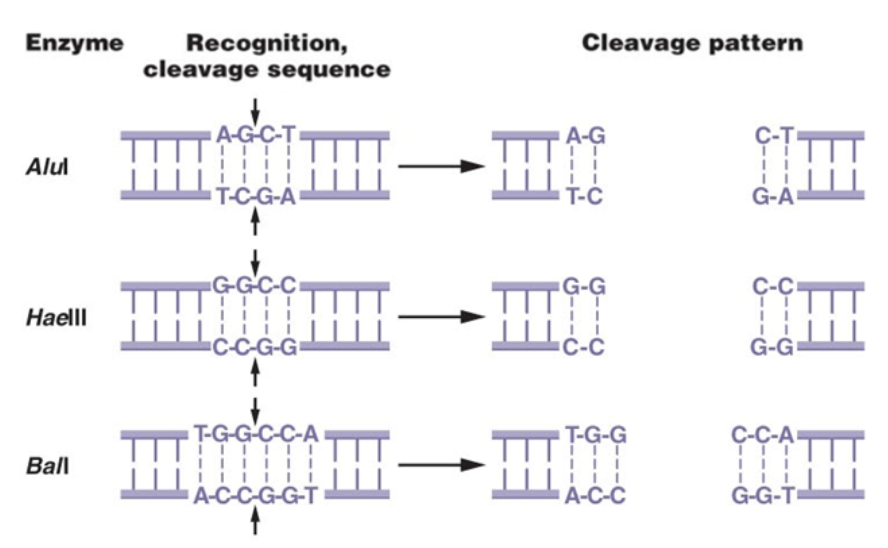
Restriction enzyme cleavage sites are useful for DNA engineering
Different Restriction enzymes can create the same sticky ends
Restriction fragments with the same sticky ends can be “ligated” back together to create useful “recombinant” DNA molecules
Can insert DNA into similarly cleaved plasmid “vectors” i.e. “cloning”
Sometimes does not recreate the restriction site

Restriction and Electrophoresis
After separation, DNA or RNA fragments are visualized by staining with the dye ethidium bromide and examination under ultraviolet light
Sanger DNA sequencing
The second method developed to sequence DNA sequencing is based on DNA synthesis.
This is the dideoxy chain termination sequencing, developed by Fred Sanger.
This method is based on in vitro DNA synthesis.
The dideoxy nucleotides lack the hydroxyl group on both the 2’ and 3’ carbons.
A radioactive nucleotide is added to the sequencing reaction.

DNA synthesis
Chain elongation occurs in the 5' to 3' direction by addition of one nucleotide at a time to the 3' end.
As the nucleotide is added, the two terminal phosphates are cleaved off as the leaving group.
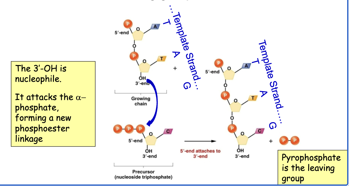
Dideoxy DNA sequencing

Autoradiogram examples
Radioactivity is provided by the primer (or a small amount of a radionucleotide)
Fragments are separated by electrophoresis using a gel system that can resolve band sizes by a single nucleotide.
Each lane contains the reaction with a single dideoxy nucleotide with the 4 dNTPs (as shown on top, G means ddG was used).
Read the sequence by starting at the bottom, which corresponds to the 5’ end closest to the primer.

Dideoxy sequencing
Large-scale genome sequencing is automated and uses fluorescent dye-labeled dideoxynucleotides.
Each ddNTP is tagged with a different color fluorescent dye.
Each sample is size separated using capillary gel electrophoresis.
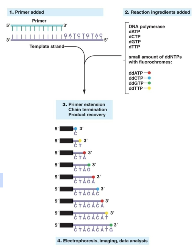
DNA sequence analysis
Example of sequence from automated machine

Massively parallel sequencing
New DNA sequencing methods are being developed to be less expensive than the automated Sanger method.
They use technologies that are “massively parallel”, i.e., millions of reactions performed on different samples in small scales at different places at the same time.
Newest methods do not rely on the slowest steps associated with the Sanger method.
Sanger method: the cloning of fragments into plasmids and their recovery in bacteria.
Method from Illumina: generation of PCR products on glass slides
Illumina Sequencing
PCR-like methods create localized clusters of millions of copies of the DNA on a glass slide. Each slide has many millions of different clusters
Uses fluorescent dyes that act like reversible ddNTPs “blocking groups”
Add the base; record image of the fluorescence over the entire slide; remove the dye/blocking group; repeat hundreds of times; Computationally record/analyze the sequence at each spot
Next Generation Sequencing: Illumina
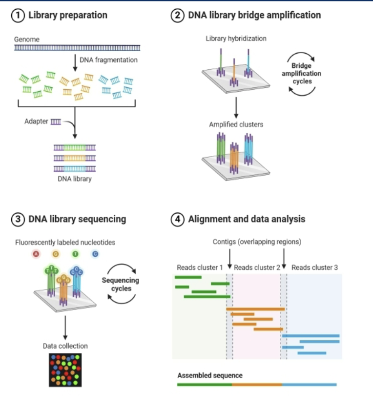
Illumina sequencing step

Illumina: new DNA sequencing methods are massively parallel

Advances in Sequencing Technology allowed >million-fold reduction in cost
