Anatomy Lab: The Brain
 Call Kai
 Learn
 Practice Test
 Spaced Repetition
 Match
 Flashcards
 Knowt Play
1/45

There's no tags or description
Looks like no tags are added yet.
46 Terms
how does Sensory function work?
use of sensory receptors, the nervous system is able to detect changes in our surrounding environment and changes that occur inside the body
how does integrative function work?
central nervous system analyzes incoming information from sensory neurons and generate appropriate responses via motor neurons.
how Motor function work?
respond to sensory input by initiating muscular contractions or glandular secretions.
if the body is too hot, how does the body return to homeostasis?
sensory receptors in the skin and brain detect this change and send that information to the control center, Integration occurs and the hypothalamus elicits a motor response, signaling to effectors, Sweat glands activate, allowing the body to lose heat as the sweat evaporates from the surface of the skin.
if the body is too cold, how does it return to homeostasis?
sensory receptors in the skin and brain detect this change and send that information to the control center, Integration occurs and the hypothalamus elicits a motor response, signaling to effectors, Smooth muscle in the walls of dermal blood vessels contracts, constricting the vessels so the body loses less heat through the surface of the skin. Skeletal muscles involuntarily contract and relax repeatedly (shiver), allowing the body to generate heat from muscle activity
what are the 5 parts of a neuron and where are they/what do they do?
Cell body - Part of the neuron that contains the nucleus and cellular organelles.
Dendrite - Multiple branching processes from the cell body. Serve as a contact to receive impulses from other neurons.
Axon hillock - Region where the axon leaves the cell body.
Synaptic knobs - Swelling at the end of an axon that forms a synapse with another neuron or effector organ
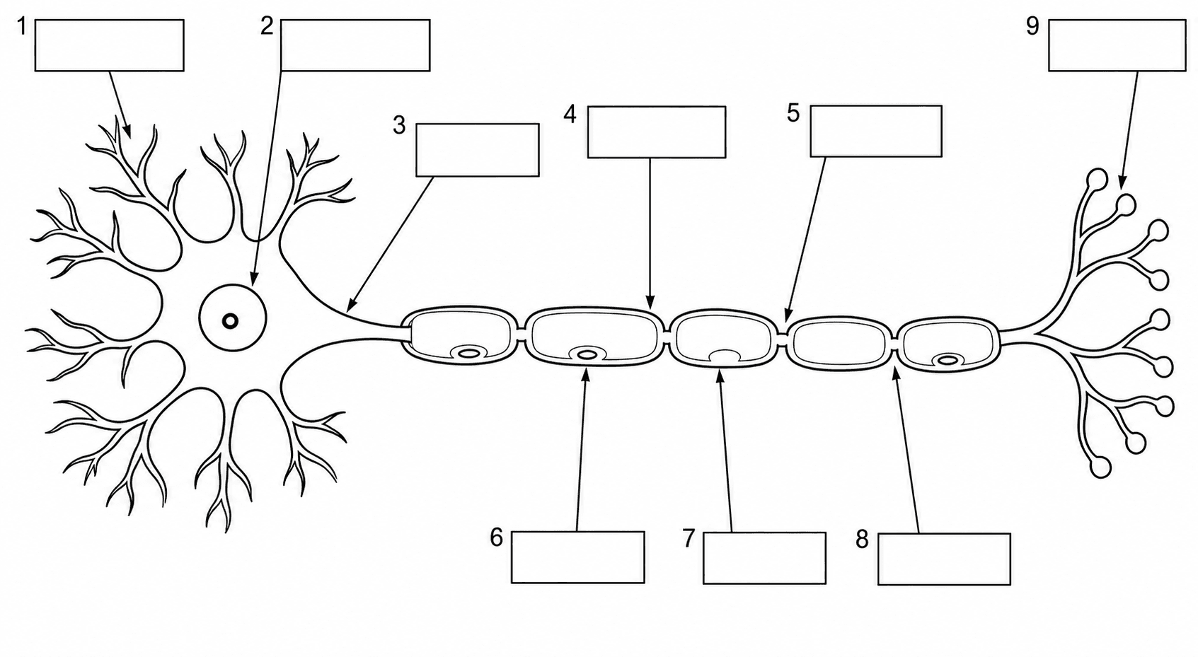
label the parts of the neuron (1,2,3,5, 7,9)
1 - dendrites
2 - nucleus
3 - axon hillcock
5 - axon
7 - myelin sheath
9 - axon terminals
what is the CNS and what are its parts and what does it do?
what is the PNS what are it’s parts and what does it do?
CNS - central nervous system; control center for all functions, incudes the brain and spinal cord
PNS - peripheral nervous system; sends sensory info from receptors to the CNS, and transmits motor information from the CNS to effector organs, includes cranial nerves and spinal nerves
what differentiates grey and white matter?
white matter has myelinated neurons and grey han unmyelinated neurons
what are the 2 divisions of the PNS and what do they do?
-sensory division consists of sensory neurons that send impulses from receptors to the CNS
-motor division consists of motor neurons that send impulses from the CNS to effector organs
what are the 3 kinds of receptors in the sensory division and what do they do?
-Somatic sensory receptors: Detect general sensations (touch, pressure, temperature, pain, and body position) in the skin, skeletal muscles, and joints.
-Visceral sensory receptors: Detect sensations in the internal organs.
-Special sensory receptors: Detect special sensations (smell, taste, vision, hearing, equilibrium).
what are the two systems of the motor divion and what do they do?
-Somatic nervous system: Contains motor (efferent) neurons going from the CNS to skeletal muscle. This impulse pathway produces voluntary actions.
-Autonomic nervous system: Contains motor (efferent) neurons going from the CNS to smooth muscle, cardiac muscle, and glands. This impulse pathway produces involuntary actions.
what is a cranial meninges?
what are the 5 parts of the Meningeal system and what are they like?
-connective tissue coverings of the brain and spinal cord that provided added protection
-Dura mater - Very tough superficial layer; composed of two layers
-Epidural space - Space between the cranial bones and the dura mater; usually filled with fat and capillaries
-Arachnoid mater - Very thin middle layer; contains numerous web-like extensions that anchor to the pia mater
-Subarachnoid space - Space between the arachnoid mater and pia mater; filled with cerebrospinal fluid
-Pia mater - Thin deep layer that connects directly to the surface of the brain and spinal cord. Follows all the contours on the surface of the brain.
what are the layers in order from the skull skin to the brain?
1 - skin
2 - aponeurosis
3 - periosteum
4 - bone
(meninges)
5 - dura mater
6 - arachnoid
7 - pia mater
what section is the brain split into? how are those two divisions split?
2 cerebral hemispheres are each split into 5 lobes, the frontal, parietal, occipital, temporal lobes, and the insula

label this chart of the brain (1,2,3,4,5,6,8,)
1 -frontal lobe
2 - parietal lobe
3 - occipital lobe
4 - temporal lobe
5 - cerebellum
6 - brainstem
8 - thalamus
where are the 5 lobes of the brain located and what do they do?
-Frontal lobe - Large lobe that makes up anterior portion of the brain; Controls conscious movement of skeletal muscles; contains Broca’s area (which controls motor speech); higher level functions include judgment and foresight (the ability to think before acting).
-Parietal lobe - Posterior to the frontal lobe; Receives sensory input from skin; higher level functions include logical reasoning (math, problem solving).
-Occipital lobe - Most posterior portion of the brain; Primary visual area and visual association area (ability to interpret visual information).
-Temporal lobe - Inferior to the parietal lobe; Primary auditory and auditory association area of the brain; conscious perception of smell.
-Insula - Deep to the temporal lobe. Involved in memory and interpretation of taste.
name the functions of each color
NOT A FINISHISHED CARD
what is a gyri?
what is a sulcus?
what is a fissure?
-ridges and grooves
-shallow grooves
-deep grooves
what are the 2 kinds of gyri , where are they and what do they do?
what are the 3 kinds of sulci and where are they?
3.what is the one fissure of the brain and where is it?
1. -Precentral gyrus - Most posterior gyrus in the frontal lobe; primary motor cortex
-Postcentral gyrus - Most anterior gyrus in the parietal lobe; primary somatosensory cortex
-Central sulcus - Divides the frontal from the parietal lobe
-Lateral sulcus - Divides the temporal from the parietal lobe
-Parieto-occipital sulcus - Divides the occipital from the parietal lobe
-Longitudinal fissure - Separates the cerebral hemispheres

label the colors and highlights of this picture
1st vertical line - Parieto-occipital sulcus
2nd vertical line - Central sulcus
lateral line - lateral sulcus
red section - precentral gyrus
blue section - postcentral gyrus
where is the brain stem located and what does it do?
it lies at the base of the brain and the top of the spinal cord. It is the structure that connects the cerebrum of the brain to the spinal cord and cerebellum.

label the parts of the brain stem
1 - thalamus
2 - midbrain
3 - pons
4 - medulla
5 - spinal cord
6 - brain stem
what is the diencephalon and where is it located? what are the 3 regians it’s split into? what does it do?
-Superior to the midbrain and inferior to the cerebrum is a centralized mass of brain tissue
-thalamus, hypothalamus, and epithalamus
-helps send signals between the brainstem, cerebellum, cerebrum, and spinal cord.
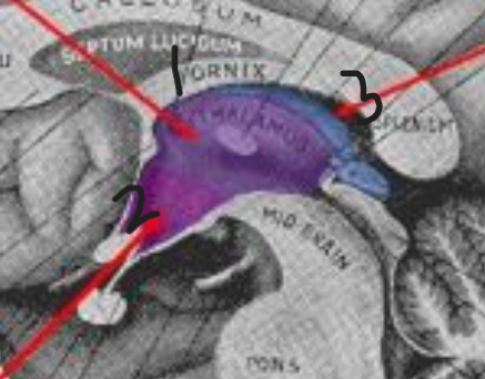
label
1 - thalamus
2 - hypothalamus
3 - epithalamus
what are Ventricles of the brain? what fluid are they filled with and how is it produced?
-a series of deep cavities within the central nervous system
-they are filled with cerebrospinal fluid (CSF) which is produced by a capillary network, called the choroid plexus, which lines the roof of each ventricle
what are the 4 kinds of ventricles, what do they look like, and where do they go?
-Lateral ventricles - Two large horseshoe-shaped cavities deep within the cerebral hemispheres and divided by a structure called the septum pellucidum
-Third ventricle - Narrow cavity located between the left and right portions of the thalamus; follows along the midsagittal plane
-Cerebral aqueduct - Connects the third ventricle to the fourth ventricle
-Fourth ventricle - Diamond-shaped cavity located anterior to the cerebellum and posterior to the pons
what is the Corpora quadrigemina and where is is located?
4 structures at the base of the brainstem underneath the cerebellum
what are the 4 structures on the Corpora quadrigemina and what they look like?
-superior colliculi (singular colliculus)—Two large superior structures that make up half of the corpora quadrigemina.
-Inferior colliculi (singular colliculus)—Two smaller inferior structures that make up half of the corpora quadrigemina.
-Pons—Large bulge on the brainstem after the corpora quadrigemina.
-Medulla oblongata—Smaller bulge after the pons before tapering into the spinal cord.

label
1 - cerebellum
2 - longitudinal fissure
3 - cerebral hemisphere
4 - gyrus
5 - sulcus

label
1 - dura mater
2 - optic nerve
3 - pituitary gland
4 - pons
5 - medulla oblingata
what are the inferior structures
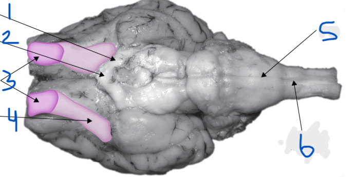
label
1 - optic tracts
2 - optic chiasm
3 - olfactory bulbs
4 - olfactory tract
5 - pons
6 - medulla oblongata