Anatomy and Physiology - Cardiovascular System
 Call Kai
 Learn
Learn Practice Test
 Spaced Repetition
 Match
Match Flashcards
 Knowt Play
1/77

There's no tags or description
Looks like no tags are added yet.
Name | Mastery | Learn | Test | Matching | Spaced | Call with Kai | Chat |
|---|
No analytics yet
Send a link to your students to track their progress
78 Terms
Blood
• Continuously regenerated connective tissue
• Moves gases, nutrients, wastes, and hormones
• Transported through cardiovascular system
Cardiovascular system
• Heart pumps blood
• Arteries transport (oxygenated) blood away from heart
• Veins transport (deoxygenated) blood toward heart
• Capillaries allow exchange between blood and body tissues; exchange with systemic cells and alveoli in the lungs; innervate all body tissue
Blood components
Consists of formed elements and plasma = whole blood
Formed elements
Erythrocytes (red blood cells) transport respiratory gases in the blood
Leukocytes (white blood cells) defend against pathogens
Platelets help form clots to prevent blood loss (colorless)
Centrifuged blood
Whole blood (plasma and formed elements) separated by centrifuge
Withdraw blood from a vein into a syringe and place it into a glass centrifuge tube.
Place the tube into a centrifuge and spin for about 10 minutes.
Components of blood separate during centrifugation to reveal plasma, buffy coat, and erythrocytes (in order from lightest to heaviest).
Erythrocytes
Bottom, red layer ~44% of sample
4.2-6.2 million/mm³
Buffy coat
Very thin (~1%) middle layer with gray-white
Consists of leukocytes and platelets
Leukocytes
Part of buffy coat
4.5-11 thousand/mm³
Neutrophils: 50-70% of leukocytes
Lymphocytes: 20-40%
Monocytes: 2-8%
Eosinophils: 1-4%
Basophils: 0.5-1%
Platelets
Part of buffy coat
150-400 thousand/mm³
Plasma
Straw-colored liquid at top of tube
~55% of sample
Water (92% - acts as solvent) + dispersed proteins (7%) + dissolved solutes (<1%)
An extracellular fluid
Similar composition to interstitial fluid, but plasma has higher protein concentration
Hematocrit
Percentage of volume of all formed elements
Clinical definition: percentage of only erythrocytes
Adult males: 42 to 56%; females 38 to 46%
Testosterone causes more erythropoietin secretion by kidney
Colloid
Blood is a colloid
An opaque mixture of water and solute (usually protein)
Plasma protein levels
Most produced in liver, others produced by leukocytes or other organs
Make up 7% of plasma
Albumin: 58% of plasma proteins
Globulins: 37%
Fibrinogen and other clotting proteins: 4%
Regulatory proteins - enzymes and hormones: <1%
Colloid osmotic pressure (COP)
Exerted by plasma proteins
Prevents loss of fluid as it moves through capillaries
Helps maintain blood volume and blood pressure
Can be decreased with diseases, resulting in fluid loss from blood and
tissue swelling
Diseases of which organs affect colloid osmotic pressure?
Liver diseases that decrease production of plasma proteins
Kidney diseases that increase elimination of plasma proteins
Albumin
58% of plasma proteins
Exerts osmotic force to retain fluid within the blood
Contributes to blood’s viscosity
Transport selected molecules (ex: ions, lipids, hormones)
Globulin
37% of plasma proteins
Alpha-globulins transport lipids and some metal ions (ex: copper)
Beta-globulins transport lipids and iron ions
Gamma-globulins are antibodies that immobilize pathogens
Fibrinogen
4% of plasma proteins
Participates in blood coagulation (clotting)
Regulatory proteins
<1% of plasma proteins
Consists of enzymes and hormones
Solutes in plasma
<1% of plasma
Electrolytes: help establish, maintain, and change membrane potentials, maintain pH balance, and regulate osmosis
Nutrients: energy source; precursor for synthesizing other molecules; participate in metabolic processes
Respiratory gases: oxygen is needed for aerobic cellular respiration; carbon dioxide is a waste product produced by cells during this process
Waste products: are only being transported to the liver and kidneys for elimination
Plasma molecules ranges
Glucose: fasting: 70-100 mg/dL; 2 hours after a meal: <145 mg/dL
Amino acids: varies, based on specific amino acid being measured
Lactate: 4.5-14.4 mg/dL
Lipids: varies; molecules that generally do not dissolve in water (cholesterol, HDL, VLDL/LDL, triglycerides, phospholipids)
Glucose (plasma molecule)
Fuel molecule for cellular respiration (primary energy source for nervous tissue)
Tightly regulated by a number of hormones, including insulin and glucagon
Amino acids (plasma molecule)
Monomers for synthesizing protein
Also regulated by some of the same hormones as glucose
Lactate
By-product of glycolysis
Cholesterol (lipid)
100-200 mg/dL
Plasma membrane component; synthesis of steroid hormones; bile salts
HDL: 40-80 mg/dL; transports lipids to the liver; “good” cholesterol
VLDL/LDL: 10-100 mg/dL; transports lipids from the liver; “bad” cholesterol
Triglycerides (lipid)
30-149 mg/dL
Fuel molecules; precursors for cholesterol synthesis
Phospholipids (lipid)
6-12 mg/dL
Molecules that form the bilayer of a plasma membrane
Functions of blood
Transportation
Protection
Regulation of body conditions (body temperature, pH, and fluid balance)
Transportation (functions of blood)
Transports formed elements, dissolved molecules, and ions through pulmonary and systemic circulation
Carries oxygen from and carbon dioxide to the lungs
Transports nutrients, hormones, heat, and waste products
Protection (functions of blood)
Leukocytes, plasma proteins, and other molecules (of immune system) protect against pathogens
Platelets and certain plasma proteins protect against blood loss
Regulation of body conditions (functions of blood)
Body temperature: blood absorbs heat from body cells (especially muscle) → released at skin blood vessels
Body pH: blood absorbs acid and base from body cells; contains chemical buffers
Fluid balance: water is added to blood from GI tract → lost through urine, skin, and respiration
→ fluid is exchanged between blood and interstitial fluid
→ blood contains proteins and ions helping maintain osmotic balance
Color (physical characteristics of blood)
Depends on degree of oxygenation
Oxygen-rich blood is bright red
Oxygen-poor blood is dark red
Volume (physical characteristics of blood)
5 L on average; ranges from 4-6 L
Viscosity (physical characteristics of blood)
4.5-5.5x relative to water
Depends on amount of dissolved and suspended substances relative to amount of fluid
Increases if erythrocyte number increases
Increases if amount of fluid decreases
Plasma concentration of solutes
0.9%
Solutes include proteins and ions
Osmotic pressure is important because it keeps the shape of blood vessels
Determines the direction of osmosis across capillary walls
For example, during dehydration, plasma is hypertonic (more solutes) → fluid is drawn from surrounding tissues
Temperature (physical characteristics of blood)
100.4 degrees F/38 degrees C
Blood is 1°C higher than measured body temperature
Warms area through which it travels
Blood pH (physical characteristics of blood)
Slightly alkaline at 7.35-7.45
Crucial for normal plasma protein shape (avoiding denaturation)

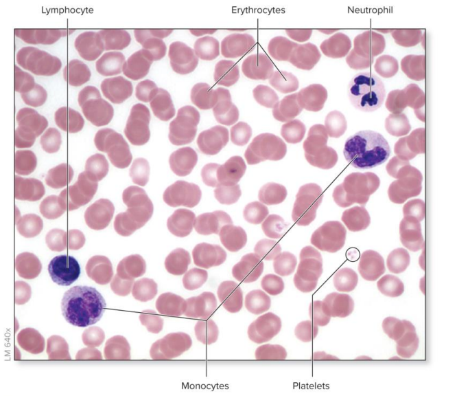
Blood smear
Thin layer of blood placed on microscope slide and stained
Formed elements differ in appearance:
Erythrocytes are most numerous: pink, anucleate, biconcave discs
Leukocytes: larger than erythrocytes, varied in form, noticeable nucleus
Platelets: small fragments of cells
Hematopoiesis
The origin, differentiation, and maturation of formed elements
Occurs in red bone marrow of certain bones
Colony-stimulating factors (CSFs) stimulate hematopoiesis
Always starts with hemocytoblasts (hematopoietic stem cell; pluripotent and undifferentiated)
Erythropoiesis (myeloid line)
Multi-CSF acts on myeloid stem cells to start erythropoiesis
Stimulation by EPO → progenitor cell
Proerythroblast
Stimulation by EPO → early erythroblast
Late erythroblast
Normoblast: nucleus ejected at this stage
Reticulocyte
ERYTHRYOCYTE
Thrombopoiesis (myeloid line)
Multi-CSF acts on myeloid stem cells to start thrombopoiesis
Progenitor cell
Megakaryoblast
Stimulation by thrombopoietin → promegakaryocyte
Stimulation by thrombopoietin → megakaryocyte
Stimulation by thrombopoietin → proplatelets on megakaryocytes break off into PLATELETS
Leukopoiesis - granulocyte line (MYELOID line)
Multi-CSF acts on myeloid stem cells to start leukopoiesis - granulocyte line
Stimulation by GM-CSF → progenitor cell
Stimulation by G-CSF → myeloblast
Promyelocytes
Eosinophilic myelocyte / basophilic myelocyte / neutrophilic myelocyte
EOSINOPHIL / BASOPHIL / NEUTROPHIL
Leukopoiesis - monocyte line (MYELOID line)
Multi-CSF acts on myeloid stem cells to start leukopoiesis - granulocyte line
Stimulation by GM-CSF → progenitor cell
Stimulation by M-CSF → monoblast
Stimulation by M-CSF → promonocyte
MONOCYTE
Leukopoiesis (LYMPHOID line)
Lymphoid stem cell → directly to NATURAL KILLER CELL
B-lymphoblast → B-LYMPHOCYTE
T-lymphoblast → T-LYMPHOCYTE
Characteristics of erythrocytes
Diameter: 7.5 µm
Function: transport oxygen and carbon dioxide
Lifespan: ~120 days
Density (avg number per mm³ of blood): ~4.8 mil for females, ~5.4 mil for males
Characteristics of leukocytes
Diameter: 1.5-3x larger than erythrocytes; 11.25-22.5 µm
Function: initiate immune response; defend against potentially harmful substances
Lifespan: varies from 12 hrs (neutrophils) to years (lymphocytes)
Density (avg number per mm³ of blood): 4,500-11,000
Characteristics of platelets
Diameter: <1/4x of an erythrocyte; ~2 µm
Function: participate in hemostasis
Lifespan: ~8-10 days
Density (avg number per mm³ of blood): 150,000-400,000

Platelet formation
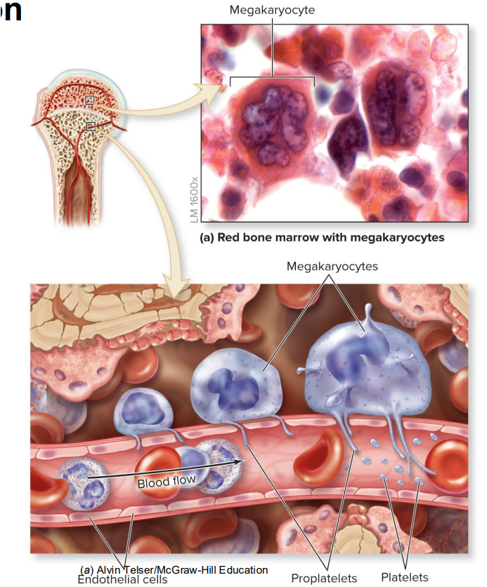
Starts in red bone marrow with megakaryocytes (lives next to blood vessels)
The capillary is lined with endothelial cells thru which lie the proplatelets
Proplatelets are spliced from the megakaryocytes by the force of blood flow → platelets
Proplatelets
Long processes/protrusions of megakaryocytes into the capillary wall that break off into platelets
Erythrocyte structure
Small, flexible formed elements
Lack nucleus and cellular organelles; packed with hemoglobin
Have biconcave disc structure and a latticework of spectrin protein providing support and flexibility
Transport oxygen and carbon dioxide between tissues and lungs

Molecular structure of hemoglobin
The red-pigmented protein found in blood
Transports oxygen and carbon dioxide (termed oxygenated when maximally loaded with oxygen and deoxygenated when some oxygen lost)
Each hemoglobin molecule is composed of four globins (two alpha chains and two beta chains)
Each chain has a heme group: a porphyrin ring with an iron ion in its center

Oxygen binding to hemoglobin
Oxygen binds to the iron ion, so each hemoglobin can bind four oxygen molecules
Binding is fairly weak; rapid attachment in lungs and rapid detachment in body tissues
Carbon dioxide binds to globin protein (not iron)
Binding is fairly weak; attachment in body tissue and detachment in lungs
Steps of erythropoietin regulating erythrocyte production
Stimulus: decreased blood oxygen levels
Receptor: kidney detects decreased blood O2
Control center: kidney cells release EPO in the blood
Effector: EPO stimulates red bone marrow to increase the rate of erythrocyte production
Net effect: increased numbers of erythrocytes enter the circulation, during which time their are oxygenated and blood O2 levels increase
Negative feedback: increased blood O2 levels are detected by the kidney, which inhibits EPO release by negative feedback
Clinical view: blood doping
Used by some athletes to enhance performance
Method 1: self-donation of erythrocytes
Blood removal prior to competition increases EPO production
Erythrocytes transfused back before competition
Method 2: pharmaceutical EPO
Dangers of blood doping
Increased blood viscosity
Heart required to work harder
May cause permanent cardiovascular damage
Banned from athletic competition