IMSK Exam 3
 Call Kai
 Learn
Learn Practice Test
 Spaced Repetition
 Match
Match Flashcards
 Knowt Play
1/400

There's no tags or description
Looks like no tags are added yet.
Name | Mastery | Learn | Test | Matching | Spaced | Call with Kai | Chat |
|---|
No analytics yet
Send a link to your students to track their progress
401 Terms

Label 1-7
1) sulcus 2) Tibial Tuberosity 3) Fibular head 4) Cranial cruciate ligament 5) caudal cruciate ligament 6) Lateral meniscus 7) Medial Meniscus
4 and 5 cruciate ligaments are named by their tibial INS
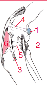
Lateral view (label 1-6)
1) fabella (lateral) 2) Lat collateral 3) long digital extensor tendon 4) femoropatellar ligamnet 5) lateral meniscus 6) infrapatellar fat pad

Medial view 1-4
1) fabella (medial) 2) medial collateral ligament 3) medial meniscus 4) intrapatellar fat pad
Femoropatellar ligamnets
continuation of the femoral fascia or at the sides of the patella
lateral - better defined
CrCL vs CdCL
cranail cruciate ligament
Caudal cruciate ligament
CrCL prevents
hyperextension, excessive internal rotation, cranial tibial translation
Screw home mechanism
some internal tibial rotation natrually occurs as you flex your knee
Meniscus - anatomy
fibrocartilage “wedges” between the femoral condyles and the tibial plateau (mostly type I collagen, they alleviate incogruity between a rounded strucutre and a flat surface)
Meniscus - funciton in stiffle
stabilization, load bearing, shock absorption, joint lubrication, mechanoreceptor
CrCL disease - etiology
biomechanical - trauma, hyperextension <10%
Biological - spontaneous degeneration >90%
CrCL etiology and presentation - traumatic
may be associated with other damage to the stiffle (collateral ligament, caudal cruciate ligament, menisci)
typically unilateral injuries
predominantly complete tears
in puppies - is likely associated with an avulsion vs midsubstance tear in the adult
CrCL disease - etiology and presentation - degenerative
spontaneous, commonly bilateral (~10% bilateral on presentation but up to50% will rupture the other side within 1 year and 60% will rupture the other side in their lifetime)
tears can be partial or complete - partials progress to full tears over time
CrCL disease - risk factors - degenerative
Breed - lab, golden, rottweiler
sex - femlae >male
neutred>male
larger>smaller
obesity implicated but not proven
Anatomy (increased tibial slope has been implicated)
other disesae (endocrinopathies)
Age - unknown if true risk facort (peak at 7-10 years) (smaller are later in life, larger are earlier)
CrCL disease - meniscal injury (medial vs lateral)
medial meniscus - #1 side injured (caudal horn)
lateral Meniscus - injuries here are much less common (Must check during sx)
CrCL disease - Hx and s/s
Hx - non-weight bearing lameness
muscle atrophy of quadriceps
medial buttress
reluctant to have the stiffle joint maximally flexed or exteneded
CrCL disease - Pe findings
gait - short stride/obvious lamenees, look for atrophy and symmertry
sit test - sit with the most painful stiffle extended out laterally and roll onto the opposite (good) limb
standing plapaiton - look for symmetry
recumbent palpation - CREPI, cranial drawer and cranial tibial thrust
CrCL disease - stiffle exam - 2 tests
Cranial drawer test (passive)
Tibial compression test (dynamic test) - mimics weight bearing
CrCL - stiffle exam - testing for cranial-caudal instability (full vs partial tears)
Full tears - cranial drawer is typically easy to palpate
partial tears - may only have mild or subtle cranial drawer so you must test in flexion and extension, test for pain on stifffle hyperextension, base a presumptive dx off of x-ray findings (secondary OA)
if still unsure - rest, NSAID, nad recheck in 2-4 weeks (repeat full exam)
CrCL band (cualda laterla nad cranial medial) reaction to flexion and extension
only loose when caudal lateral is in flexion
CrCL disease - x-rays signs of secondayr OA
Scerlosis
Osteophytes - proximal and distal patella, trochlear ridges, fabella
Efussion - cranila displacment of the infrapatellar fat pad, caudal displacment of gastrocnemius fascial plane
NO
NO
CrCL disease - sx options - Lateral sutures - objectives of sx
placing a prosthetic sutreu to mimic the CrCL
goal is to neutralize the effects of cranial tibial thrust and cranial drawer by replacing the damaged CrCL with lateral suture
CrCL disease - sx lateral suture - stability
provides static and dynamic stabilization
craniocaudal instability is neutralized by extra-capsular support
short term stability - suture material
long term stability - periarticular fibrosis
after sx, the cranial cruciate ligament is not neccessary to stiffle stabilization
tfibrosis replaces the suture and creates the stability
CrCL - sx options - lateral suture - clinical concerns/benefits
recent evidence is less supportive - instability presensent in many dogs
suture will break over time and relies on soft tissue healing
if P has cushing they don’t have fibrious laying down and it won’t work
CrCL - disease - TTA - objective of this sx
to neutralize the effects of crnail tibial thrust
the tibial tuberosity is osteotmized and advanced forward
the stiffule is redesigned and cranial tibial thrust is neutralized by placement of the patellar tendon perpendicular to the tibial plateau
during CrCL disease sx and you cut the bone that means it is what kind of sx and provides what kind of stabilization
it is a dynamic surgery and provides dynamic stabilization
CrCL - TTA sx - stabilization
dynamic
when weight bearing - stiffle is stable
when not weight bearing - cranial drawer may be palpable
Since it is dynamic the cranial cruciate ligament is not necessary for stiffle stabilizaiton and active wieght bearing
CrCL - TTA - clinical conerns/benefits
less evidence for efficacy - results appear similar to lateral suture
removal of any inected impalnts can be challenging
CrCL disease - TPLO sx - objectives
to neutralize the effects of cranial tibial thrust
the stiffle is redesigned and the cranial tibial thrust is neutralized by roating the tibial plateau to be “level”
CrCL disease - TPLO - stabilization
dynamic stabilization
when weight bearing - stable stiffle
when not weight bearing - cranial drawer may be palpable
after sx the CrCL is not necessary for stifle stabiliatoin under active weight beairng “dynamic stabilization”
TPLO - clinical concerns/Benefits
best evidence for efficacy - dogs use limb quickly after surgery
dogs indistringusihable from normal 6-12 months after sx
Caudal crucitae function
primary stabilizer agaisnt caudal drawe (caudal tibial subluxation) function with cranial crucitate ligament to - prevent internal rotation and prevent hyperextension
Caudal cruciate injury - prevalence, breeds, etiology, concurent injuries
Prevalence - rare in isolation
Breeds - any dogs
etiology - trauma (tibia expeirences blow to the cranial cortext forcing it caudally)
concurrent injuries - often combined with medial collateral/ crnail crucitate ligmaent injury
LA x-rays, marker is always placed
lateral or dorsal (the one depends on what x-rays you are doing)
if doing a side angle should always be lateral (lateral trumps dorsal)
Typical x-rays for fetlock in LA and what to lock for
LM, DP
proximal sesamiod bones (camera is angled down a little to evalute)
condyles, sagital ridge, condyles, sagitatal goove
also flexed LM (common for OCD)
4 common abnormalities in fetlock LA
OA, trauma/repetitive stress injuries, OCD, sesamodiitis
LA fetlock OA - s/s
fetlock joint effusion (variable)
lameness localized to the fetlock region (or joint)
degree of lameness variable
LA fetlock OA - readiographic signs
osteophytes, supracondylar lysis, soft tissue swelling, narrowed joint space (marked)
LA fetock OA - ostephophyte formation location
Dorsomedial and dorsolateral aspect of proximal P1
proximal and distal aspects of sesamoid bones
palmar processes of P1

What is wrong with these horses
Fetlock OA due to osteophyte formation
Feltock LA oA supracondylar lysis
bone resorption due to chronic effusion/synovitits
Normal A=B
abnormal A>B

LA fetlock - repetitive stress injuries etiology
sclerosis of carpal bone
- cyclic loading (additional bone laid down = sclerosis, bone beocmes less elastic and compromised bow flow, microfracutres = stress riser, predisposed to gross fracture)
- wolff’s law
LA fetlock subchondral bone cysts - etiology, x-ray, tx
etiology - likely traumatic (sit leison in cartilage, hydraulic theory)
x-rays - may need to take steeper DP to see in MC/MTIII, will be surrounded by sclerosis
Tx - surgical stabilizaiton (subchondral screw)
LA fetlock POD - x-rays s/s
sclerosis and flattening between P2 and P1
occurs in racehorses, maladaptive remodeling
LA fetlock POD - tx
rest, injection of the joint
if severe, significantly reduced athletic use
Dorsal P1 chip fractures - etiology and location
etiology - in TB racehores, trauma, hyperextension of fetlock, clinical significance variable
locaiton - most commonly dorsomedially in forelimbs
LA Dorsal P1 chip fracture - tx
arthroscopic removal
Palmar/Plantar P1 chip fracutres - etiology, location, tx
etiology - more common in hindlimbs of SB racehorese may be developmental or avulsion fracture
may need special x-ray to see
tx - arthroscopic removal
LA Metacarpal(tarsal) III condylar fractures - etilogy, x-rays, tx
etilogy - common in racehores
x-ray - medial tend to spiral and may ned special view
tx - surgical stabilization
LA - proximal sesamoid bone fractures - etiology, types, dx, tx
etiology - common race horse injruy
types - apical (proximal 1/3), midbody, basilar (distal 1/3)
dx - U/S to evalutate suspensory ligament
tx - surgical stabilization if large, treat the suspensory
LA fetlock OCD location
saggital ridge
LA fetlock sesamoiditis - dx
x-rays but also u/s suspensory branches
Metacarpus/tarsus - routine views and what can you see
LM, DP
MTII anda IV (split bone)
McIII/MTIII (canon bone)
no
no
Periostitis (bucked shins) - etiology, location
racehorses, stress remodeling in response to training, microfractures and subperiosteal hemorrhage leads to subperiosteal callus
can lead to stress fractures/saucer fractures
generally occurs in forelimbs
Metacarpus/tarsus LA stress fractures - etiology, location, x-rays, tx
racehorses in training (2-3), middle or distal 1/3rd of dorsal cortex, acute on chronic,
may need DMPLOS at various angles
may need bone scan to diagnose
tx - rest
Don’t confuses stress fractures with
nutritent formane
is in the proximal third of the bone and has uniform width and tube like apperance
Sequestrum LA metacarplas - locatoin, causes, x-rays
common in metacarpus/tarsus beucase of lack of soft tissue coverage
loss of periosteal blood supply, death of outer 1/3 cortex
takes 10-14 days to show up on rds
compotnets - squestrum (piece of bone), involucrum (lucent zone around bone), cloaca (may not have if early)S
Sequestrum tx
surgical removal of sequestrum
LA metacarpus/tarsus - split exostosis - s/s, location, tx
s/s - lamness when first occur, often indicdental ulness impinging on suspensory ligament
location - most common in medial splint bone in FLs
tx - non if not imping on suspensory, if they are shockwave, loca steroids helpful
Suspensory ligament - OR and INS
or - proximal third metacarpal/tarsal bone
INS - proximal sesamoid bones
lA Splint bone fractures - locaiton,etiology, dx, tx
common
proximal ½ - blunt trauma
distal - supensory injruy
dx - U/S suspensory
tx - rest, surgical removal/stabilziation
LA Carpal Pathology exam findings (4)
decreased ROM, pain during passive range of motion, effusion present on palpation, positive flexion exam
during a flexion test the carpus shoudl touch the
metacarpus
LA Osteoarthritis in carpus - begins as and progresses to, s/s on r-xray
beings as capsulitis, synoviits and when marked leads to medial collapse of joint with risk of fracture or lage osteophytes
s/s - osteophytes
LA Carpal Chip fractures - etiology, s/s, locaiton
Racehorses, secondary to hyperextension of carpus
s/S - acute lamness, occ crepitus, same as OA
location - raidal carpla bone, plamar translation (proximal row)
LA Carpal chip fractures are commonly found in what bone
radial carpal
lA Carpal slab fractures - etiology, location, S/s
racehores
locaiton - 3rd carpla bone, radial carpal bone, intermediate carpla bone
s/s - same as OA, occ crepitus, likely very reactive to flexion, x-ray BEFORE blocking
LA Carpal fractures - tx and prognosis
sx - remove the fragment, stabilize the fracture
sequela if you do nothing (OA)
prognoisis for athelets - good for small, chips, nondisplaced slap fractures if caught acutely, dependent on degree of cartilage damage
LA Carpal - catastrophic fractures - origin, tx
traumatic in origin
acute tx = stabilization - lateral/palmar splint
longer term stabilization - carpal arthrodeiss, performed for preservation of life
LA carpal hygromas - what is it, origin, s/s
isolated dorsal, fluctuant swelling over the carpus
synovial fistula = communicaiton between adjacent synovial strucutres (may appear as two separate areas of swelling)
traumatic in origin
horses may not be lame if no other pathology present
Carpal hygromas LA - dx, tx, prognosis
dx - u/s, contrast x-rays, arthroscopy
tx - antiinflammatories/bandaging early, surgical resection of hygroma, none
prognosis ofr athletic use - excellent, cosmetic issue
LA Carpal extensor tenosynovitis - what is it, location, origin, lameness?
acute or chronic flutuant swelling dorsal carpus
often at distal aspect of carpus/proximal metacarpus
traumatic in origin
horses usually sound
LA carpal extensor tenosynovitis - dx, tx, prognosis
dx - u/s, synoviocentesis if hx of wound
tx - local anti-inflammatories, sx (if septic), none
prognosis for atheltic use - excellent (cosmetic)
LA diseases of proximal forelimb (3)
ulnar/olecranon fractures, radial fractures, elbow/shoulder OA
LA ulnar/olecranon fractures - origin, s/s, dx
trauamtic in origin
acute, makred lameness, may have dropped elbow apperacne (due to lack of tricepts function w/ dispalced fractures), swelling/pain on palpation of the area
dx - radiographs, nuclear scintigraphy
LA Ulnar/olecranon fractures - tx, prognosis
tx - surgical repiar, strict stal rest (for non-displaced, non-articular fractures)
prognosis for athletic use - closed fracture (good if non articular), open fracutre (guarded due to high risk of infection)
LA radial fractures - origin, s/s, dx
traumatic in origin
S/S
Subsolar Abscess etiology
defect in sole/wall (microcrack, white line)
trauam (puncture, bruise)
environment (moisture, wet-dry condition)
trasnlocation of bacteria
Subsolar abscess - s/s
Acute severe lameness - lame at walk → non-weight bearing
single leg
What is the most common cause of severe lamness in equine
subsolar abscess
DDx for severe acute lameness in equine
Fractures/dislocation
sepsis (intrasynovial, celluitits)
severe soft tissue injury
laminitis
Subsolar abscess - dx
PE - increase digital pulse and hoof capsule temp, coronary band/heel bulb (swelling, painful, purulent discharge), hoof tester response
Visual exam, explore suspect area
± peri-neural ax (treatmetn), x-rays
Subsolar abscess - tx
main goal = drainage
facilitate ventral drainage (can rupture at coronary band)
foot bandage - hyperosmotic
tetanus prophylaxis can happen due to open foot wound - check vx status
± pain management and antibotics
Subsolar abscess - prognosis
Uncomplicated - good
involvement of other structures - depnds on what structures
Thrush -what is it, etiology, locaiton, environment
degerative keratolytic condition of the frog with specific etiologic agent is unknon (anaerobic)
sulci of frog (superficial tissues)
enviroment - poor foot care, stable hygiene
Thrush - s/s, ddx
none to minimal lameness, foul odor and discharge, 1 to multiple limbs
ddx - canker
Thrush - tx, prognosis
tx - routine cleaning/picking out hoof, topical tx - disinfectant, hardener, enviromental managment is key
prognosis - good recurrence
Canker - what is it, etiology,
Proliferative pododermatitis (epidermis and dermis) - frog central or collateral sulci (heel bulbs, sole, hoof wall, coronary band)
etiology - exact unknown, anaerobic bacteria, spirochete, bovine papillomavirus
Canker - predisposing factors, s/s
predisposing - poor hygeiene, enviromnet (warm/moist), breed
s/s - lameness (depends on severity), 1 to multi limbs, painful
Canker -ddx and dx
ddx - thrush, proud flesh
dx - presumptive, biopsy/histopath
Canker - tx
surgical debridemetn
topical therapy - antiseptic/antimicrobial, desiccating agents
hoof bandage
pain managemetn
environmental management
Canker - prognosis
inital - guarded with complete recovery possible
recurrence - enviromental factors
While line disease - what is it, etiology, can lead to
crumpling/decomposition of white line (separation of hoof wall)
etiology - unknown, anaerobic bacteria, fungi
1 or multible limbs
distal phalanx displacment possible
White line disease - s/s, ddx
incidental finding
lameness - debris->pressure, distal phalanx displacmetn (hoof wall pulls away)
ddx - abscess, laminits
White line disease - dx/tx
dx - soft/chalky horn, x-rays
tx - hoof wall resection, topical rx, hoof wall stabilization
White line disease - prognosis
fair to good - depends on extent of disease, distal phlanx displacmetn, hoof wall regrowth
recurrence
Hoof crakcs are classififed by
vertical or horizontal fissures, orientation, location, depth, length
Hoof cracks - predisposing factors, s/s
predisposing factors - hoof wall imbalance, trauma (coronary band), hoof quality, infrequent/poor trimming
s/s - visual exam, ± lameness
Hoof cracks - tx and prognosis
tx - id and treat underlying cause, stabilize crack, unload wall under crack
prognosis - depend on locaiton/depth/cause