Comparative Female Reproductive Anatomy
 Call Kai
 Learn
 Practice Test
 Spaced Repetition
 Match
 Flashcards
 Knowt Play
1/83

Earn XP
Description and Tags
Year 2 Repro
84 Terms
What is a gravid uterus?
a uterus containing a pregnancy
Which structures attach mammary tissue to the body?
lateral and median suspensory ligaments
Which blood vessels drain the mammary glands?
milk vein/ subcutaneous abdominal vein
cranial and caudal superficial epigastric veins
Which 3 different uterus types are found in mammals?
simplex (no uterine horns)
bicornuate (2 uterine horns)
duplex (2 uterine horns and 2 cervices)
How can bicornuate uteri differ between different species?
they can have varying degrees of development of uterine horns
Which species has very poorly developed uterine horns?
horse
Which species has moderately developed uterine horns?
cattle
Which species have highly developed uterine horns?
cats
dogs
pigs
Which species have a duplex uterus?
marsupials
rabbits
mice (sometimes)

Which species does this uterus belong to?
cow

Which species does this uterus belong to?
ewe

Which species does this uterus belong to?
sow

Which species does this uterus belong to?
mare

Which species does this uterus belong to?
bitch
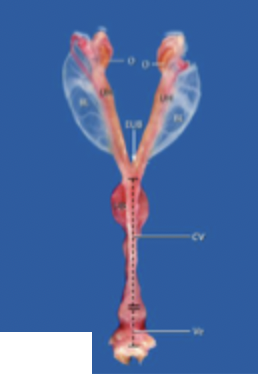
Which species does this uterus belong to?
queen
Why are you unable to pick up the cervix during a rectal exam of a mare?
it has a tight dorsal attachment of the mesometrium
Which ovarian structure are you able to palpate during an exam?
corpus luteum (although not in the mare)
however cannot palpate the follicles
What is the structure of ovarian follicles?
thin walled
fluid filled
contain granulosa cells which synthesise oestrogen
What happens when a dominant follicle produces lots of oestrogen?
if there is no progesterone present, the follicle will ovulate
if there are level of progesterone, the follicle will die back
What is the structure of the corpus luteum?
filled with luteal cells
sometimes contains a central cavity
grey-yellow colour
yellow cut surface
What are the differences between cow and mare ovaries?
mare ovaries are larger
mare are bean shaped, cow are not
mares have larger ovulatory follicles
cow corpus luteum protrudes so is palpable but the mare does not
mares have a distinct ovulatory fossa but cows ovulate from different places
How do ovarian follicles appear on an ultrasound?
little clear black holes (no specks as this indicates pus/ other material)
How does the corpus luteum appear on an ultrasound?
large, more granular structure located in the ovary
How does the appearance of an ovulatory follicle in a mare ultrasound differ from a cow?
it is much larger
How does the appearance of follicles on ultrasound differ according to their development stage (selected, mature, dominant)
progress from lots of very small black dots to one very large, clear one
What is the structure of the infundibulum?
funnel-shaped opening at the ovarian end of the oviduct
externally has ruffled appearance
What is the structure of the ampulla?
the area of the oviduct with a relatively large diameter and lots of cilia
What is the structure of the isthmus?
area of the oviduct with a smaller diameter and thicker muscular walls
connected to the uterus at the uterotubal junction
Why is the infundibulum less developed in the mare?
she always ovulates from an ovulatory fossa so needs less area to capture the oocyte
What are the 3 layers of non-gravid uterine tissue?
perimetrium (serosa)
myometrium
endometrium (mucosa and submucosa)
Which species have distinct maternal caruncles?
cattle and sheep
What is the structure of the cervix?
thick-walled
non compliant
cervical canal is surrounded by folds/rings
acts as a barrier to sperm transport or to isolate the conceptus from the external environment during pregnancy
What are the identifying features of the bitch and queen cervix?
it is relatively small and smooth
What are the identifying features of the cow and ewe cervix?
it has distinct protrusions called cervical rings
What are the identifying features of the sow cervix?
it has interdigitating prominences to accept the male cork-screw penis
What are the identifying features of the mare cervix?
it has no cervical rings but has longitudinal folds which are continuous with endometrial folds
What are the 3 main reasons why you might introduce a catheter through the cervix?
to perform AI
to perform embryo transfer
to treat a uterine infection
Which embryological germ layer does the caudal vagina develop from?
ectoderm
Which embryological germ layer does the cranial vagina and uterus develop from?
mesoderm

Which species does this uterus belong to?
a freemartin cow
Which structure forms the inner foetal sac?
amnion
Which structure forms the outer foetal sac?
chorioallantois

Does this image show foetal or maternal tissue?
foetal
What are the chorionic villi?
small finger-like protrusions from the foetal placenta which protrude towards the uterine endometrium to increase the surface area for exchange with the dam
What type of placenta do cattle and sheep have?
cotyledonary
epitheliochorial
What is the key feature of a cotyledonary placenta?
discrete button-like regions of chorionic villi
What type of placenta does the sow and mare have?
diffuse
epitheliochorial
How many layers does the placenta in cattle, sheep, pigs and horses have?
6
How many layers does the placenta in dogs and cats have?
5
What type of placenta do dogs and cats have?
zonary
endotheliochorial
What are the 2 key differences between a sheep and a cow placenta?
cow is much larger
sheep has concave maternal caruncles, while cow has convex maternal caruncles
How does a zonary placenta appear?
a pigmented stripe zone which contains chorionic villi, maternal haemorrhage and necrosis is normal here
What colour is the pigmented zone of the placenta in the bitch?
green
What colour is the pigmented zone of the placenta in the queen?
brown
What is the pathway of the umbilical arteries?
travel from left and right internal iliac arteries in the foetus to the foetal-maternal blood exchange at the chorionic villi
What is the pathway of the umbilical vein?
from foetal-maternal blood exchange at the chorionic villi to the foetal ductus venosus and vena cava
What is the pathway of the urachus?
from the foetal bladder to the allantoic cavity
Which 3 key changes happen to foetal circulation at birth?
amniotic membrane of the umbilical cord is broken
umbilical arteries retract back
the umbilical vein and urachus close and temporarily remain outside the body
What do the umbilical arteries become after birth?
round and lateral ligaments of the bladder
What does the umbilical vein become after birth?
round ligament of the liver
What does the urachus become after birth?
median ligament of the bladder

1
external abdominal oblique muscle
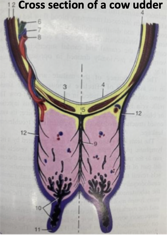
2
internal abdominal oblique muscle
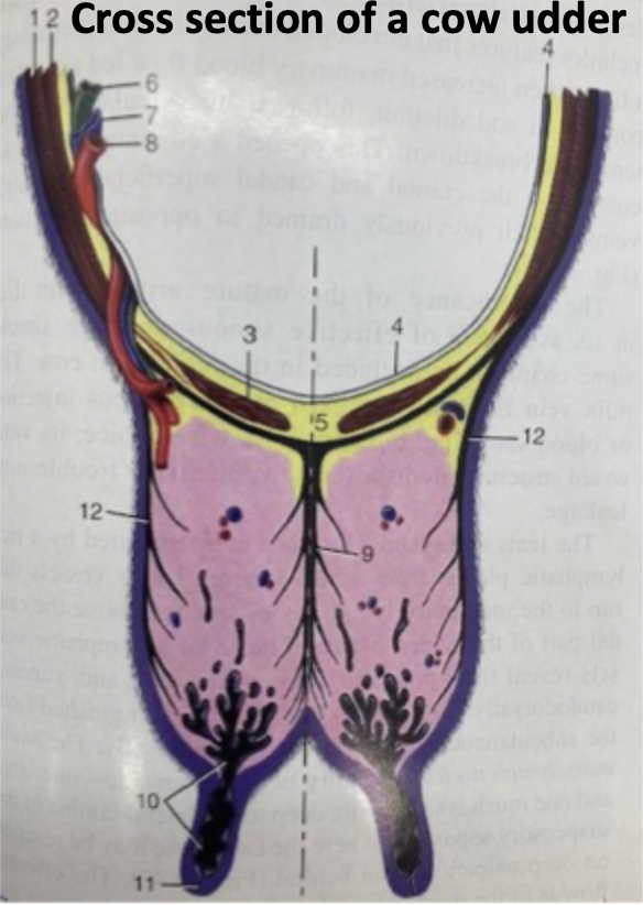
3
rectus abdominis muscle

4
peritoneum

5
linea alba

9
median suspensory ligament

10
lactiferous sinus

11
papillary duct
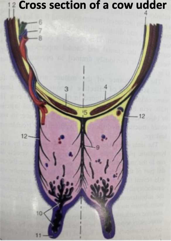
12
lateral susponsory ligament
How does the uterus sit in situ in the cow?
the uterine horns fold over the pelvic rim

How does the uterus sit in situ in the mare?
sits horizontally in the abdominal cavity

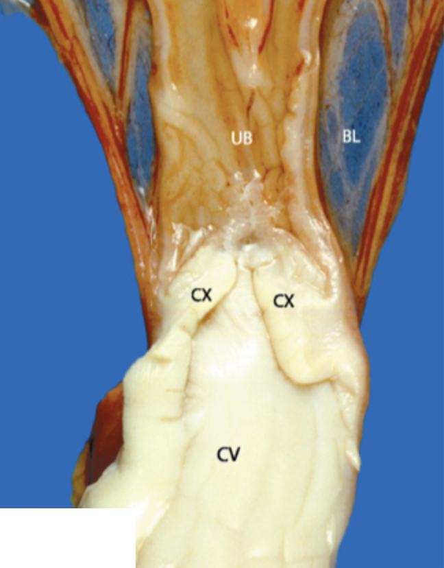
Which species does this cervix belong to?
queen or bitch
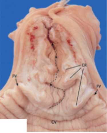
Which species does this cervix belong to?
cow

Which species does this cervix belong to?
ewe

Which species does this cervix belong to?
sow

Which species does this cervix belong to?
mare

X
chorioallantois

Y
amnion

Which species does this placenta belong to?
cow

Which species does this placenta belong to?
ewe

Which species does this placenta belong to?
bitch or queen

Which species does this placenta belong to?
mare
What is the difference between a caruncle and a cotyledon?
the caruncle is the maternal side of the placental connection, and the cotyledon is the foetal complimentary side of the placental connection, these two structures join to form a placentome