Anaemia
 Call Kai
 Learn
Learn Practice Test
 Spaced Repetition
 Match
Match Flashcards
 Knowt Play
1/31

There's no tags or description
Looks like no tags are added yet.
Name | Mastery | Learn | Test | Matching | Spaced | Call with Kai | Chat |
|---|
No analytics yet
Send a link to your students to track their progress
32 Terms
clinical presentation
» Lethargic or collapsed
» Pale mucus membranes
• Jaundiced?
» Tachycardia
» Bounding or weak peripheral pulses
» Hypotension
» Altered respiration?
severity of anaemia
» Depends on the species normal for Hct (hematocrit. It is the percentage of total blood volume made up of red blood cells (erythrocytes))/PCV (packed cell volume)
» Mild – may reflect other disease process, worth monitoring, but unlikely to be primary investigative focus
• Canine between lower reference interval (~37%) and 30% (may be differences for greyhounds/sighthounds)
• Feline below lower reference interval (~27%) and 20%
• Equine – depends on breed (Reference interval usually ~37-58% in ‘hotbloods’; ~32-46% warmbloods; ~24-44% in ‘coldbloods’ / native breeds
» Moderate- likely to be a primary concern, investigation may indicate the underlying disorder
• PCV below 20%
» Marked- significant clinical concern- requires investigation, and rapid initiation of treatmentsupportive if underlying cause is not yet known. Pathophysiology of the anaemia is likely the primary cause of illness, or a consequence of the primary cause.
• PCV below 12-15%
acute or chronic blood loss
Acute blood loss
» Blood is lost over a few hours
» Anaemia results from dilution of remaining erythrocytes
» Recovery
• Reabsorption of erythrocytes (if internal haemorrhage)
• Tissue hypoxia stimulates production of erythropoetin
Chronic blood loss
» Blood is lost over days or weeks or months
» If external loss then Fe deficiency biggest factor in development of anaemia
cytopenia-a lower-than-normal number of blood cells circulating in the body
haemorrhage = loss
haemolysis = destruction


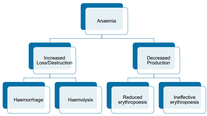
anaemias

increased loss/decreased production anaemia
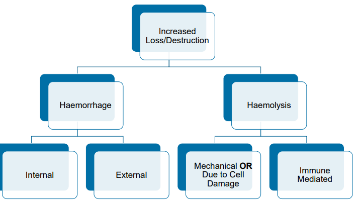
haemorrhage = loss
haemolysis = destruction
Increased Loss
» Predominantly regenerative
» Red cell morphology extremely helpful in
refining likely causes
» Red cell parameters may be useful in
determining possible underlying mechanisms
Decreased Production
» Non-regenerative
» Red cell morphology might provide clues, but
often may be unremarkable
anaemia
going a step further
anaemia- haemorrhage
Internal Haemorrhage
» Haemorrhage into cavities e.g.
• Thoracic
• Pericardial
• Peritoneal
• Joints
» Extravasated (a fluid, such as blood, lymph, or an intravenous medication, has leaked or been forced out of a blood vessel or natural channel into the surrounding tissue) blood is broken down and
recycled – iron is available for new red cell
production
» Regenerative
External Haemorrhage
» Haemorrhage outside the body e.g.
• External trauma
• Epistaxis -nose bleed
• Haematemesis - vomiting of blood - e.g. digestive blood like coffee granules, bleeding in intestine/stomach
• Haematuria - urinating with blood
• Melena - black tarry faeces - smelly
» Blood (and hence iron) is lost outside the body.
If rate of loss is in excess of the ability to absorb
iron from the diet
» —> Iron deficiency anaemia
» May be regenerative- depends on stage of
process
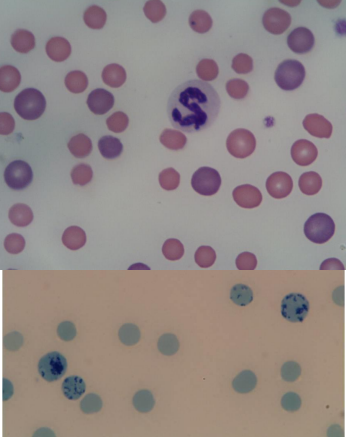
evidence of regeneration
» May take 3-5 days to be seen in peripheral blood
» Polychromatophils see on standard stain
• Purple staining
• Usually larger in size
» New Methylene Blue stain
• Ribosomes and Polyribosomes stained by supravital stain
» Younger erythrocytes
• Increased MCV (Mean Corpuscular Volume) stands for mean corpuscular volume, which measures the average size of your red blood cells)- macrocytic
• Decreased MCHC (mean corpuscular hemoglobin concentration, which is a measurement of the average concentration of hemoglobin inside your red blood cells)- hypochromic
• Increased RDW (red blood cell distribution width)- anisocytosis (cell size variation)
Evidence of Iron (Fe) Deficiency- (Chronic Blood loss)
» Hypochromic cells, with pronounced central pallor
» Decreased MCV- microcytic
» Decreased MCHC- hypochromic
» With progression – microcytic, hypochromic, anaemia
• But one may develop before the other.
» May have other morphologic changes
• Red cell fragments, schistocytes, elliptocytes
» Initially will be regenerative
• Over time – when Fe deficiency is severe- progress to non-regenerative
» Most often chronic external blood loss
• May be dietary deficiency (particularly young animals on a milk –fed diet)

anaemia - haemolysis
Mechanical or Due to Cell Damage or Defects
» Morphology often extremely helpful
• Heinz Body Haemolytic Anaemia
• Heinz bodies- oxidative damage
• Eccentrocytes- oxidative damage
• Schistocytes- shear damage
» Morphology often indicates underlying
pathophysiology -> suggests likely differentials
Immune Mediated Destruction
Immune mediated haemolytic anaemia (IMHA)
» Morphology can be helpful- additional testing
required
• Spherocytes (without schistocytes!)
» May be idiopathic (dont know the cause)/primary autoimmune
» May be secondary e.g. to infectious disease,
lymphoid neoplasms, histiocytic disorders
» ‘Other’ – e.g. Neonatal Isoerythrolysis
Significant Anaemias often present with a mix of both processes in haemolysis
Intravascular
» Lysis of blood cells within vessels
• Does not include sinuses of liver, spleen or bone marrow
» Severe or rapidly falling anaemia
» Present over hours or days
» Haemoglobinaemia (but may not be grossly visible)
» Haemoglobinuria
» Hyperbilirubinaemia- variable
» Bilirubinuria- variable
Extravascular
» Erythrocyte lysis outside of blood vessels
• Within macrophages- (not due to clearing haemorrhage)
» Mild to marked anaemia
» Presents over days or weeks
» Haemoglobinaemia and haemoglobinuria not present
» Hyperbilirubinaemia and hyperbilirubinuria usually present
• In blood unconjugated bilirubin> conjugated bilirubin
Haemolytic icterus (jaundice) – Pre-hepatic
» Pathologic haemolysis -> increased Hgb degradation -> increased bilirubin formation
» Bu (unconjugated bilirubin) travels to liver via tissue macrophages
• Generally liver has large capacity for uptake and conjugation
» Rate limiting step is usually the transport of Bc (conjugated bilirubin) to the biliary system for excretion
• If this is exceeded then Bc is ‘regurgitated’ out into plasma
» Bc then competes for uptake with Bu- so both forms increase in circulating plasma

oxidative injury
» Heinz bodies and Eccentrocytes
» Overwhelming of reductive pathways that keep Hgb in reduced state
» Hgb-Fe2+ undergoes spontaneous conformational change-> hemichromes
» Hemichromes of heme depleted Hgb precipitate -> Heinz bodies
» Erythrocytes are less deformable
• Trapped in spleen and lysed
» Structural damage caused by oxidation -> fragile cells
» Redistribution of band 3 proteins (Heinz bodies)->
• Antibody binding
• Removal by macrophages in spleen and liver

erythrocyte fragmentation
» Schistocytes-fragmented pieces of red blood cells that form when cells are physically torn apart by mechanical damage or tight fibrin strands in blood vessels
» Keratocytes (helmet cells)- can be seen in vitro in feline samples
» Acanthocytes
» Spherocyte-like cells
» Multiple pathogenesis suspected
• Trauma by relatively rigid structures e.g. fibrin
• Rheologic forces

uncommon
» Defects in ATP generation
• PK deficiency (Pyruvate kinase deficiency)
• Moderate anaemia, moderate to extreme reticulocytosis, mild to moderate icterus
• May progress to myelofibrosis -> pancytopenia
• PFK deficiency
• First noted in English Springer Spaniels, now reported in other breeds including American Cocker Spaniels
• Anaemia, haemoglobinaemia, haemoglobinuria after hyperventiliation (producing a respiratory alkalosis) and haemolytic icterus
• When not actively haemolysing then mild regenerative anaemia
» Hypophosphataemic haemoglobinuria in cattle
• Postparturient- 3-8 weeks after calving.
• Defective mobilisation of phosphorus from bone -> defective erythrocyte ATP production -> haemolysis
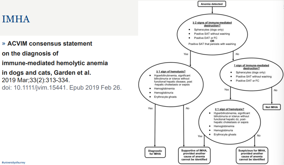
IMHA
» Idiopathic (presumptive primary autoimmune)
» Drug induced
» Vaccine induced
» Alloimmune
• Neonatal isoerythrolysis
• Blood transfusion reactions
» Secondary to increased Ig production eg lymphoma
IMHA blood smear
» Haemolysis
» Erythrocytes coated in erythrocyte surface associated immunoglobulin ESAIg and/or C3 undergo extravascular haemolysis (removed)
» Erythrocytes coated in erythrocyte surface associated immunoglobulin ESAIg and/or C3 -> Spherocytes due to macrophage removing portion of membrane
• Spherocytes may undergo
• Intravascular haemolysis (fragility) OR
• Extravascular haemolysis (rigidity)
» ESAIg may also bind complement -> activation of complent cascade
• Intravascular haemolysis

Haemolysis – Infectious–IMHA and/or other mechanisms
» Mycoplasma spp.
» Leptospira spp.
» Anaplasma spp.
» Clostridium spp.
• Bacillary haemoglobinuria
• Yellow lamb disease (Clostridium perfringens)
• Clostridial infections in horses
» EIAV – Equine Infectious Anaemia Virus- Swamp fever- NOTIFIABLE
» FeLV – Feline Leukaemia virus
» Ehrlichia spp.
» Babesia spp.
» Theileria spp.
» Trypanosoma spp.

IMHA in saline
» Persistence of agglutination
» Despite dilution with saline
» Dilution factors can be variable
» Often start at 1:2 -> 1:4
» Confirm on 1:10 dilution
• 1 part blood 9 parts saline
agglutination

drug induced IMHA
» Not common- three major mechanisms considered
• Drug adsorption- e.g. penicillin binds covalently to erythrocytes and stimulates hapten-dependent antibodies
• Autoantibody induction- induces formation of autoantibodies – antibodies can bind to normal erythrocytes in absence of drug
• Drug-dependent antibody induction- induces antibodies only when soluble drug is present.
» Penicillin – horses
» Cephalosporins- dogs
» Methimazole (rarely reported)– cats
Neonatal Isoerythrolysis-a life-threatening, immune-mediated blood disorder where a newborn's red blood cells are destroyed by maternal antibodies absorbed through colostrum
» Dam (or Queen) negative for blood factor mates with Sire positive for blood factor
» Foetus is positive for blood factor
» Mare- Leakage of blood across the placenta during pregnancy leads to development of antibodies to the blood factor during pregnancy (and persistent beyond this pregnancy)
» Queen- alloantibodies already present
» Equine Qa and Aa factors most often implicated.
» Feline Group B Queen to A or AB kittens
» Dogs usually only an issue if DEA 1.1. negative bitch has had a blood transfusion with DEA 1.1 +ve blood and had sufficient time to produce antibodies prior to colostrum intake
presentation
» Typically normal at birth
» Signs may develop as early as 5 hours- more usually 12-48 hours
» Subacute to peracute
» Lethargy, weakness, tachypnoea, icterus, pigmenturia, haemodynamic shock,
can be fatal
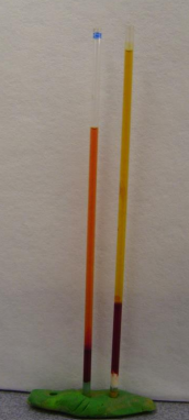
» Spun PCV from a Neonatal isoerythrolysis foal
• Initial sample on left- haemolysed/haemoglobinaemia
• Follow up sample 5 days later- icteric/bilirubinaemia
• NB neonates often have higher levels of bilirubin
EIAV- Equine Infectious Anaemia Virus
» Confirmed infected animals – destroyed
» In contacts restricted and tested twice in a 90 day period
» Coggins test – gold standard (Agar gel immunodiffusion AGID)
• ELISA test also available – possible false positive- confirm positives with Coggins Test
» Can get a false negative in acute stage- consider retesting 10-14 days after presentation
» Presentation can be variable, acute, subacute, chronic and carrier
EIAV- Equine Infectious Anaemia Virus
» Confirmed infected animals – destroyed
» In contacts restricted and tested twice in a 90 day period
» Coggins test – gold standard (Agar gel immunodiffusion AGID)
• ELISA test also available – possible false positive- confirm positives with Coggins Test
» Can get a false negative in acute stage- consider retesting 10-14 days after presentation
» Presentation can be variable, acute, subacute, chronic and carrier
Mycoplasma spp.
» Feline
• Mycoplasma hemofelis (not be confused with Mycoplasma felis)
• Mycoplasma haemominutum
• Mycoplasma turicensis
» Alpaca
• Mycoplasma haemolamae
» Cattle
• Mycoplasma weyonii
» Pigs
• Mycoplasma haemosuis, and Mycoplasma parvum
» Dogs
• Mycoplasma haemocanis
» PCR often more sensitive than blood smear examination
» Numbers of circulating organisms can be variable.

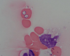
babesia
» Babesia canis reported in non-travelled UK dogs

Anaemias
Decreased Production
Reduced erythropoesis
» Decreased erythropoietin
» Refractory to erythropoietin
» Primary bone marrow disorder
• Damage
• Displacement/Replacment
Ineffective erythropoesis
» Destruction of erythrocyte precursors
» Defective erythrocytes
• Defective heme synthesis
• Abnormal erythroid maturation
• Defective nucleic acid metabolism

non-regenerative anaemias
» Whether anaemias are due to decreased or ineffective erythropoiesis –> non-regenerative
• Absence of reticuolcytes or polychromatophils in peripheral blood (and within bone marrow samples)
• Usually normocytic, normochromic, non-regenerative (anaemia present >5 days)
» Bone marrow disorders e.g.
• Haematopoetic neoplasia- damage or replacement of bone marrow by neoplastic population
• -Pure Red Cell Aplasia (PRCA)- possible immune mediated component -?destruction of early erythroid
• Myelofibrosis- can be a sequalae of PK deficiency
• Damage following inflammation, necrosis, toxicosis (e.g. oestrogen),
Anaemia of Chronic or Inflammatory Disease
» Mild, normocytic, normochromic, non-regenerative anaemia
• Most common cause of anaemia in veterinary patients
• Requires treatment of the underlying disease to resolve the anaemia
• Anaemia likely does not require further investigation, unless persistent, progresses to moderate/marked
» Inflammation -> shortened erythrocyte lifespans
» Inflammation and/or other -> sequestration of Fe – so unavailable for erythrocyte production
• Hepcidin production increased in response to IL-6 -> hepcidin binds to ferroportin -> internalises ferroportin so no longer in membranes -> macrophages cannot export Fe
• Alterations in ferritin production and transferrin receptors -> increased Fe storage and decreased availability
• Number of other cytokines have effects on Fe kinetics
» Inflammation -> cytokines reduce erythroid cell response to EPO and may reduce amount of EPO produced
non-regenerative anaemia - reduced erytropoeisis
» Normocytic, normochromic, mild to moderate -> severe if persistent
» Renal disease- chronic kidney disease
• Decreased production of EPO
» Endocrine
• Hypothyroidism
• Hypoadrenocorticism
• hypoandrogenism
» Liver disease or failure
Non-regenerative anaemia – Ineffective erythropoesis
» Nutritional deficiencies
• Fe – microcytic, hypochromic
• Folate or Cobalamin (B12) – normocytic, normochromic
• In humans macrocytic- not usually seen in veterinary species
• Dog breeds eg Giant Schnauzer – inherited malabsorption of B12
• Cattle – cobalt deficient soil grazing
• Copper- functional Fe deficiency- microcytic, hypochromic
» Inherited diseases
• Dyserythropoesis of English Springer Spaniels
• Congenital dyserythropoesis of polled Hereford calves
» FeLV- may cause hypoplasia (reduced) or erythroid neoplasia (ineffective) – macrocytic, normochromic
anaemias summary

Polycythemia or Erythrocytosis
» Increased red cells- if severe/persistent then may have sluggish movement of blood through vessels
» May be relative erythrocytosis- increased PCV in response to reduction in water e.g dehydration
» May be splenic contraction (physiologic erythrocytosis)– excitement
» May be pathologic
• Primary- polycythemia vera
• Secondary
• Appropriate- response to cardiac or respiratory disease, hyperthyroidism
• Inappropriate- e.g. Renal tumour-> hypoxia of renal tissue-> increased erythropoietin production, or other non-renal neoplasms