BKE4 1 Human Vision and Fibre Optic Endoscopy
 Call Kai
 Learn
 Practice Test
 Spaced Repetition
 Match
 Flashcards
 Knowt Play
1/23

There's no tags or description
Looks like no tags are added yet.
24 Terms
Accommodation
The process of changing the shape (or focal length/thickness) of the lens to form a sharp image on the retina as the object distance changes.
Accommodation process for viewing a near object
Ciliary muscles contract -> tension in suspensory ligaments decreases -> lens becomes thicker under its own elasticity -> focal length decreases (refracting power increases) -> light is refracted more to converge on the retina.

Accommodation process for viewing a distant object
Ciliary muscles relax -> tension in suspensory ligaments increases -> lens is pulled thinner -> focal length increases (refracting power decreases) -> light is refracted less to converge on the retina.

Range of accommodation, Near point and Far point
Near point: The closest point an object can be placed and still form a sharp image (typically 25 cm).
Far point: The furthest point an object can be placed and form a sharp image (infinity for a normal eye). Range of accommodation: The range from the near point (25 cm) to the far point (infinity) over which the normal eye forms a sharp image.
Rod cells (characteristics & functions)
Distributed throughout the retina (except yellow and blind spots). More numerous than cones. Work best in low light conditions (dim light). Provide only black and white (greyscale) vision. Most sensitive to wavelength around 500 nm.
Cone cells (characteristics & functions)
Concentrated at the yellow spot. Less numerous than rods. Active only in high brightness (daylight). Responsible for colour vision and distinguishing fine details. Three types: red (~564 nm), green (~534 nm), and blue (~420 nm).
Yellow spot vs Blind spot
Yellow spot: Near the centre of the retina, contains only cones, gives highest resolution for objects directly in front.
Blind spot: Where the optic nerve leaves the eye, contains no light-sensitive cells, forms no image.
The iris controls the _______, typically ranging from 2 mm in bright light to 6 mm in dim light.
In dim light, _______ increases, which decreases minimum angular separation (θ) and INCREASES _____________ based on the Rayleigh criterion.
The iris controls the pupil size (diameter d), typically ranging from 2 mm in bright light to 6 mm in dim light.
In dim light, pupil size d increases, which decreases minimum angular separation (θ) and INCREASES theoretical resolving power based on the Rayleigh criterion.
angular separation of objects formula

Rayleigh Criterion formula for minimum angular separation: θ = 1.22λ / d
θ = 1.22λ / d (where θ is minimum angular separation in rad, λ is wavelength of light in m, d is diameter of pupil/aperture in m).
Two points can be resolved if their angular separation is > θ.
[IN DSE FORMULA SHEET]

TRAP: Factors affecting Resolving Power
A larger pupil (d) or shorter wavelength (λ) DECREASES the minimum angular separation (θ), which INCREASES the resolving power, allowing finer details to be resolved. Common HKEAA mistake: Confusing a smaller θ value with lower resolving power.
Power of a lens (P)
P = 1 / f (where f is focal length in metres). Unit is dioptre (D). Convex lens has positive power (+ve), concave lens has negative power (-ve). For lenses in contact: Peffective = P1 + P2. [IN DSE FORMULA SHEET]
Short-sightedness (Myopia)
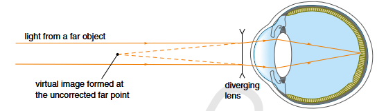
Cannot see distant objects clearly. Far point is closer than infinity. Causes: Eyeball is too long OR lens is too thick (refracting power too high) even when ciliary muscles fully relax. Light from a far object converges IN FRONT OF the retina.
Correction for short-sightedness
Wear spectacles with diverging (concave) lenses. The lens forms a virtual image of a distant object at the uncorrected far point of the eye.
Long-sightedness (Hypermetropia)
Cannot see near objects clearly. Near point is further than 25 cm. Causes: Eyeball is too short OR lens is too thin (refracting power too low) even when ciliary muscles contract maximally. Light from a near object converges BEHIND the retina.
Correction for long-sightedness
Wear spectacles with converging (convex) lenses. The lens forms a virtual image of a near object (usually at 25 cm) at the uncorrected near point of the eye.

Old-Sightedness (Presbyopia) Complications & Bifocals
Mimics long-sightedness (near point > 25 cm) due to decreased lens elasticity from ageing. A person can suffer from both short-sightedness and old-sightedness simultaneously. Corrected using bifocal spectacles: upper diverging part corrects short sight (far objects), lower converging part corrects old sight (near objects).

TRAP: Applying lens formula to spectacles correction
Common mistake: Forgetting the negative sign for virtual images. When correcting vision, the image formed by the spectacle lens acts as the object for the eye. Therefore, image distance (v) MUST be negative (e.g., v = -0.8 m for an uncorrected far point of 0.8 m). Formula: 1/u + 1/v = P. [IN DSE FORMULA SHEET]
Characteristics and principle of an optical fibre
Made of a glass/polymer core surrounded by a cladding. Light travels along the core by a series of total internal reflections. Requirement: Refractive index of core (n1) MUST BE GREATER THAN refractive index of cladding (n2).
Critical angle formula for optical fibre (Core & Cladding)
n1 sin C = n2 sin 90 (where n1 is core refractive index, n2 is cladding refractive index, C is critical angle). [NOT IN DSE FORMULA SHEET - MUST MEMORIZE/DERIVE]
Fibre Cladding (Functions & effect of thickness)
Has a lower refractive index than the core for total internal reflection. Protects the core from scratches. Crucially, it prevents light escaping into adjacent fibres at points of contact, allowing tight packing into bundles without signal leakage. A THINNER fibre allows more bending without light escaping.
Coherent vs Incoherent bundle fibres in endoscopy
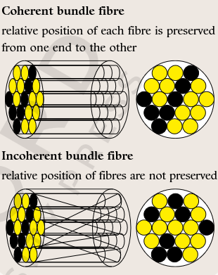
Coherent bundle: Relative positions of fibres are preserved at both ends; used for IMAGE transmission. Higher resolution if thinner fibres are used. Incoherent bundle: Relative positions are NOT preserved; cheaper/easier to make; used for ILLUMINATION (transmitting light into the body).
Advantages of fibre optic endoscopy over X-rays
Direct observation of organ's surface in real original colours; DOES NOT involve ionizing radiation (safer); allows simultaneous minimally invasive surgery or tissue sampling (biopsy) via instrument channels.
Limitations of fibre optic endoscopy
Limited field of view; examines inner surfaces of hollow organs only (cannot see internal solid structures); requires fasting/anaesthesia; risk of perforation/infection; fibre breakage with repeated bending degrades image quality.