All Equine Medicine
 Call Kai
 Learn
 Practice Test
 Spaced Repetition
 Match
 Flashcards
 Knowt Play
1/560

There's no tags or description
Looks like no tags are added yet.
561 Terms
Describe Colic in terms of:
1. Definition
2. Signs of Mild Colic Pain
3. Signs of Moderate Colic Pain
4. Signs of Severe Colic Pain
5. Causes of Colic
1. Blanket term used to describe any disease process that causes abdominal pain.
2. Inappetence, restless, weight shifting, stretching out, posturing to urinate, laying down, intermittent pawing
3. Continuous pawing, head tossing, penile protrusion, yawning, flehmen response, Up and down, dog sitting, kicking at abdomen
4. Violent pawing, violent rolling, collapsing/throwing themselves on the ground, refusal to stand
5. Abdominal cavity pain OR extra-abdominal disease misinterpreted as abdominal pain

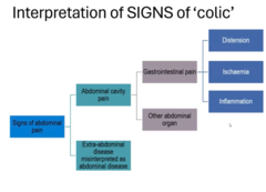
Describe Causes of Abdominal Pain in Horses in terms of:
1. Gastrointestinal Pain (Stomach → Rectum)
2. Abdominal organ Pain
3. Extra-Abdominal Pain
1. a. Distention (abnormal gas production, impaired motility, mechanical, impaction, compression)
b. Ischaemia (Strangulation, loss of blood flow to an area)
c. Inflammation
2. Hepatic, Renal, Urinary, Peritoneal, Repro system pain
3. Laminitis, fracture, rhabdomyolysis, Hendra, Botulism, hepatic encephalopathy, pleuropneumonia, heart failure, Vasculitis, hypoglycaemia
Describe History Taking in a Colic Patient in terms of:
1. Signalment
2. Primary Complaint
3. History
1. a. Age: Parascarius

Describe Physical Exam in a Colic Patient in terms of:
1. Parameters to Assess
2. Baseline Values to Monitor Response to Treatment
3. Degree of Cardiovascular Compromise
4. Assess for Secondary Complications
5. Abdominal Contour
6. Abdominal Auscultation
7. Percussion & Succussion
1. HR, GIT Sounds, RR, MM, Temp, demeanour, Peripheral pulse
2. Record baseline parameters (Esp. HR + GIT Sounds) prior to treatment/diagnostics but if horse is in pain manage the horse first
3. Clinical estimate of dehydration (Decreased water intake/sweating). Shock
4. Endotoxemia (MM colour), Wounds, Eyes, Aspiration pneumonia
5. Gas distension, Asymmetrical (displacement)
6. Abnormal Sounds (Sand Sounds)
7. Ping for gas distended viscous

Describe Normal Horse Parameters in terms of:
1. Temp
2. HR
3. RR
1. 37-38.5℃
2. 36 +/- 10
3. 12 +/- 4
Describe Colic Diagnostic Testing in terms of:
1. Limitations
2. Safety
3. Nasogastric Intubation
4. Nasogastric Intubation Parameters / Assessment
1. Patient condition, client finance, where horse is located
2. Use appropriate restraint and medication
3. Primarily indicated for Gastric decompression for gastric overload, look for refluxing (if significant reflux keep tubed). Caution if using pump (can rupture stomach),
4. Assess Solids (feed, grain, bedding), odour, reflux volume (1-2L normal - gastric accumulation vs. SI obstruction), parasites, pH (acidic = gastric, Alkaline = small intestine), Alleviations of signs of Pain
Describe Abdominal Palpation Per Rectum as Colic Diagnostic Testing
Identifies bowel distension, Presence of tight bands (LC displacement/volvulus), Impactions, masses/hernias, ruptures viscus

Describe Abdominocentesis as a Colic Diagnostic Testing in terms of:
1. Abdominocentesis Definition
2. Abdominocentesis Technique
3. Sample Collection
4. Abdominocentesis Complications
1. Aseptic collection of peritoneal fluid. Changes in health of organs esp. intestine reflected as physical and cytological changes in fluid. Used to assess for intestinal injury (inflammation, Ischaemia) + Peritoneal cavity disease (Peritonitis, Bowel rupture)
2. Most ventral aspect of linea alba → Aseptic prep + technique → LA → Stab incision needed for teat cannula (#15 Blade) → Teat cannula/18G needle
3. Plain Tube (Culture) > Lithium Heparin (lactate/Biochem) > EDTA (Cytology). In that order
4. Enterocentesis (seeds bacteria into abdomen), Amniocentesis (pregnant mares), Splenic tap (Can cause splenic rupture), Omental evisceration, Haemorrhage (underlying coagulopathy)

Describe Abdominocentesis as a Colic Diagnostic Testing in terms of:
1. Red Colour
2. Brown/Green Colour
3. Straw Coloured
4. Dark Yellow
5. Serosanguinous (Light Red)
6. Yellow/Orange
1. Blood. Iatrogenic contamination, Splenic tap, Hemoabdomen)
2. Ingesta. Enterocentesis, Bowel rupture)
3. No compromise
4. Mild intestinal injury
5. Marked intestinal injury/Devitalised bowel
6. Peritonitis

Describe Abdominocentesis as a Colic Diagnostic Testing in terms of:
1. Transparency
2. Protein
3. Cytology
4. Microbiology
5. Early Strangulating Lesion Considerations
1. Normal is clear, Turbidity caused by increased WBCs = inflammation
2. High Protein = Intestinal injury. Normal
3. Look for neutrophilia, bacteria, RBCs
4. Bacterial C+S. Plain red tube
5. Early strangulating lesion can have normal abdominal fluid

Describe Abdominocentesis as a Colic Diagnostic Testing in terms of:
1. Lactate Sampling
2. Lactate Uses
1. Indicates tissue ischaemia. Normal
Describe Abdominocentesis as a Colic Diagnostic Testing in terms of:
1. Transudate
2. Modified Transudate
3. Exudate
1. Yellow, clear, 0-5000 nucleated cells, <20 g/L protein, Mononuclear cells, non-degenerated neutrophils, low RBCS
2. Yellow, Turbid, 5000 - 10 000 Nucleated Cells, <20g/L Protein, Macrophages and non-degenerate neutrophils predominate
3. Yellow/red, Cloudy, >5000 nucleated cells, >25g/L Protein, Non-degenerate or degenerate neutrophils predominate

Describe Ultrasound as a Colic Diagnostic Testing in terms of:
1. What Can be Seen
2. Limitations
3. FLASH Scan
4. Abdominal X-Ray
1. Excess peritoneal fluid, adhesions, masses, small intestine distension, lack of intestinal motility, intussusceptions and position of structures (Nephrosplenic entrapment)
2. Depth of penetration, gas filled organs
3. Protocol for fast localised abdominal sonography of horses.
4. Uncommon. Can see sand colic and enteroliths

Describe Clinical Pathology as a Colic Diagnostic Testing in terms of:
1. PCV + TP
2. Haematology
3. Azotemia
4. Muscle Parameters (CK + AST)
5. Hepatic Parameters (AST, GGT, Bilirubin)
1. Increase in mortality associated with PCV at admission, Total protein losses common due to PLE. Interpret with caution as estimated after correction of dehydration
2. RBCs (Haemoconcentration), WBCs (Leukocytosis, Leukopenia - due to endotoxemia)
3. Creatinine ↑ due to decrease glomerular filtration rate
4. Muscle damage secondary to colic, CK sensitive for myolysis
5. Inappetence + GIT toxin translocation, ↑ Serum GGT in horses with right dorsal displacement of large colon (direct pressure on bile duct)
Describe Clinical Pathology as a Colic Diagnostic Testing in terms of:
1. Glucose
2. Electrolyte Abnormalities
3. Blood Gas
4. Coagulation Tests
1. Increased due to adrenalin (Common in colic), Associated with degree of recovery
2. Multiple electrolyte abnormalities common. Na ↓ with losses or ↑ with dehydration. Hypocalcaemia very common (predisposes to ileus), Hypophosphatemia/hypomagnesaemia common
3. Metabolic acidosis common
4. Subclinical or clinical DIC if severe
Describe Medical Therapy of Colic in terms of:
1. Main Goals
2. Analgesia
3. Fluid Therapy
4. Husbandry
1. Relieve pain and re-establish GIT function
2. NSAIDs, Sedatives, Opiates
3. Laxatives, IV fluids, Enteral fluids
4. Fasting, preventing self injury
Describe Analgesia Treatment for Colic in terms of:
1. Flunixin meglumine (NSAID)
2. Meloxicam (NSAID)
3. Phenylbutazone (NSAID)
4. Xylazine, Romifidine, Detomidine (a2 Agonists)
5. Butorphanol, Methadone, Morphine (Opiod)
6. Lignocaine (Sodium Channel Blocker)
7. N-Butylscopolammonium Bromide + Dipyrone (Anti-Spasmodics)
1. Analgesic, anti-pyretic, anti-inflammatory, anti-endotoxic. Never give IM (causes clostridial myonecrosis)
2. Licensed in foals, preserves small intestine mucosa after ischaemic injury
3. Best for musculoskeletal pain, Increasing dose does not increase analgesic effect bur prolongs duration until elimination
4. Sedative with short duration analgesia. Useful for immediate control of signs of abdominal pain and allow procedures to be performed safely. Causes decreased GIT motility, CVS + Resp. depression.
5. Analgesia, significant excitability/agitation (Administer with a2 Agonist), Inhibits GIT motility
6. Analgesia, Anti-inflammatory if given systemic. Reduces factors that inhibit intestinal motility. Can have toxicity (tremors, ataxia, seizures)
7. Only used for spasmodic colin (increased gut sounds)
Describe Fluid therapy Treatment for Colic in terms of:
1. Fluids
2. Laxatives (Magnesium sulphate, Dioctyl sodium succinate, Paraffin Oil)
1. CVS support. Addresses dehydration and electrolyte disorders
2. Increases water content of ingesta
Describe Colic Prognosis in terms of:
1. Response to Treatment
2. Surgical Criteria
1. Mild colics will respond well to medical therapy. Horses that fail to respond, have recurrence of Colic signs, ort increase in severity require more intensive management, referral, or surgery
2. Unrelenting pain of first visit, repeated ineffective treatment, grossly abnormal rectal palpation, Large volume of gastric reflux, progressive abdominal distension. RElieve obstruction (sand, enteroliths, FBs, remove devitalised intestine, reposition intestines). Diagnosis of Problem often only possible by Ex-Lap
Define the Following Terms:
1. Endotoxemia
2. Sepsis
3. Systemic Inflammatory Response Syndrome (SIRS)
4. Multiple Organ Dysfunction (MODS)
1. Presence of endotoxin within the bloodstream and also used to refer to the clinical syndrome associated with it. Causes INFLAMMATION
2. Systemic inflammatory response to infection
3. General systemic inflammatory process independent of cause
4. Insufficiency of 2+ organ systems evident by clinical or clinicopathologic changes

Describe Endotoxin Lipopolysaccharide (LPS)
Structural components of outer layer of gram -ve bacteria cell wall. Lipid A portion of LPS responsible for toxic princibles

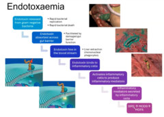
Describe the Pathogenesis of Endotoxemia
Endotoxin released from gram -ve bacteria (Rapid bacterial replication + rapid bacterial death)
→ Endotoxin absorbed across gut barrier (facilitated by damaged gut barrier)
→ Endotoxin free in blood stream (overwhelms liver extraction)
→ Endotoxin binds to inflammatory cells
→ Activates inflammatory cells to produce inflammatory mediators
→ Inflammatory mediators secreted by inflammatory cells (Protective AND destructive, Mass activation throughout the body)
→ SIRS, MODS, MOFS
Describe Causes of Equine Endotoxemia
1. GIT
2. Other
1. Acute GIT injury (Colic) increases GIT permeability to LPS (LPs detected in 40% of colic cases). GIT permeability also caused by GIT rupture, Local ischaemia, inflammation, systemic acidosis
2. Confined to specific area (e.g lung), Bacteraemia

Describe Events of Endotoxemia in terms of:
1. Endothelial Dysfunction + Damage
2. Neutrophil activation, migration and transmigration
3. Plasma Activation
4. Coagulopathy
1. Decreased responsiveness to vasoactive agents, increased vascular permeability and tendency towards clot formation
2. Stimulation of phagocytosis and respiratory burst to release lysosomal enzymes,inflammatory mediators and expression of adhesion molecules
3. Initiates enzymatic reactions on proteins
4. Coagulopathy and DIC, Complement activation, Acute Phase Response (IL1, IL6) - synthesis of proteins by hepatocytes to suppress and contain inflammatory responses, Hemodynamic changes, development of shock, Organ failure

Describe Clinical Signs of Equine Endotoxemia in terms of:
1. Acute Clinical Signs
2. Acute Signs Progression
3. Hypodynamic Shock Phase
4. Haemostatic Abnormalities
5. Organ Failure
1. Anorexia, lethargy, hyperaemic state, tachypnoea/tachycardia, discomfort, sweating, muscle fasciculation, recumbency
2. Exacerbation of existing signs, Diarrhoea, hyper/hypothermia, toxic brick red/purple MM
3. Low temp, weak pulses, diffuse scleral reddening
4. Thrombosis, Haemorrhage, Mucosal peteachiation
5. Renal failure, laminitis, liver failure, CVS failure

Describe Clinical Pathology of Equine Endotoxemia in terms of:
1. Haemogram
2. Biochemistry
3. Coagulation Profile
Reflect primary disease +/- Organ Failure
1. Leukopenia due to neutropenia. Toxic changes in neutrophils. Rebound neutrophilia on recovery. Toxic changes in neutrophils
2. Hypocalcemia as direct/indirect effect.
3. Disseminated Intravascular coagulopathy (DIC)

Describe Endotoxemia Treatment in terms of:
1. Inhibition of Endotoxin Release into Circulation
2. LPS Scavenging
3. NSAIDs, Antioxidants, Lignocaine CRI
4. Supportive Care
1. ABs. Beware that
2. Polymyxin B (Binds to lipid A), Immunotherapy, vaccination, antiserum
3. Inhibits inflammatory mediators
4. Fluid therapy and CVS support, Acid-base balance, Monitoring
Describe Reperfusion Injury in terms of:
1. Definition
2. Part 1 - Ischaemia
3. Part 2 - Reperfusion
1. Damage to tissue caused when blood supply returns to tissue after a period of ischaemia
2. Decrease in the blood supply by constriction or obstruction of the blood vessels → O2 is critical for ATP generation → Dysfunction of membrane pumps (Entry of Ca, Na, H20 into cell) → Mitochondrial dysfunction → cell membrane failure → cell necrosis → Reactive O2 species production + proinflammatory cytokine production → Increased tissue vulnerability on reperfusion
3. Leukocyte chemotaxis and activation → Inflammatory mediators, cytokine release → activated leukocytes release Reactive oxygen species (oxidative burst) → damage cellular membranes → vascular permeability, oedema, thrombosis, cell death

Describe Implications/Consequences of Reperfusion Injury in terms of
1. Overall Pathogenesis of Gut Reperfusion Injury
2. Small Intestine Consequences Pathogenesis
3. Large Colon Consequences Pathogenesis
1. Ischaemia → neutrophil activation + Mucosal injury → bacteria in gut lumen translocate into blood through mucosa → Neutrophils already activated → Liver overwhelmed → inflammatory mediators, ROS, endotoxins in systemic circulation → MODS → MOFS → death
2. Fluid sequesters in subepithelial space. → epithelium loosens from underlying attachments → sloughing of sheets of cells → villi denuded of epithelium by 3h → necrosis of mucosal epithelium extending to crypt base → Ileus + loss of absorption +/- increased secretion
3. Necrosis of clumps of surface epithelial cells → cells then loosen from attachments at their base + neighbouring cells → sloughing of small clusters of cells (rather than sheetsI → leus + loss of absorption +/- increased secretion
Describe Implications/Consequences of Reperfusion Injury in terms of
1. Endotoxemia
2. Sequestration of Fluid into GIT lumen
3. Prognosis
1. Mucosal epithelial injury → disruption of mucosal barrier → migration of luminal bacteria and endotoxin into systemic circulation
2. Hypovolaemia, hypotension, impaired perfusion
3. Dependant on the degree and duration of ischaemia before surgical intervention. Despite surgical correction, intense medical therapy + supportive care perioperative mortality is high. If horse survives initial period of mucosal necrosis + sloughing and viable enterocytes present mucosa can regenerate

Describe Treatment of Reperfusion Injury in terms of
1. Objectives of Treatment
2. Treatment Agents
1. PRevent formation of ROS, stop cytokines, prevent endothelial cell damage, stop neutrophil chemotaxis ir activation, treat inflammation
2. a. Flunixin (NSAID)
b. Lignocaine (Stabilises membrane, analgesia, anti-inflammatory)
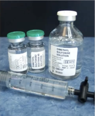
c. Dimethyl Sulfoxide - DMSO (scavenges ROS)
d. Carolina Rinse: Decreases cell injury during reperfusion
Describe Peritonitis in terms of:
1. Peritoneum Anatomy
2. Peritonitis Definition
3. Pathophysiology
1. Lies in abdominal cavity, parietal and visceral. Contains peritoneal fluid that allows sliding of viscera, produce cellular mediators, fluid resorption and diffusion
2. Inflammation of the mesothelial lining of the peritoneum (Diffuse/localised, acute/chronic, septic/sterile, primary/secondary)
3. Infectious/Toxic/Mechanical Insult
→a. Peritoneal mesothelial cell death → loss of fibrin action + thromboplastin release → coagulation pathways activated → Peritonitis
→b. Peritoneal mast cells → inflammatory mediators → vasodilation → increased permeability → ↑ fibrin, macrophages, neutrophils → Peritonitis

Describe Peritonitis in terms of:
1. Clinical Signs
2. Diagnosis
3. Treatment
4. Prognosis
1. Colic, Inflammation +/- sepsis +/- CVS dysfunction
2. a. Haemogram: (Inflammation = Fibrinogen, Hypo/hyperproteinemia)
b. Abdominocentesis: WBC, TP (↓ glucose + ↑ lactate = septic bacteria)
c. US: Fluid volume + nature
d. Ex-Lap
3. Identify aetiology (Surgical exploration), ABs, Fluids, Lavage, Analgesia, Anti-inflammatory, nasogastric decompression
4. Depends on Primary cause. Early aggressive therapy is best

Describe Abdominal/Peritoneal Adhesions in terms of:
1. Definition
2. Causes
3. Post-Trauma Inflammatory Response
1. Abnormal fibrous bands between organs, tissues or both in the abdominal cavity that are normally separated. Congenital or Acquired
2. Sequelae to peritonitis, Surgical trauma, surgical FB (Cutting, abrasion, desiccation, gauze, hair, suture)
3. Fluid exudate released from injured peritoneal surfaces → rich in plasma proteins esp. Fibrinogen → tacky substance that causes adjacent organs/injured serosal surfaces to coalesce → adhesions

Describe Abdominal/Peritoneal Adhesions in terms of:
1. Peritoneal Healing
2. Normal Hemostasis/Pathophysiology of Adhesions)
3. Sequelae
1. Peritoneal damage → Neutrophils (d1-2) → Macrophages (peak d5) → mesothelial cells proliferate throughout the wound base and form islands of cells (d4-7)
2. Formation of fibrin matrix is normally temporary → fibrinolytic system usually removes adhesions within 72h. If this does not occur within 5-7d OR fibrinolytic activity is reduced → fibrin matrix persists and fibroblasts migrate into the fibrin and lay down collagen = Adhesions
3. Present in 30% of post-operative cases needing repeat laparotomy → Adhesion formation may not be associated with the site of primary lesion or resection

Describe Prevention of Abdominal/Peritoneal Adhesions in terms of:
1. Prophylactic Treatments
2. Preventing Adhesions in Surgery/ Inflammation Reduction
3. Mechanical Separation of Injured Areas
1. Heparin (Anticoagulant actions, inhibition of thrombin mediated conversion from fibrinogen to fibrin)
Tissue Plasminogen Activator (Pro Fibrinolytics)
2. Atraumatic tissue handling, hemostasis, minimise contamination, use of non-reactive suture, prevent tissue ischemia, ABs, NSAIDs
3. Peritoneal lavage, Polymer solutions, Gels, Omentectomy

Describe Abdominal/Peritoneal Adhesions in terms of:
1. Clinical Signs
2. Risk Factors
3. Diagnosis
4. Treatment
5. Prognosis
1. Acute severe colic 5-7d post op, Postoperative Ileus, REcurrent colic, poor BCS
2. Foals/miniatures, Pain on admission, abnormal peritoneal fluid, site of intestinal obstruction, Ischaemia + distension, resection/anastomosis, post-op ileum, septic peritonitis, repeat laparotomies
3. Risk factors, CS, US, Ex-Lap, Necropsy
4. Mild/Recurrent Colic: Medical/diet change
Severe: X-Lap, adhesiolysis, resection of adhere bowel, Intestinal bypass. Euthanasia
5. Guarded. Repeat Celiotomy associated with poor prognosis.

Describe Dysphagia in terms of:
1. Definition
2. Pathophysiology
1. Difficulty in Eating. Describes a set of clinical signs rather than a mechanism or a location of a problem
2. Evaluated by examining each individual phase of eating (Prehension, Mastication, 2 phases of Deglutition (Swallowing)). Classified as pain induced, neurogenic, or obstructive.

Describe Prehension in terms of:
1. Control
2. Structures Involved
3. Clinical Signs if Dysfunctional
4. Causes of Prehension Difficulty
1. Voluntary. CN 7 + Muscles of facial expression (Lips)
2. Lips, incisors, TMJ, buccal muscles, Nerves
3. Ptyalism, dropping feed, head tossing, exaggerated head movements, submerging head in feed/water
4. Trauma of lips/buccal muscles, inflammation, fractured/loose teeth, Malocclusion, brachygnathia/prognathous, CNS damage, PNS damage

Describe Mastication in terms of:
1. Control
2. Structures Involved
3. Clinical Signs if Dysfunctional
4. Causes of Mastication Difficulty
1. Voluntary. CN 5 + Masticatory muscles
2. Teeth, TMJ, tongue, Masticatory muscles, nerves
3. Ptyalism, prehension with no chewing, exaggerated chewing, reluctance to open mouth, holding/packing feed into the oral cavity, dropping feed
4. Fractured teeth/jaw, tooth abscess, sharp teeth/hooks, inflammation of TMJ, Masticatory muscle atrophy/spasm/inflammation, tongue inflammation/FB, Stomatitis

Describe Swallowing (Deglutition) in terms of:
1. 1st (Pharyngeal) Phase Control/Anatomy
2. Causes of Phase 1 Difficulty
3. 2nd (Oesophageal) Phase Control/Anatomy
1. Involuntary Reflex. CN 9+10+11. Larynx dislocated from intrapharyngeal ostium, soft palate elevated, epiglottis retroverts, arytenoids/vocal folds adduct → ingresta bolus propelled by constrictor muscles of pharynx into relaxed upper esophageal sphincter
2. Space occupying lesion (neoplasia, LN enlargement), Neuro disorders, inflammation, stomach tube damage
3. Involuntary reflex. CN 10. Primary peristaltic waves initiated by closure of cricopharynx

Describe Dysphagia Clinical Evaluation + Diagnosis in terms of:
1. Clinical Exam
2. Nasogastric Tube
3. Haematology (CBC) + Biochem
4. Endoscopy
5. Radiography
1. History, Clinical exam (incl. Neuro), IDentify if dysphagia is due to problem with prehension, mastication, or swallowing, Observe in feeding environment, oral cavity exam
2. Establishes patency
3. Checks for systemic illness with secondary disfunction (Hepatic encephalopathy, encephalopathy)
4. Nasal passages, pharynx, guttural pouches oesophagus. Can use water/air/[physical contact to evaluate swallow reflex
5. Head + Neck. Soft tissue abnormalities
Describe Oral Tumours in terms of:
1. Types of Tumours
2. Diagnosis
3. Treatment
1. Teeth (odontogenic), Bone (Osteogenic), Soft tissue (SCC, melanoma, sarcoids)
2. Radiography/histology
3. Based on type/location/size. Prognosis variable

Describe Cleft Palate in terms of:
1. Definition
2. Clinical Signs
3. Diagnosis
4. Treatment
5. Prognosis
1. Aetiology unknown. COngenital. Soft palate + rarely hard palate
2. Typically foals: milk at nostrils, coughing after nursing, ill-thrift, Aspiration pneumonia common
3. CS, oral exams , endoscopy
4. Surgical, require multiple surgeries, dehiscence common due to food passage
5. Almost all have complications. pharyngeal function compromised precluding athletic function

Describe Tongue Lacerations in terms of:
1. Prevalence
2. Healing
3. Surgical Treatment
4.
1. Traumatic lesions common
2. Excellent blood supply and healing often rapid. Extensive lesions may still heal with adequate function, some loss of control may be present. Excessive tension on the tongue can tear the frenulum or fracture the hyoid bone which may result in transient/permanent flaccidity
3. GA → exteriorise tongue and debride wound with scalpel → close laceration bottom up using multiple layers to close dead space (dorsal tongue holds suture well) → absorbable sutures

Describe the Oesophagus in terms of:
1. Musculature
2. Sphincters
3. Developmental Abnormalities
1. Striated muscle larynx to mid thorax, smooth muscle mid thorax to cardia
2. Upper pharyngeal + lower cardiac sphincters (Prevents gastric reflux
3. Rare. Congenital cysts, dilation, cricopharyngeal laxity, Strictures, hypertrophy of esophageal musculature. Diagnoses on Radiography +/- barium study, endoscopy. Manage via diet/surgery

Describe Oesophageal Obstruction (Choke) in terms of:
1. Prevalence
2. Most Common Cause
3. Other Causes
1. Most common disorder of equine oesophagus
2. Most common cause is ingesta of abnormal consistency (Poor quality hay, beet pulp, hay cubes). Horses that are fast eaters, have dental disease, shetland ponies. Sedation is a predisposing factor
3. a. Intraluminal: FB, Feed
b. Extraluminal: Neoplasia, vascular ring anomaly, granuloma
c. Intramural: Abscess, granuloma, neoplasia cyst, stenosis
d. Functional: dehydration, exhaustion, pharmacutical, megaoesophagus, neuropathy
Describe Oesophageal Obstruction (Choke) in terms of:
1. Clinical Signs
2. Common Sites for Obstruction
3. Diagnosis
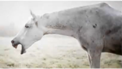
1. Green discharge from nares, Acute onset after eating, Marked signs of distress, Retching/Gagging, profuse salivation, coughing, palpable mass in oesophageal region. Tell owner NPO until examination (some horses will try to keep eating)
2. Post pharynx (Top 1/3), thoracic inlet, base of heart, cardia of stomach (Due to anatomical narrowing/decreased distensibility
3. CS, palpation, inability to pass stomach tube, endoscopy, Radiography +/- contrast

Describe First Stage (Field) Medical Treatment of Oesophageal Obstruction (Choke) in terms of:
1. Initial considerations
2. Heavy Sedation
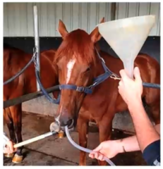
3. Nasogastric Intubation +/- Oesophageal Lavage
4. AB Therapy
5. Pharmacological Assistance
1. Many cases resolve spontaneously. Will be lots of coughing + epistaxis and will be distressing. NPO
2. a2 agonist + butorphanol. Head lower than thoracic inlet to prevent aspiration
3. Confirm obstruction + location, Water +/- obstetrical lubricant carefully to dislodge obstruction (don't push with tube-use flush), Don't use paraffin oil, can rupture oesophagus
4. High change of aspiration pneumonia
5. Oxytocin (stimulates smooth muscle contraction), Guaifenesin (Skeletal muscle relaxant)
Describe Second Stage (Hospital) Medical Treatment of Oesophageal Obstruction (Choke) in terms of:
1. IV fluids
2. Acid-Base Status
3. Other treatment
4. Further Treatment
1. For deficit, maintenance and ongoing losses. Attempt to rehydrate ingesta in oesophagus → reattempt NGT every 3h and flush to try and dislodge obstruction
2. Assess and correct acid-base status. Equine saliva contain high Conc. K, Cl-, HCO3-. Salivary loss due to choke results in initial hyponatremic metabolic acidosis → as disease progresses metabolic alkalosis develops due to progressive hypochloremia
3. NSAIDS + broad spectrum AB
4. If still unsuccessful → GA → Secure airway with cuffed ET tube → repeat endoscopy + stomach tubing. Surgery rarely required

Describe Oesophageal Obstruction (Choke) in terms of:
1. Complications
2. Prognosis
1. Oesophageal Ulceration, Inflammation, Perforation, and rupture. Cellulitis, Abscessation, Diverticuli, motility disorders, oesophageal reflux, laryngeal/pharyngeal trauma when passing stomach tubes, aspiration pneumonia, megaesophagus, recurrent choke episodes
2. Prognosis good if treated within 24h. Time obstructed reflects prognosis. Often sequelae/complications are what causes problems
Describe Oesophageal Obstruction (Choke) in terms of:
1. Management + Prevention
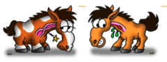
1. Proper dental care, good quality hay, slow down fast eaters. In horse with previous choke avoid dry pelleted feed + beet pulp which expand when wet (or soak prior to feeding), Avoid hay cubes
2.

Describe Other Disorders of the Oesophagus in terms of:
1. Clostridial Infections
2. Intramural Abscesses
3. Oesophageal Rupture
4. Neoplasia
1. Caused by long-term use of indwelling feeding tubes. Characterised by extensive myonecrosis and gas infiltration of tissue
2. Caused by FB
3. Caused by kicks, neck trauma, NGT
4. Rare. Primary SCC most common
Describe Stomach Anatomy in terms of:
1. Internal Structure
2. Cardiac Sphincter
3. Functions of the Stomach
1. 1/3 squamous, 2/3 glandular separated by margo plicatus
2. Well developed, prevents retrograde flow into oesophagus
3. - Temporary store of rapidly ingested feed
- Initiates chemical + enzymatic digestion (HCL + Pepsin)
- Liquification of fluid (Muscular grinding)
- Protection against ingested microorganisms (HCL sterilises)

Describe Gastric Physiology in terms of:
1. Mucous Cells (Bicarbonate Rich Mucous)
2. Parietal Cells (HCL)
3. Enterochromaffin-like cell (Histamine)
4. G Cells (Gastrin)
5. Chief Cells (Pepsiongen)
1. Protects gastric glandular mucosa from acid
2. Sterilises food, Initiates protein digestion
3. Stimulates HCL production
4. Prokinetic, stimulates HCL production
5. Inactive pepsin

Describe Gastric Physiology in terms of:
1. Continuous HCL Secretion
2. Eating Stimulating Release of HCO3 Rich saliva
3. Roughage Absorbs Gastric Secretions Reducing Contact with Mucosal Surface
4. Gastric Secretions Contain Compounds Harmful to Gastric Mucosa
1. Independent of eating. Gastric pH remains at 2 when not eating
2. Neutralises gastric acidity
3. Once horse stops eating gastric pH rapidly falls
4. HCL, pepsin, bile acids, VFAs
Describe Gastric Mucosa Protective Mechanisms in terms of:
1. Squamous Region
2. Glandular Region
1. Stratified Squamous Epithelium. Common place of ulcers
2. Mucous layer, bicarbonate secretion into mucous, extensive mucosal blood supply, rapid cellular restitution, Proteases secreted as zymogens (Only activate in acid

Describe Equine Gastric Ulcer Syndrome (EGUS) in terms of
1. Definition
2. Prevalence
3. Lesions
4. Equine Squamous Gastric Disease (ESGD)
5. Equine Glandular Gastric Disease (EGGD)
1. Inflammation, erosion & ulceration of the stomach of the horse.
2. 70-100% of TB/SB racehorses in active training, less common in pleasure horses
3. Inflammatory, Erosions (superficial layer affected), or Ulcers (penetrates to submucosa)
4. 80% of Adult Cases. Squamous portion lacks defences of glandular portion
5. 20% of Adult cases (more predominant in foals). Defences are bicarbonate, Mucous, extensive capillary network
Describe Pathophysiology of EGUS in terms of:
1. Equine Squamous Gastric Disease (ESGD)
2. Equine Glandular Gastric Disease (EGGD)
1. Ulcers result from excessive exposure to mucosal aggressive agents. Gastric Acid (HCL), VFAs (produced by bacteria from suger in stomach, synergistic with H+)
2. Ulcers result from impaired protective mechanisms. Gastric fluid bathing glandular mucosal consistently at pH 1-3

Describe Equine Gastric Ulcer Syndrome (EGUS) in terms of:
1. Adult Clinical Signs
2. Foal Clinical Signs
3. Diagnosis
4. Common Sites of Ulceration
1. Bruxism, Colic (esp. around feeding, more severe on CHO), poor appetite, poor performance, weight loss, rough hair coat, behaviour changes
2. Bruxism, salivation, recumbency, infrequent nursing, diarrhoea, pot belly, rough hair coat, may not show signs until severe
3. GASTROSCOPY. >16h fast before scoping
4. Above macro plicatus along the outflow tract on the lesser curvature

Describe ESGD Severity Scoring in terms of:
0. Grade 0
1. Grade 1
2. Grade 2
3. Grade 3
4. Grade 4
5. EGGD Severity Scoring
0. Normal gastric mucosa
1. Intact mucosa, evidence of reddening or hyperkeratosis
2. Small single or multifactorial areas
3. Large single or multifocal lesions or extensive superficial lesions
4. extensive lesions with areas of apparent deep ulceration
5. No system, description based

Describe ESGD Risk Factors in terms of:
1. General Diet
2. CHO Diet Metabolism
3. Excersise
4. Other Risk Factors
5. EGGD Risk Factors
1. Feed deprived lowers stomach acid pH (no saliva), racehorse diet (high conc. low roughage), Intermittent feed and water deprivation results in progressive squamous ulceration
2. VFAs produced from CHO metabolism in the stomach → able to penetrate intact gastric mucosa → acidification of ICF. Synergistic effect of VFAs with H+
3. Marked rapid decrease in gastric pH with onset of excersise. Marked decrease in stomach volume when at gait higher than a trot (increases exposure of squamous mucosa to acid
4. Stall Confinement, Transport stress
5. Unknown
Describe EGUS Treatment in terms of:
1. Measurement of Treatment Effectiveness
2. Histamine Antagonist (Acid Suppressive Therapy)
3. Proton Pump Inhibitors (Acid Suppressive Therapy)
1. Healing of gastric ulcers is highly correlated with gastric pH. Ability to maintain gastric pH >4 is measure of treatment effectiveness
2. Ranitidine. Competitive inhibition at H2 receptor on gastric parietal cell. needs to be given at strict 8h intervals
3. Omeprazole. Blocks secretion of H+ from parietal cell membrane by binding to pumps. More effective than H2 agonist

Describe EGUS Treatment in terms of:
1. Antacids (Buffer of Excess Acids)
2. Mucosal Protectants (Prostaglandin E2 Anologue)
1. No practical use in adults as large volume needed
2. Misoprostol. Requires acidic environment to activate → administer prior to acid suppression therapy. Stimulates mucous secretion + inhibits gastric acid secretion
Describe EGUS in terms of:
1. Management
2; Complications
1. Address risk factors, reduce excersise, free-choice access to grass/hay (helps buffer acid), preventative treatment at times of greater risk, decrease stress
2. Uncommon. Chronic ulceration, delayed gastric emptying, oesophageal reflux, oesophagitis, perforation, fibrosis and contracture of stomach

Describe Gastric Impaction in terms of
1. Primary
2. Secondary
3. Clinical Signs
4. Diagnosis
5. Treatment
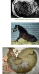
1. Grain engorgement/improper mastication, indigestible material (bedding, poor hay), Feeds that swell (wheat, barely, beet pulp)
2. Intestinal ileus, Gastric outflow obstruction (FB, Mass)
3. Acute severe colic. Can result in gastric rupture
4. US, endoscopy, or at surgery
5. Pain relief, gastric lavage, ex-Lap to remove
Describe Gastric Parasitism in terms of
1. Definition
2. Clinical Signs
3. Treatment
1. Gasterophilus spp. Habronema spp.
2. Rarely cause clinical signs. Can cause gastritis
3. Ivermectin

Describe Small Intestine Anatomy in terms of:
1. Structures
2. Secretions
3. Attachments
4. Crypts of Lieberkuhn
5. Villi
6. Function
1. Duodenum → jejunum → ilium
2. 130L of fluid secreted into SI/day
3. Mesentery, duodenocolic ligament, hepatoduodenal ligament, ileocaecal fold
4. Stem cells located at base which divide and migrate up to villi. Secrete fluid
5. Mature cells with absorptive function. injury to villi allows secretion to continue while absorption is disrupted → hypersecretion
6. Digestion+absorption, movement of water, absorption of bile acids + vit. B12

Describe Small Intestine Anatomy in terms of:
1. Peristalsis Motility
2. Segmentation Motility
3. Enteric Nervous System
4. Enteric Endocrine System
1. Wave of contraction passing along the length of the GIT
2. Non propulsive. Alternating contraction + relaxation causing mixing of food and absorption
3. Motility, secretion, microcirculation
4. Digestion functions (secretes gastinm, secretin, motilin ect.

Describe Small Intestine Disease in terms of:
1. Clinical Signs
2. US examination
3. Rectal exam
1. Colic, Hypomotile abdominal borborygmi (ileus), gastric reflux, SIRS
2. Found between liver and kidney on right side. Distension, hypomotile SI +/- increased mural thickness, Peritoneal fluid, gastric size. Should Normally be

Describe Small Intestine Obstructions in terms of:
1. Causes
2. Simple Non-Strangulating Obstruction
3. Strangulating Obstruction
1. Caused by mechanical or functional ileus.
2. Intestinal lumen is partially or completely occluded, but blood flow is preserved
3. Lumen blockage and concurrent compromise of blood supply → ischaemia with eventual necrosis + perforation. Not responsive to medical therapy and needs surgery

Describe Strangulating Lesions of the Small Intestine in terms of:
1. Pedunculated Lipoma
2. Epiploic foramen Entrapment
3. Herniations
4. Ligamentous Rents
1. Most common strangulating SI lesion
2. Opening in omental bursa where SI can get entrapment
3. Umbilical/ Inguinal/ diaphragmatic/ incisional. Herniation and strangulation of SI
4. Pathological opening due to defects/tears is mesentery, omentum, and ligaments → Herniation and strangulation of SI

Describe Strangulating Lesions of the Small Intestine in terms of:
5. Intussusception
6. SI Volvulus
5. Invagination of adjacent SI. Diagnosed as "Target like lesion" on US. caused by hypermobility. Can be chronic
6. SI rotates along mesenteric axis, very common in foals. Diagnosed on US and Ex-lap

Describe Non-Strangulating Lesions of the Small Intestine in terms of:
1. Mechanical (Simple) Obstructions
2. Functional Obstructions (Ileus)
1. Conditions resulting in occlusion of the intestinal lumen without initially/primarily compromising its blood supply → progressive development of compromised mural integrity
2. Disruption of the normal propulsive GI motor activity from non-mechanical mechanisms
Primary: Inflammation, Ischaemia
Secondary: Electrolyte, Pain, systemic disease, anesthesia/post-op
Describe Mechanical (Simple) Obstructions of the SI in terms of:
1. Ascarid Impaction (Parascaris equorum)
2. Ileal Impaction
3. Ileal Hypertrophy
4. Extraluminal Compression
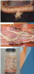
1. Caused by treating heavy worm burdens causing sudden death of many parasites. Causes impaction, rupture, peritonitis, intussusception. Treat via surgery
2. Caused by certain hay/tapeworms. Diagnosis via rectal or Surgery. Can treat medically/surgically
3. Secondary to functional obstruction causing increase in muscle mass and subsequent obstruction
4. Adhesions, Masses
Describe Anterior Enteritis (Duodenitis-proximal jejunitis) as a Functional Ileus of the SI in terms of:
1. Definition
2. Consequences/Pathophysiology
3. Causes
4. Clinical Signs
1. Inflammation + oedema of the duodenum + proximal jejunum
2. Inflammation + oedema → Excess fluid and electrolyte secretion of the SI → Intestinal hypersecretion + impaired motility → severe Reflux
3. Unknown. Suspected Clostridium, Salmonella, Fungal. Feeding conc. is risk factor
4. Pain relieved by gastric decompression, ↑ HR + RR, fever, Dilated SI +/- increased SI wall thickness
Describe Anterior Enteritis (Duodenitis-proximal jejunitis) as a Functional Ileus of the SI in terms of:
1. Peritoneal Fluid Analysis
2. Clinical Pathology
3. Ultrasound
4. Rectal
1. Yellow, turbid, serosanguinous if severe. Mild increased in total blood count with an increase in TP
2. Increased PCV/TP, Electrolyte/acid-base imbalances, secondary hepatopathy causing increased liver enzymes
3. Dilated, non-motile SI, thickened walls, peritoneal fluid
4. Distended loops of SI

Describe Anterior Enteritis (Duodenitis-proximal jejunitis) Treatment in terms of:
1. General Treatment
2. Prokinetic Drugs
3. Prognosis
4. Complications
1. Frequent gastric decompression, fluid/electrolyte replacement, ABs, Anti-endotoxic treatment, Parenteral nutrition
2. Lignocaine, Bethanechol. However usually ineffective
3. Good, may require surgery from manual decompression
4. Laminitis, adhesions, infarction of SI, peritonitis, cholangiohepatitis, renal failure

Describe Equine Proliferative Enteropathy as a Functional Ileus of the SI in terms of:
1. Definition
2. Consequences/Pathophysiology
3. CS
4. US
5. Clin Path
1. Disease of weanling foals. Caused by Lawsonia intracellularis (obligate intracellular bacterium)
2. Bacteria found in the cytoplasm of proliferative crypt epithelial cells of the SI → villous structure lost and replaced with crypt cells (hypertrophy) → hypersecretory + PLE
3. Oedema, variable fecal inconsistency, ill-thrift, colic
4. Thickened small intestine
5. Hypoptoeineamia (due to PLE)

Describe Equine Proliferative Enteropathy as a Functional Ileus of the SI in terms of:
1. Diagnosis
2. Treatment
3. Prognosis
1. Faecal PCR (specific but lacks sensitivity), Serum antibodies, Silver staining of tissue biopsy, culture
2. ABs, Supportive therapy (colloids, IV fluids, nutrition)
3. Good

Describe Inflammatory Bowel Disease (IBS) as a Functional Ileus of the SI in terms of:
1. Definition
2. Granulomatous Enteritis
3. Lymphocytic-Plasmacytic Enterocolitis
1. Collection of disorders characterised by profound cellular infiltration of the Si + LI
2. Chronic wasting condition in 2-3y horses. Causes partial/complete malabsorption, non-responsive to corticosteroids
3. Partial/complete malabsorption, not responsive to corticosteroids, Poor prognosis
Describe Inflammatory Bowel Disease (IBS) as a Functional Ileus of the SI in terms of:
1. Multisystemic Eosinophilic Epitheliotropic Disease
2. Eosinophilic Enterocolotis
1. Eosinophilic Infiltrative disease of the GIT + LN/Liver/Pancreas/Skin. Includes many eosinophilic disorders. Causes colic, skin conditions, liver disease (HEC), malabsorption, maldigestion
2. Segmental lesions in the small and large intestine. Not systemic
Describe Inflammatory Bowel Diseases (IBS) in terms of:
1. Clinical Signs
2. Pathophysiology
3. Treatment
4. Glucose Absorption Test
5. Other Diagnostics
1. Weight loss, colic, Oedema (PLE), diarrhoea, dermatitis (MEED)
2. Related to interaction of multiple immune functions → lack of immunity to normal flora
3. Corticosteroids, Immunosuppressants
4. Detects malabsorption by monitoring peak glucose levels in blood. IBS will have severely lower blood glucose c/w normal horse
5. Rectal biopsy, Laparoscopy, Carbohydrate absorption test,

Describe Alimentary Lymphosarcoma as a
IBS Functional Ileus of the SI in terms of:
1. Definition
2. Clinical Signs
3. Clin Path
4. Other Diagnostics
5. Treatment
1. Neoplasia of GALT with progressive SI involvement. in young horses
2. Anorexia, lethargy, peripheral oedema (PLE), +/- colic
3. Anaemia neutrophilia, lymphocytosis, hypoalbuminemia
4. Rectal palpation of enlarged LNs, abdominocentesis, biopsy
5. Corticosteroids + Supportive care
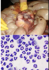
Describe Diarrhoea in terms of:
1. Definition
2. Pathogenesis
3. Causes
4. Acute Diarrhoea
5. Chronic Diarrhoea
1. Passage of faecal material with increased water content
2. Disruption of the normal balance of fluid + electrolyte secretion, and absorption in the GIT. Colon is the major site of fluid absorption in the GIT
3. In large animals Due to Large Intestine Disease. Can be caused by SI disease in foals
4. Severe dehydration and electrolyte abnormalities
5. Compensation for faecal losses occurs
Describe Diarrhoea in terms of:
1. Pathophysiology
2. Infectious Causes
1. Malabsorption, Increased Secretion, Decreased transit time, Osmotic overload, Increased hydrostatic pressure from blood to lumen
2. Bacteria: Salmonella, Clostridiums
Viruses: Coronavirus, rotavirus (Foals)
Parasites: Strongyles
Describe Diarrhoea in terms of:
1. Non-Infectious Causes
2. Non-Inflammatory Causes
3. Acute Colitis/Typhlitis
1. Toxicity (NSAIDs), AB induces, sand irritation, diet changes, peritonitis, IBS
2. Increased interstitial hydrostatic Pressure (CHF, Liver disease), Increased intraluminal osmolarity, Dietary, Sand
3. Active inflammation within the colon is associated with local & systemic pathophysiology events (Coagulopathy, impaired cardiopulmonary function, sequelae of inflammatory mediator cascade and sepsis

Describe Diarrhoea in terms of:
1. Signalment
2. History
3. Main Clinical Signs
4. GI Auscultation
5. Rectal Palpation
1. Young Horses (Clostridial Infections), Performance horses (NSAIDs)
2. Recent feed change, no. animals involved (Toxic/infectious), Prior AB administration
3. Diarrhoea of variable volume/consistency, colic (if severe/abdominal distension), fever, signs of endotoxemia (Compromised GIT wall), dehydration, Hgh HR + RR, Ventral oedema (hypoproteinemia)
4. Hypermotile, absent/hypomotile in early disease, abdominal distension may be present
5. Fluid filled colon+caecum, gas distention variable

Describe Diarrhoea Diagnosis in terms of:
1. Haematology Clin Path
2. Biochemistry Clin Path
3. Abdominal Rads
4. Abdominal US
1. Dehydration, leukopenia (infectious +/- endotoxin, hyperfibrinogenemia
2. Metabolic/Lactic Acidosis, Hypocalcaemia, Hypoalbuminemia, Azotemia (pre-renal), Increased liver enzymes
3. Sand, enteroliths, mineral FB
4. Peritoneal fluid, SI distension, LI wall thickness/fluid content

Describe Diarrhoea Diagnosis in terms of:
1. Abdominocentesis
2. Rectal Mucosal Biopsy
1. Infarction of the caecum/colon. PEritonitis
2. Culture, cyathostomes, histopath (IBD)

Describe Faecal Diagnostics in Diarrhoea in terms of:
1. Gross Appearance
2. Aerobic Culture
3. PCR
4. Faecal Toxin Tests
5. Microscope Analysis
1. Consistency, Parasites
2. Salmonella
3. Salmonella, Coronavirus, Rotavirus
4. Enterotoxin, Clostridiales
5. Gram +ve/-ve imbalance, presence of faecal leukocytes
Describe Diarrhoea Treatment in terms of:
1. Fluid Therapy
2. Anti-Endotoxin Therapy
3. AB Therapy
4. Toxin-Binding
5. Prophylactic Digital Cryotherapy
1. IV. Colloids beneficial (Replenishes protein + may restore oncotic pressure)
2. NSAIDs, Aspirin, Polymyxin B
3. Avoid except for Clostridiosis, substantial leukopenia, and peritonitis
4. Di-Tri-Octahdralmsmectite (intestinal adsorbent), activate charcoal, Bismuth subsalicylate, Mineral oil
5. Prevents activation of matrix metalloproteinases that disrupt laminar attachment and subsequent laminitis

Describe Diarrhoea Treatment in terms of:
1. Re-Establishment of Normal GIT Flora
2. Supportive Care
3. Nutritional Support
1. Faecal Transplantation (Faces from healthy horse administered via NGT in warm fluids)
2. Isolation, Tail-wrap + perineal care, Heavy Bedding +/- leg wraps
3. Severe catabolic disease. Need easily digestible frequent meals. Minimise CHO, SCFAcids for enhanced mucosal repair

Describe Complications of Diarrhoea
- Laminitis
- Haemodynamic renal failure
- Cholangiohepatitis
- Peritonitis

Describe Salmonellosis as a Causes of Diarrhoea in terms of:
1. Bacterial Characteristics
2. Risk Factors
3. Clinical Presentations
1. Gram -ve anaerobe, intracellular, Extremely Infectious
2. Change in Gut Flora (Transportation, ABs, Diet, Surgery, NGT, Concurrent Gi disease)
3. a. Abortion
b. Subclinical Carrier
c. Mild clinical disease
d. Severe Clinical Disease

Describe Salmonellosis as a Causes of Diarrhoea in terms of:
1. Severe Clinical Disease CS
2. Severe Clinical Disease Prognosis
3. Mangement
1. Watery foul smelling diarrhoea, dehydration, metabolic acidosis, electrolyte losses, Acute onset anorexia, depression +/- colic, Profound neutropenia + haemoconcentration, PLE, Bacteraemia
2. Dependant on serotype (differ in pathogenicity). Typically good with early intervention
3. Zoonotic! Isolate (Animals shed for 30d), Biosecurity (wash hands, footbaths), Thorough cleaning of areas of faeces and organic debris

Describe Clostridium spp. as a Causes of Diarrhoea in terms of:
1. Characteristics
2. Clostridium Perfringens
1. Gram +ve anaerobe, spore forming rods, secrete endotoxins, found in soil, environment water, feed
2. Normal Flora in GIT (Usually controlled by other gut flora). More common with enterocolitis in neonate, rare in adults. Endotoxins cause acute diarrhoea and types of C. perfringens secrete different endotoxins (alpha, beta, epsilon, iota). Effects previously healthy vigorous foals, High case fatality

Describe Clostridium spp. as a Causes of Diarrhoea in terms of:
1. Clostridium Difficile Characteristics
2. Clostridium Difficile CS
3. Clostridium Difficile Treatment
4. Clostridium Difficile Prognosisv
1. Rarely isolated from normal horses, found in 40% of horses with diarrhoea. Risk factors include AB therapy, Management factors, macrolide treatment, hospitalisation, Age, geography.
2. Produces cytotoxin + endotoxin. green-brown diarrhea, abdominal distention, scant faeces (+/- haemorrhage), colic, fever, depression.
3. Metronidazole/Penicillin drug of choice
4. 25% mortality, better prognosis c/w C. perfringens
Describe Larval Cyathostomiasis as a Causes of Diarrhoea in terms of:
1. Definition
2. Clinical Signs
3. Life Cycle/Pathogenesis
1. Small strongyles, may cause hypobiosis cysts in the LI wall
2. Acute diarrhea, recurrent diarrhoea, rapid weight loss + oedema, non-specific Colic, intussusceptions
3. L3 hypobiotic in LI cysts (can secrete noxious chemicals promoting inflammation and diarrhoea) → synchronous emergence of L4 larvae → mucosa disruption → inflammatory mediatiors → shedding/ulceration of intestinal mucosa/blood loss/oedema → increases active/passive secretion of electrolyes protein and fluid → diarrhoea
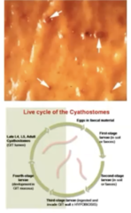
Describe Larval Cyathostomiasis as a Causes of Diarrhoea in terms of:
1. Diagnosis
2. Treatment
3. Prevention/Management
1. Gross examination of faeces, Faecal wet preparation, Biopsy. FEC don't correlate with larval burden
2. Anthelmintic (Fenbendazole, Moxidectin). Corticosteroid: (For inflammation)
3. Correct deworming programs, maintain refugia, only treat high egg shedders, pasture management (remove manure)