Anatomy of Articulation and Resonance
 Call Kai
 Learn
 Practice Test
 Spaced Repetition
 Match
 Flashcards
 Knowt Play
1/149

There's no tags or description
Looks like no tags are added yet.
150 Terms
Vocal tract
nasal passages, oral cavity, larynx
What connects the nasal passages, oral cavity, larynx?
Pharynx


Articulation
process of joining or moving two elements together
Articulation for speech
process of bringing two or more structures together to form the sounds of speech, combinations of movements form differently shaped spaces in the vocal tract, phoneme quality is determined by the size and shape of those space
Mobile articulators
mandible (lower jaw including teeth), lips, tongue, velum, vocal folds
Immobile articulators: maxillae (upper jaw including upper teeth), hard palate, alveolar ridge
Bones of the face
mandible, maxillae, nasal bones, palatine bones, vomer, zygomatic bone
Bones of the cranium
frontal, parietal, occipital, temporal, ethmoid, sphenoid


Mandible
considered unpaired.
important features: ramus, coronoid process, condylar process, mylohyoid line, mental symphysis, dental alveoli (alveolar process)


Maxillae
bones of upper jaw and parts of nose, palate, etc. Processes are named for the structures with which they articulate.
Identify: palatine process, incisive foramen, canine fossa, intermaxillary suture, premaxillary suture


Nasal bones
bridge of your nose, make up superior anterior nasal surface (form upper margin of nasal cavity)

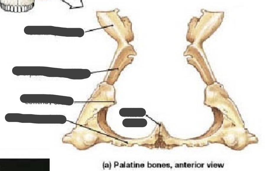
Palatine bones
posterior to maxillae, perpendicular/vertical plate (posterior wall of nasal cavity), horizontal plate (make up rest of pard palate posterior ¼ section of hard palate)

Vomer
inferior/posterior to nasal bone, makes up part of nasal septum


Zygomatic bone
forms the cheekbones, anterior portion attaches muscle that elevate the upper lip.
(Zygomatic arch: temporal process of zygomatic bone and zygomatic process of temporal bone)


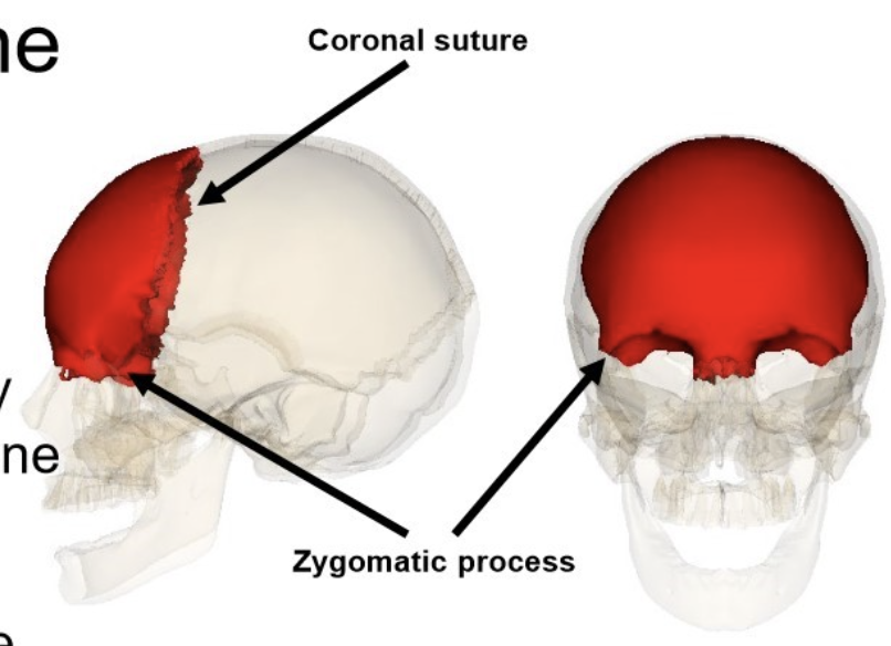
Frontal bone
unpaired.
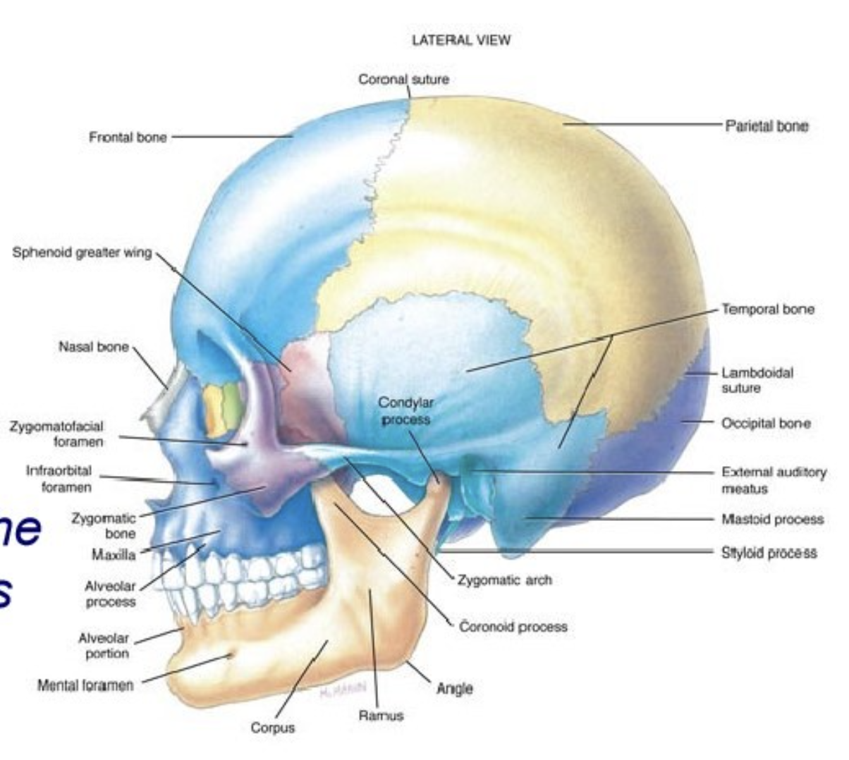
Coronal suture forms boundary with parietal bone.
Articulates with zygomatic bone at zygomatic process.

Parietal
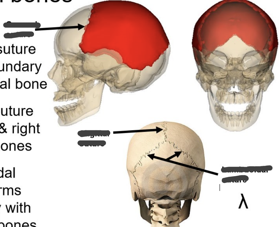
paired.
Coronal suture forms boundary with frontal bone.
Sagittal suture joins left & right parietal bones.
Lambdoidal suture forms boundary with occipital bones.


Occipital
unpaired.
Forms the skull base.
Lambdoidal suture forms boundary with parietal bones.
Foramen magnum allows medulla/spinal cord to exit skull.
Condyles are the surfaces that articulate w/ atlas bone.


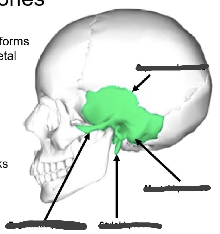
Temporal
Squamous portion (lower margin includes EAM, zygomatic process)
Tympanic portion (includes anterior and inferior walls of EAM, styloid process)
Mastoid portion (posterior part of the bone, mastoid process)
Petrous portion (houses cochlea and semicircular canals, internal auditory meatus)



Ethmoid
[ethmos = strainer]
unpaired bone
Cosre of skull & face
Cribiform plate separates nasal cavities from brain
Has many processes (nasal conche, etc)
Perpendicular plate part of bony nasal septum


Sphenoid bone
unpaired bone.
Butterfly shaped [corpus, greater wings, lesser wings, pterygoid plates (fossa between medial & lateral house attachment points for muscles of mastication and tensor veli palatini)]

Temporomandibular Joints
synovial (mobile) joint, condyle of mandible fits into cavity in temporal bone separated by an articular disk, joint is surrounded by fibrous capsule and lubricated with synovial fluid
Oral cavity
lips to faucial pillars, floor of mouth to hard/soft palate
Faucial pillars
transition between oral cavity & pharynx, two sets: anterior & posterior, palatine tonsils between ant. & post.
(shadow monster)


Buccal cavity/sulci
space between teeth and cheeks
role in oral resonance
phoneme (ex: lateral fricative)
collects food in disordered feeding
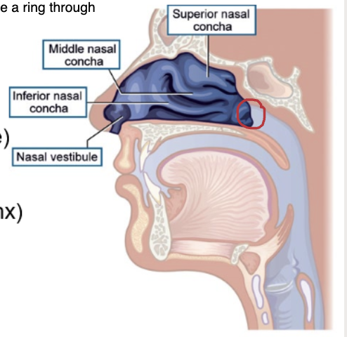
Nasal cavities/passages
formed by maxillae, palatine bones, nasal bones, vomer, perpendicular plate of ethmoid (nasal septum), ethmoid (roof of nasal cavity, and mucosa)
Anterior, mid-portion, posterior
Functions: filter/warm/humidify air, immune system, speech-related (necessary for nasal sounds, natural part of resonance in non-nasal)
Anterior cavity (nasal)
nares
Mid-portion cavity (nasal)
nasal conchae aka turbinates
nasal meatus: passage between conchae
Posterior cavity (nasal)
nasal choanae connection to nasopharynx

Pharyngeal cavity (pharynx)
Vertical tube ~12 cm long (lower boundary = vocal folds)
Shape altered: velopharyngeal muscles, pharyngeal constrictor muscles, muscles for laryngeal elevation and depression
Includes nasopharynx, oropharynx, laryngopharynx
Nasopharynx
from velum choanae, houses pharyngeal tonsils (adenoids)
Oropharynx
from hyoid to velum
laryngopharynx
from VFs to hyoid


Micrognathia
developmental condition of small mandible or retrognathia, not enough room for tongue on floor of mouth
may cause tongue to move up into the maxillary/palatine are during development resulting in cleft palate, even if not cleft it may still result in functional articulation problems
Cleft lip cause/risk
premaxillary suture fails to fuse in utero (weeks 6-12), may or may not include alveolar process of the maxilla
Cleft lip effect of articulation/swallowing
infant feeding (difficulty making seal around nipple of breast/bottle. articulation affected mildly compared to cleft palate, good outcomes with surgical repair, orthodontics, etc.
resonance not usually affected.
Maxillary deficiency/hypoplasia causes/risk
repaired cleft palate
fetal alcohol syndrome
Crouzon syndrome
Maxillary deficiency/hypoplasia treatment
maxillary advancement surgery post-adolescent
Bell’s Palsy define
facial paralysis (usually temporary) resulting from damage or trauma to the facial nerve (CN-VII)
Bell’s Palsy cause/risk
unknown, possibly due to virus
Cleft lip with/without cleft palate: twice as many ____ as ____
twice as many males as females
Isolated cleft palate:
____ as common as lip
twice as many ____ as ____
half as common as lip
twice as many females as males
CL +/- CP are more common in ____ than Black or Latino pop
White
Cleft Palate cause/risk
no single cause.
Genetic factors (20% of cases).
Environmental in utero: smoking, maternal diabetes, medications while pregnant
Cleft Palate Treatment
numerous surgeries, orthodontics
Furlow's Palatoplasty:
"double–opposing z-plasty, used to lengthen tissue by borrowing tissues from width of velum to add length
Pharyngeal Flap:
most common procedure used for VP insufficiency, effective for midline gaps, flap is raised from the posterior pharyngeal wall and sutured to velum
Pharyngeal Wall Augmentation
filler is injected ton the VP opening (filler could be hydroxyapatite, cartilage, fascia, fat, silicone, and protoplast)
Sphincter Pharyngoplasty
Narrowing the VP opening by rearranging myomucosal flaps raised from the posterior tonsillar pillars. Posterior pharyngeal walls are attached, Patient with poor PPW movement and smaller VP ports benefit more from this procedure
Occlusion:
the process of bringing upper and lower teeth (maxillary and mandibular into alignment)
Class 1 Occlusion
normal orientation of mandible and maxillae, 1st molar of mandibular arch is one-half tooth advanced relative to 1st maxillary motor, needed for mastication and articulation

Class 2 Malocclusion
relatively retracted mandible, first mandibular molars are RETRACTED at least one half tooth from first maxillary molars

Class 3 Malocclusion
relatively advanced mandible, first mandibular molars are ADVANCED more than one half tooth from first maxillary molars


Overbite:
How far your upper front teeth vertically overlap your lower front teeth when your jaw is closed
Overjet:
How far your upper front teeth horizontally protrude or stick out forward past your lower front teeth
Palatal lifts:
helps a weak or paralyzed soft palate move into proper position

Palatal Obturator:
closes and opening or gap in the palate

Speech Bulb:
Fills the space between the soft palate and the pharyngeal walls (used when soft palate too short)

Velopharyngeal Dysfunction (VPD):
general term for disorder of the VP valve that implies faulty VP closure
Velopharyngeal Insufficiency (VPI):
abnormal structure.
Anatomical cause: Short palate, deep pharynx, post-adenoidectomy or pharyngeal tumor resection.
Treatment includes surgical reconstruction
Velopharyngeal Incompetence (VPI):
abnormal function.
Physiological cause: Reduced movement of soft palate, pharyngeal hypotonia, velar paralysis or paresis.
Treatment includes surgery, prostheses, or speech therapy
Resonance disorders:
opening, inconsistent movement, or obstruction that changes the way air flows through nasal and oral cavities during speech
Hypernasality:
too much nasal resonance (ex: submucous cleft)
Hyponasality:
too little nasal resonance (ex: hypertrophic adenoids)
Cul-de-sac resonance:
sound "trapped" in one of the cavities (oral, nasal, pharyngeal)
How velopharyngeal dysfunction can affect oral pressure consonants and produce compensatory articulation patterns?
Speech sounds requiring build-up of intraoral pressure most affected (stops, fricatives, affricates).
Place of articulation – all paces possible
Hypernasality and/or nasal air emissions
Replacement of oral consonants with
mid-palatal or glottal stops
pharyngeal or nasal fricatives
Sinuses:
air filled cavities lined with respiratory epithelial cells (mucosal membranes). Produce mucus, cleared through ciliary action.
Drains into nasal passages
What are the four sinuses?
Frontal
Maxillary (largest)
Ethmoid
Sphenoid
Function of Sinuses
Mechanical protection/filter
Immune function
Humidify and warm inhaled air
Lighten skull wight
Provide a cushion against facial trauma
Enhance voice resonance


Eustachian tube:
connects nasopharynx with middle ear (courses slightly up from nasopharynx to ear, but more horizontal course in children)
Closed in relax state, can be opened with pharyngeal/velar muscles (tensor veli palatini)
Eustachian Tube Function:
aeration/air pressure equalization of middle ear
Surface features of the lips and nose
Columella
Philtrum
Philtril ridge
Cupid's bow


Orbicularis oris
Function: brings corners of lips together, contributes to: lip rounding, constricts oral opening, protrusion of lips, press lips against teeth
Attachments: corners of lips
Course: around the lips
Nerve: CN VII (Fascial nerve)
Risorius
Function: retracts lips at corners, supports feeding & smiling/grinning
Attachments: fascia of the masseter muscles, into corner of the moth and lower lip
Course: forward (post/ant)
Nerve: CN VII (Fascial nerve)
Buccinator
Function: flattens cheek, assists in retraction of corners of mouth (keeps food on molars, helps constrict oropharynx)
Attachments: pterygomandibular ligament, corners of mouth (orbicularis oris)
Course: forward, deep to the risorius
Nerve: CN VII (Fascial nerve)
Muscles that elevate the upper lip:
levator labii (superioris and superioiris alaque nasi), Zygomatic minor, Zygomatic major, Levator anguli oris
Lavator labii (superioris and superioiris alaque nasi)
Function: elevated upper lip
Attachments: Maxilla (superioris: infraorbital margin, superioiris alaque nasi: frontal process), mid-lateral region of upper lip
Course: down
Nerve: CN VII (Fascial nerve)
Zygomatic minor
Function: elevates upper li
Attachments: facial surface of zygomatic bone, mid-lateral upper lip
Course: down and medial
Nerve: CN VII (Fascial nerve)
Zygomatic major
Function: elevates and pulls bac (retracts) angles of mouth
Attachments: lateral to zygomatic minor, corner of lips
Course: down and medial
Nerve: CN VII (Fascial nerve)
Levator anguli oris
Function: draws corners of lips up and medial
Attachments: from canine fossa of maxilla to insert into corners of upper and lower lips
Course: deep to levator labii sup., down and lateral
Nerve: CN VII (Fascial nerve)
Depressor labii inferioris
Function: pulls lower lip down
Attachments: mandible at oblique, lower lip
Course: up into lower lip
Nerve: CN VII (Fascial nerve)