BIO Lab 6 - 2.6, 2.7, 2.8 (synovial joints, range of motion, knee joint, shoulder joint)
 Call Kai
 Learn
Learn Practice Test
 Spaced Repetition
 Match
Match Flashcards
 Knowt Play
1/56

There's no tags or description
Looks like no tags are added yet.
Name | Mastery | Learn | Test | Matching | Spaced | Call with Kai | Chat |
|---|
No analytics yet
Send a link to your students to track their progress
57 Terms
synovial joints
freely movable joints (diarthrosis)

joint cavity
the space between two connecting bones

articular cartilage
covers the surfaces of bones where they come together to form joints
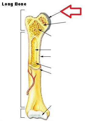
hyaline cartilage
Most common type of cartilage; it is found on the ends of long bones, ribs, and nose
joint capsule
fibrous tissue that encloses bone ends and other joint surfaces
dense irregular collagenous connective tissue
Protein fibers arranged in a randomly oriented network which provides strength and structural reinforcement for the joint
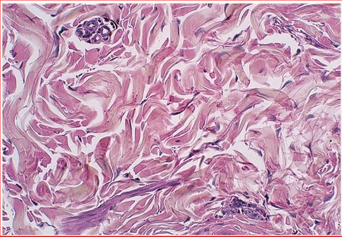
synovial membrane
connective and epithelial tissue, lining of a joint that secretes synovial fluid into the joint space.

synovial fluid
viscous, joint-lubricating fluid secreted by the synovial membrane

what are the functions of synovial fluid
fills the join cavity, reduces friction, exchanges oxygen, nutrients, and wastes with the cells of articular cartilage
ligaments
attach bone to bone

dense regular collagenous connective tissue
A type of connective tissue proper that contains collagen fibers arranged in regular bundles; found in locations such as tendons.

extrinsic ligaments
external to joint capsule
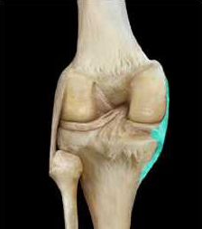
intrinscic ligaments
embedded inside the joint capsule
articular discs
Another term for menisci (singular = meniscus), the fibrocartilage pads between opposing surfaces in a joint, prevent dislocation
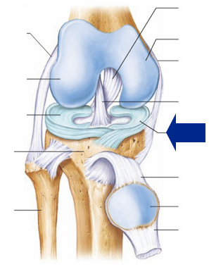
tendons
surround synovial joints, made up of dense regular collagenous connective tissue, attach muscle to bone

bursa
fluid-filled sac that allows for easy movement of one part of a joint over another, located between joints, reduces friction

tendon sheaths
Elongated bursa wrapped completely around tendon subjected to friction
inflammation of bursa
bursitis
joint capsule
what is this?

joint cavity
what is this?

synovial membrane
what is this?
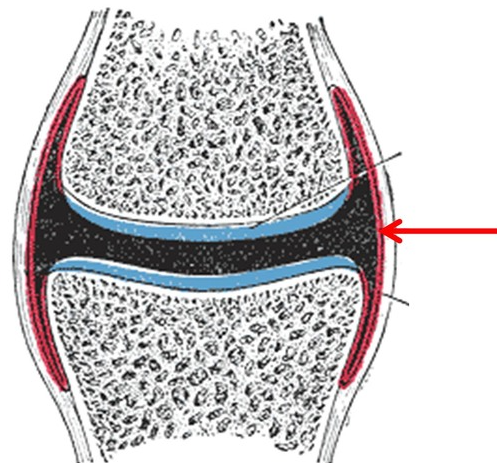
synovial fluid
what is this?

extrinisc and intrinsic
what are each of these ligaments?

articular discs, menisci
what is this?
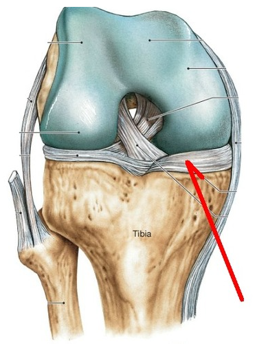
tendon sheath
what is this?

the range of motion of a synovial joint is described in terms of an invisible ___________
axis
synovial joint movements are classified according to the ____________
number of planes of motion in which the bones can move
nonaxial joints
allow motion to occur in one or more planes but do not move around an axis, simply glide past eachother

uniaxial joints
only permits flexion and extension, hinge, pivot

biaxial joints
allow motion around two axes, flexion extension and abduction adduction

multiaxial joints
greatest range of motion, shoulder, hip

plane/gliding joint
both surfaces of the articulating bones are either flat or slightly concave and convex

condylar joint
one articular bone has an oval convexed surface that fits into a similarly shaped depression on the other bone, outward curve sliding on a depression that matches its shape

saddle joint
both articulating bones have saddle like surfaces that concave in one direction and convex in the other

hinge joint
one surface of the articulating bone is convexed and the others is concave

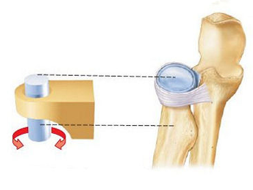
pivot joint
bone spins or rotates in a longitudional axis. the moving bone rotates within a ligament and the bone it is articulating with
ball and socket joint
one bone has a smooth hemispherical surface that fits into a cuplike socket, only found in the shoulder and hip joints

what is the knee bone made up of
medial and lateral femoral condyles and the tibial plateau
how is the knee joint stabilized
two extrinsic ligaments (tibial and fibular collateral ligaments) and two intrinsic ligaments (anterior and posterior cruciate ligaments)

cruciate ligaments
controls the front and back movement of the knee joint, secures from any unwanted movement, means shaped like a cross

Anterior Cruciate Ligament (ACL)
extends from the anterior tibial plateau to the posterior part of the lateral femoral condyle, prevent hyperextension of the knee
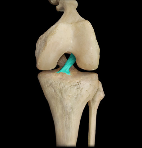
Posterior Cruciate Ligament (PCL)
extends from the posterior tibial plateau to the anterior part of the medial femoral condyle, it crosses under the ACL to form an X, PCL prevents posterior displacement of the tibia bone on the femur bone

collateral ligaments
controls the side to side movement of the knee joint and secures it from any unwanted movements, collateral means they are found on the sides of the joint.

tibial collateral ligament
medial collateral ligament (MCL), extends from the medial tibia bone to the medial femur bone, resists stresses that pull the tibia bone laterally on the femur bone

fibular collateral ligament (LCL)
lateral collateral ligament extends from the lateral fibular bone to the lateral femur bone, resists stresses that pull the tibia bone medially on the femur bone

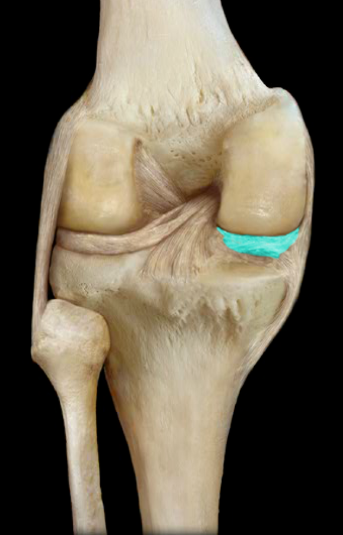
medial and lateral menisci
half-moon shaped articular discs are located on the outer edges of the tibial plateau and they improve the fit of the tibia and femur bones, the tibial collateral ligament attaches to the medial meniscus
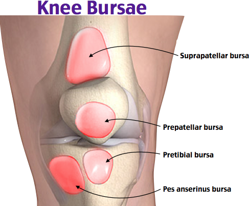
suprapatellar, deep infrapatellar, subcutaneous prepattelar bursae
concentrated around the patella bone, bone generates a great deal of friction over the femur bone during motion, they can become inflamed as a result of repetitive activities
what kind of joint it the glenohumeral joint?
multiaxial ball and socked joint that consists of the humeral head and glenoid cavity
glenoid labrum
fibrocartilaginous ring along the rim of the glenoid cavity that improves the fit of the humeral head in the glenoid cavity

3 glenohumeral ligaments
reinforces the anterior join capsule, they are weak and sometimes absent
coracohumeral ligament
located in the anterior articular capsule between the greater tubercle and coracoid process

coracoacrmial ligament
located between the coracoid process and acromion

transverse humeral ligament
extends between the greater and lesser tubercles which forms a tunnel that houses one of the tendons of the biceps brachii muscle

biceps brachii tendon
- stabilizing force found in the anterior arm
- passes through the articular capsule of the shoulder joint on its way to its attachment point on the scapula
-also a tendon sheath wrapped around one of its tendons
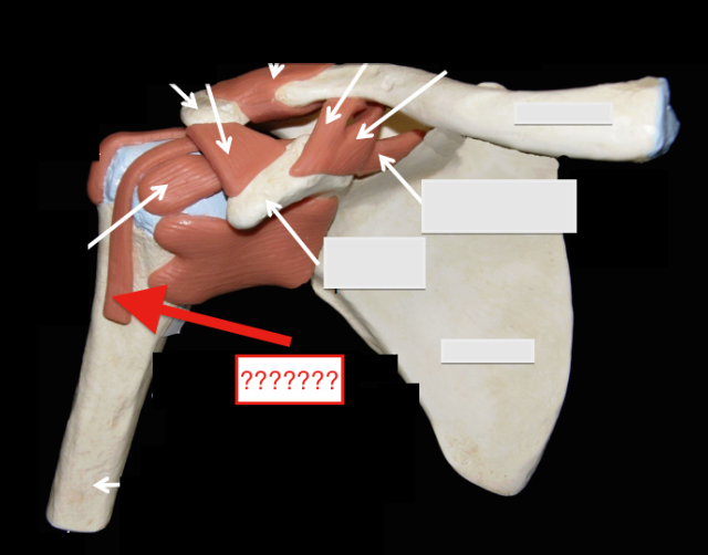
rotator cuff
a major stabilizing force of the shoulder joint, 3 posterior supraspinatus, infraspinatus, teres minor, 1 anterior subscapularis,

subacromial bursa
one of the largest bursae located just below the acromion

subscapular bursa
bursa that prevents rubbing of the subscapularis muscle tendon against the scapula
