2.1.1. Cell structure

0.0(0)
Studied by 0 people
call kaiCall Kai
learnLearn
examPractice Test
spaced repetitionSpaced Repetition
heart puzzleMatch
flashcardsFlashcards
GameKnowt Play
Card Sorting

1/29

encourage image

There's no tags or description

Looks like no tags are added yet.

30 Terms

1
New cards

what things must be ensured for biological drawings

Biological drawings are line drawings that focus on particular features of a specimen:

  • Drawings should be accurate, reflecting the proportions of the observed structures as closely as possible.

  • drawing is centrally placed on the page, with sufficient space for labels.

  • Use as much of the paper as possible.

  • Include a title / heading.

  • Using a sharp pencil, draw using continuous lines with no shading, on plain paper.

  • Draw clearly defined structures.

  • Include a scale bar.

  • State the magnification used.

  • Label lines should be drawn with a ruler and should not overlap each other.
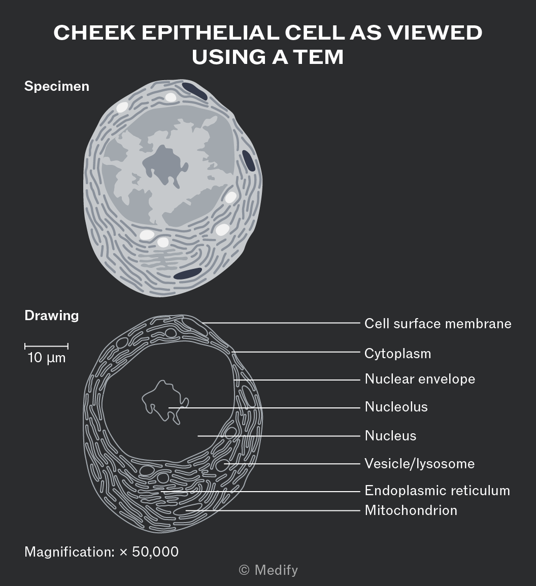
The image shows a cheek epithelial cell as viewed using a TEM. At the top, there is a labeled 'Specimen' which is an illustration of a cell with visible internal structures. Below is a labeled 'Drawing' of the same cell with detailed annotations. The drawing includes a scale bar indicating 10 µm and labels pointing to specific cell parts: Cell surface membrane, Cytoplasm, Nuclear envelope, Nucleolus, Nucleus, Vesicle/lysosome, Endoplasmic reticulum, and Mitochondrion. The magnification is noted as × 50,000.


2
New cards

anatomy of nucleus

The nucleus is usually the largest organelle

  1. containing the cell’s genetic material (DNA).

  2. The nucleus controls cell activities, including growth and metabolism, by regulating gene expression and protein synthesis.

  3. It contains chromatin, consisting of linear DNA wrapped around histone proteins, which condenses to form chromosomes during cell division.

  4. The nucleolus is a dense, spherical region within the nucleus that synthesises ribosomal RNA (rRNA) and assembles ribosomal subunits.


3
New cards

anatomy of nuclear membrane

The nuclear envelope is a double membrane

  1. surrounding the nucleus

  2. containing nuclear pores

  3. that regulate the movement of substances such as mRNA and proteins between the nucleus and cytoplasm.
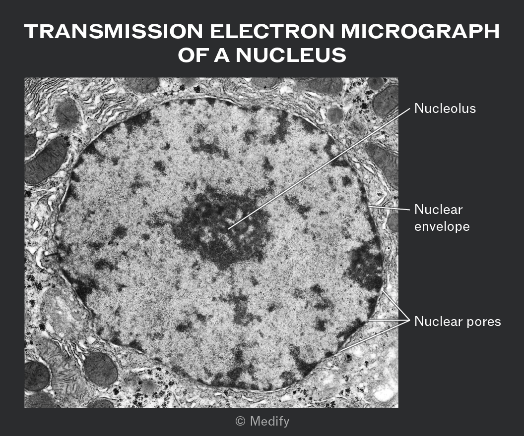

A transmission electron micrograph of a cell nucleus is shown. The image features a large, circular structure occupying the center, labeled with three annotations. The first annotation points to a dense, dark area within the circle labeled 'Nucleolus.' The second annotation points to the boundary of the circle labeled 'Nuclear envelope.' The third annotation points to small openings along the boundary labeled 'Nuclear pores.' The background shows a textured, grainy appearance typical of cell ultrastructure. The title above the image reads 'TRANSMISSION ELECTRON MICROGRAPH OF A NUCLEUS.'


4
New cards

anatomy of RER, function

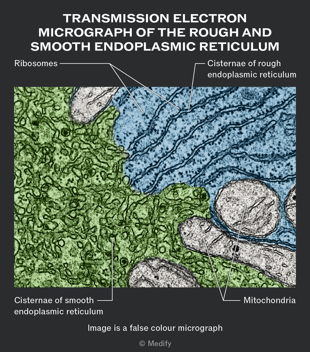
  1. The rough endoplasmic reticulum (RER) is a network of interconnected, flattened membrane sacs

  2. called cisternae,

  3. which are continuous with the nuclear envelope.

  4. Ribosomes attached to its surface give it a rough appearance.


The RER is the site of synthesis and transport of proteins destined for secretion or insertion into cell membranes.

  1. Proteins enter the cisternae,

  2. where they are folded and modified

  3. before being transported to the Golgi apparatus

  4. for further processing.


  5. A false color transmission electron micrograph of the rough and smooth endoplasmic reticulum. The image is labeled with 'Ribosomes' pointing to small dark dots, 'Cisternae of rough endoplasmic reticulum' pointing to a blue shaded region with dark lines, 'Cisternae of smooth endoplasmic reticulum' pointing to a green shaded region with a textured pattern, and 'Mitochondria' pointing to oval-shaped structures with a granular interior. The image description at the bottom reads 'Image is a false colour micrograph'.


5
New cards

anatomy of SER

The smooth endoplasmic reticulum (SER) is a network of membranous tubules and sacs which extends throughout the cytoplasm.

It is similar in structure to the rough endoplasmic reticulum, but lacks attached ribosomes and therefore appears smooth.

The SER is involved in the synthesis and transport of lipids, including phospholipids, cholesterol and steroid hormones.

6
New cards

anatomy of Golgi, function

  1. The Golgi apparatus is a stack of flattened, single membrane–bound sacs called cisternae.

  2. It modifies proteins and lipids received from the endoplasmic reticulum, for example, by adding carbohydrate chains.

  3. These molecules are then sorted and packaged into vesicles

  4. for transport to the cell surface

  5. for exocytosis or to other organelles, such as lysosomes.
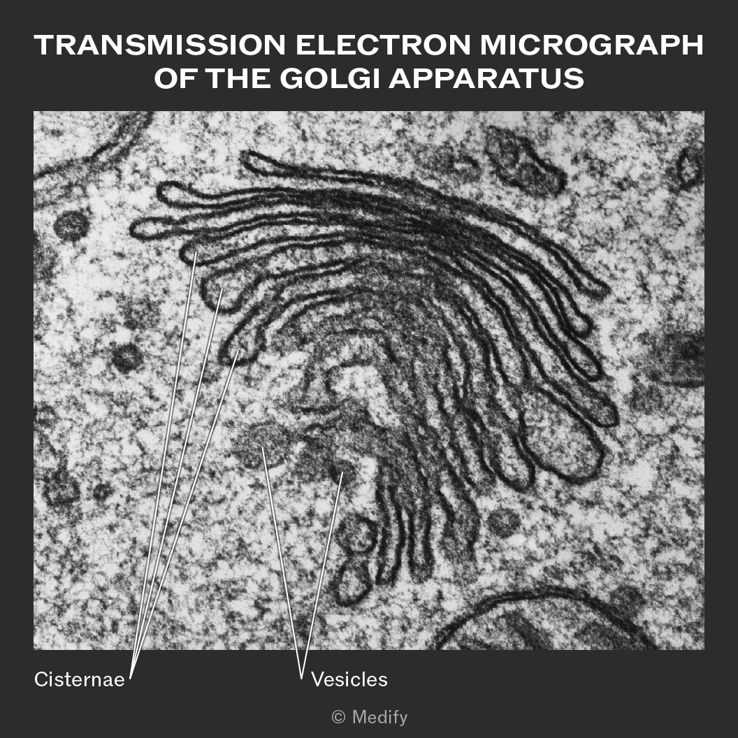


A transmission electron micrograph of the Golgi apparatus is shown. The image features a series of stacked, curved, and flattened membrane-bound structures labeled as 'Cisternae' with lines pointing to them. Smaller round structures next to the cisternae are labeled as 'Vesicles.' The surrounding area shows a textured background typical of cellular environments. The title at the top reads 'TRANSMISSION ELECTRON MICROGRAPH OF THE GOLGI APPARATUS.'


7
New cards

anatomy structure of ribosome

  1. Ribosomes are small,

  2. non–membrane–bound organelles made of ribosomal RNA (rRNA) and protein.

  3. They are found free in the cytoplasm or attached to the rough endoplasmic reticulum and are the site of protein synthesis.
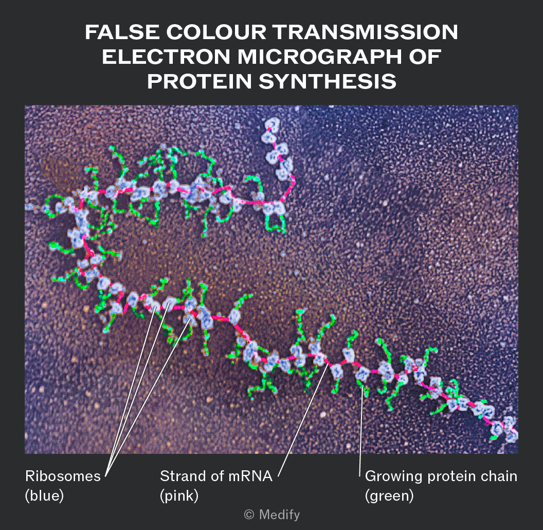

False colour transmission electron micrograph of protein synthesis. The image shows a complex structure with colors indicating different components. Ribosomes are highlighted in blue, a strand of mRNA is shown in pink, and the growing protein chain is in green. The background has a textured appearance. Labels point to each component: 'Ribosomes (blue)' pointing to blue structures, 'Strand of mRNA (pink)' pointing to the pink strand, and 'Growing protein chain (green)' pointing to green segments. © Medify.


8
New cards

anatomy structure of mitochondria

  1. Mitochondria are double membrane–bound organelles

  2. that are the site of the later stages of aerobicrespiration and ATP production.

  3. Cells with high metabolic activity, such as muscles and neurones, contain many mitochondria.

  4. The inner membrane is folded into cristae, increasing the surface area for respiratory enzymes and electron carriers.

  5. Mitochondria with more densely folded cristae have a greater capacity for aerobic respiration and ATP production.

  6. The fluid-filled interior is called the matrix and contains enzymes, mitochondrial DNA and ribosomes.
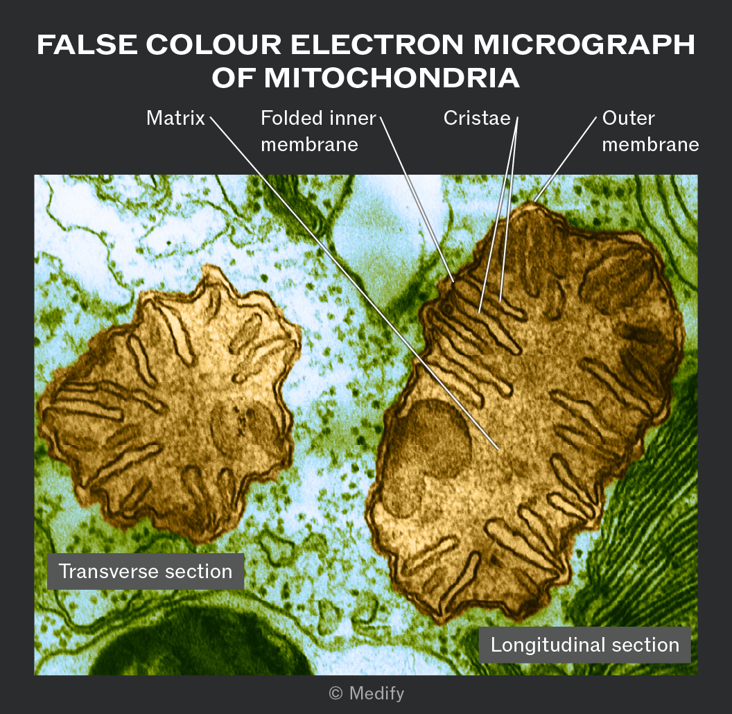

A false color electron micrograph of mitochondria is shown with two sections labeled. The left section is labeled 'Transverse section' and the right section is labeled 'Longitudinal section.' Annotations point to different parts of the mitochondria: 'Matrix' is labeled on the left side, 'Folded inner membrane' is labeled near the center, 'Cristae' is labeled towards the right, and 'Outer membrane' is labeled on the far right. The image is dominated by brown and greenish hues, showing the internal structure with folds and membranes. The title at the top reads 'FALSE COLOUR ELECTRON MICROGRAPH OF MITOCHONDRIA.'


9
New cards

structure anatomy of lysosome

  1. Lysosomes are single membrane–bound vesicles

  2. formed from the Golgi apparatus

  3. that contain hydrolytic (digestive) enzymes, including lysozymes enzyme


  4. They digest ;

  5. cellular debris

  6. worn-out organelles

  7. pathogens such as bacteria,

  8. and also play a role in apoptosis (programmed cell death).

Lysosomes are particularly abundant in phagocytic cells such as macrophages and neutrophils.
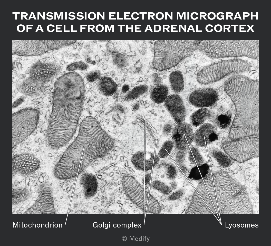

Transmission electron micrograph of a cell from the adrenal cortex. The image shows various cellular structures labeled with lines pointing to them. A mitochondrion is labeled on the left, characterized by an elongated, oval shape with distinct internal ridges. A Golgi complex is labeled in the center of the image, appearing as a stack of flattened membrane sacs. Several lysosomes are labeled on the right, identified as small, roughly spherical structures. The background shows a dense network of cellular material. © Medify is noted at the bottom.


10
New cards

antaobmy function of cell surface membrane

  1. The cell–surface membrane, also called the plasma membrane, separates the cell’s contents from the environment surrounding the cell.

  2. The partially or selectively permeable membrane controls the movement of substances

  3. and is involved in cell signalling and recognition.

  4. The membrane consists of a phospholipid bilayer

  5. containing proteins, cholesterol and carbohydrate-containing molecules.
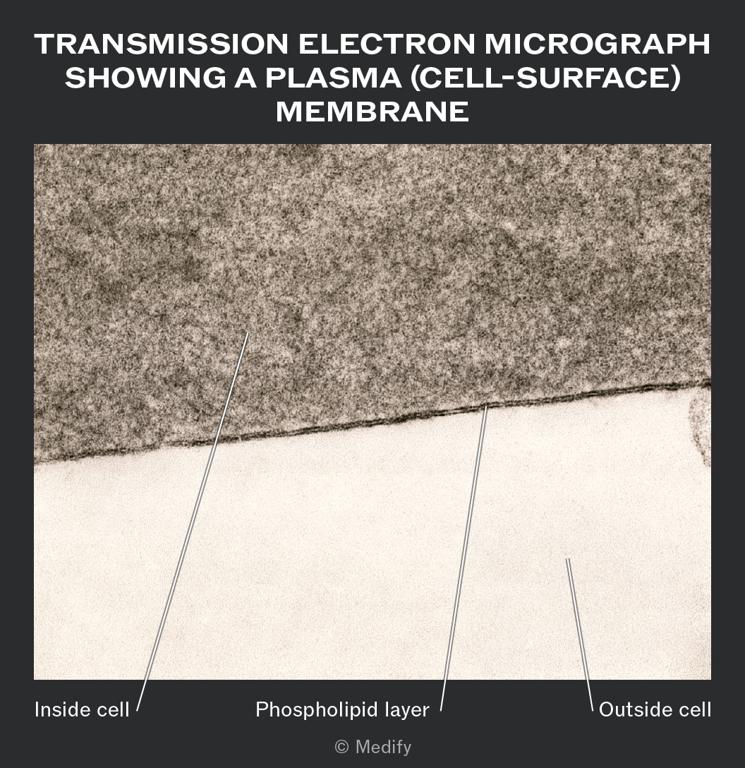

Image titled 'Transmission Electron Micrograph Showing a Plasma (Cell-Surface) Membrane.' The micrograph displays a section of a cell membrane, with labeled parts: 'Inside cell' pointing to a darker area, 'Phospholipid layer' pointing to a thin line between the dark and light areas, and 'Outside cell' pointing to a lighter area. © Medify


11
New cards

anatomy function of centrioles

  1. Centrioles are small hollow cylinders

  2. found in pairs in animal cells,

  3. but absent from most plant and fungal cells.

  4. Each centriole consists of a ring of nine microtubules formed from protein subunits.


Centrioles are involved in ;

  1. spindle formation during nuclear division (mitosis and meiosis)

  2. and help organise microtubules within the cytoplasm. (Microtubules are tiny protein tubes that form part of the cell's cytoskeleton.

    Centrioles help organise these tubes, which:

    • Help maintain cell structure.

    • Help move materials around inside the cell.

    • Help organise the cell during division.)


  1. They also hold cilia and flagella in place;


  2. Cilia are tiny hair-like structures that move substances across the cell surface.

  3. Flagella are longer, whip-like structures that help a cell move (e.g. sperm cells).

  1. Centrioles are small cylindrical structures made of microtubules, found in the cytoplasm of animal cells
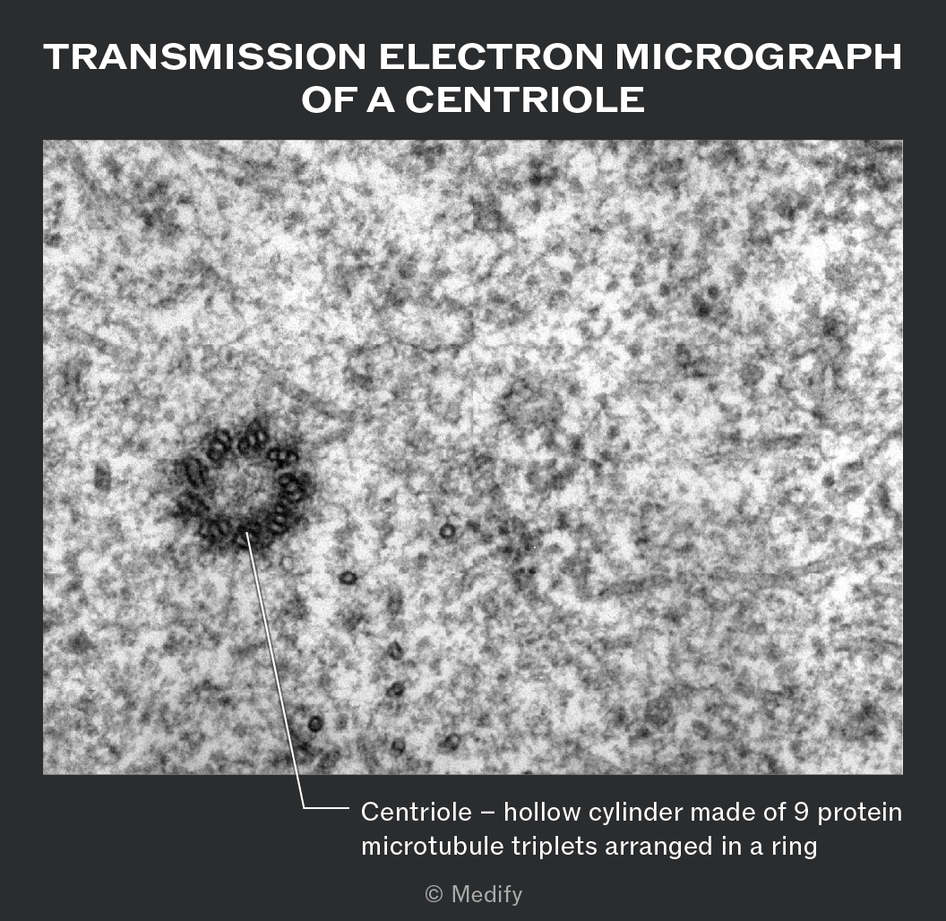

The image is a transmission electron micrograph of a centriole. The background shows a granular texture with a circular structure on the left side, which is the centriole. A line points to the centriole with a label that reads: 'Centriole – hollow cylinder made of 9 protein microtubule triplets arranged in a ring.' The top of the image has the text: 'TRANSMISSION ELECTRON MICROGRAPH OF A CENTRIOLE.' At the bottom right, there is a copyright notice: '© Medify.'


<ol><li><p><strong>Centrioles</strong> are small <strong>hollow</strong> <strong>cylinders</strong></p></li><li><p>found in pairs in <strong>animal</strong> <strong>cells</strong>,</p></li><li><p>but absent from most plant and fungal cells.</p></li><li><p>Each centriole consists of a ring of <strong>nine</strong> <strong>microtubules</strong> formed from protein subunits.</p></li></ol><p></p><p>Centrioles are involved in ;</p><ol><li><p><strong>spindle</strong> <strong>formation</strong> during <strong>nuclear</strong> <strong>division</strong> (mitosis and meiosis)</p></li><li><p>and help <strong>organise</strong> <strong>microtubules</strong> within the cytoplasm. (Microtubules are tiny protein tubes that form part of the cell's cytoskeleton.</p><p>Centrioles help organise these tubes, which:</p><ul><li><p>Help maintain cell structure.</p></li><li><p>Help move materials around inside the cell.</p></li><li><p>Help organise the cell during division.)</p></li></ul></li></ol><p></p><p></p><ol start="3"><li><p>They also hold cilia and flagella in place;  </p></li><li><p></p></li><li><p></p></li><li><p><strong>Cilia</strong> are tiny hair-like structures that move substances across the cell surface.</p></li><li><p><strong>Flagella</strong> are longer, whip-like structures that help a cell move (e.g. sperm cells).</p></li></ol><ol><li><p><span><strong>Centrioles</strong> are small cylindrical structures made of microtubules, found in the cytoplasm of animal cells</span></p><p></p></li></ol><img src="https://d3nzlfkeryzt6j.cloudfront.net/miia30qqe6zh8dn7zlzxeu8dnqk1" data-width="75%" data-align="center" alt="The image is a transmission electron micrograph of a centriole. The background shows a granular texture with a circular structure on the left side, which is the centriole. A line points to the centriole with a label that reads: 'Centriole – hollow cylinder made of 9 protein microtubule triplets arranged in a ring.' The top of the image has the text: 'TRANSMISSION ELECTRON MICROGRAPH OF A CENTRIOLE.' At the bottom right, there is a copyright notice: '© Medify.'" style="display: block; width: 75%; margin-left: auto; margin-right: auto;"><p></p>
12
New cards

anatomy and function of chloroplast

  1. is a double membrane–bound organelles found in plant

  2. and algal cells that are the site of photosynthesis.


Chloroplasts contain;

  1. flattened membrane sacs called thylakoids, which contain chlorophyll and

  2. are stacked into grana

  3. connected by lamellae.

  4. These membranes are the site of the light-dependent reactions.

  5. The fluid-filled stroma contains enzymes involved in the light-independent reactions.

  6. Chloroplasts also contain circular DNA, ribosomes and starch grains for temporary carbohydrate storage.
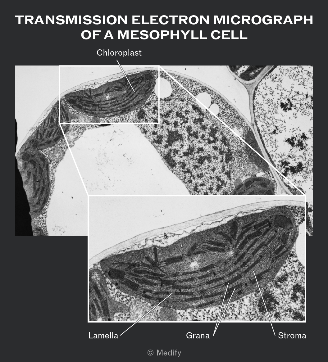

The image is titled 'Transmission Electron Micrograph of a Mesophyll Cell'. It shows a detailed view of a chloroplast within a mesophyll cell. The chloroplast is highlighted with a rectangular inset. Within this inset, three parts are labeled: 'Lamella', 'Grana', and 'Stroma'. The 'Lamella' is indicated as a structure running along the inside of the chloroplast, the 'Grana' is shown as stacked, disk-like structures, and the 'Stroma' is the fluid-filled space surrounding the grana. The image provides a close-up view of the internal structure of the chloroplast.


<ol><li><p>is a <strong>double</strong> <strong>membrane–bound</strong> organelles found in <strong>plant</strong> </p></li><li><p>and <strong>algal</strong> <strong>cells</strong> that are the <strong>site</strong> <strong>of photosynthesis</strong>.</p></li></ol><p></p><p></p><p>Chloroplasts contain; </p><ol><li><p> <strong>flattened</strong> <strong>membrane</strong> <strong>sacs</strong> called <strong>thylakoids</strong>, which contain <strong>chlorophyll</strong> and</p></li><li><p>are stacked into <strong>grana</strong></p></li><li><p> connected by <strong>lamellae</strong>. </p></li><li><p>These membranes are the site of the light-dependent reactions. </p></li><li><p>The fluid-filled <strong>stroma</strong> contains <strong>enzymes</strong> involved in the light-independent reactions.</p></li><li><p>Chloroplasts also contain <strong>circular</strong> <span><strong>DNA</strong></span>, <strong>ribosomes</strong> and <strong>starch</strong> <strong>grains</strong> for temporary carbohydrate storage.</p></li></ol><p></p><img src="https://d3nzlfkeryzt6j.cloudfront.net/w3j6vxsj8oct62ppq6w8jbdw9eka" data-width="50%" data-align="center" alt="The image is titled 'Transmission Electron Micrograph of a Mesophyll Cell'. It shows a detailed view of a chloroplast within a mesophyll cell. The chloroplast is highlighted with a rectangular inset. Within this inset, three parts are labeled: 'Lamella', 'Grana', and 'Stroma'. The 'Lamella' is indicated as a structure running along the inside of the chloroplast, the 'Grana' is shown as stacked, disk-like structures, and the 'Stroma' is the fluid-filled space surrounding the grana. The image provides a close-up view of the internal structure of the chloroplast." style="display: block; width: 50%; margin-left: auto; margin-right: auto;"><p></p>
13
New cards

anatomy and structure of cell wall

  1. A cell wall is a freely permeable,

  2. rigid outer layer

  3. surrounding the cell–surface membrane in plants, algae and fungi.

  4. Plant and algal cell walls are mainly made of cellulose

  5. , whereas fungal cell walls are made of chitin.

The cell wall provides;

  1. mechanical strength

  2. and support,

  3. helping maintain cell shape

  4. and preventing the cell from bursting when water enters by osmosis.

  5. The resistance of the cell wall to expansion allows turgor pressure to develop, making plant cells rigid.
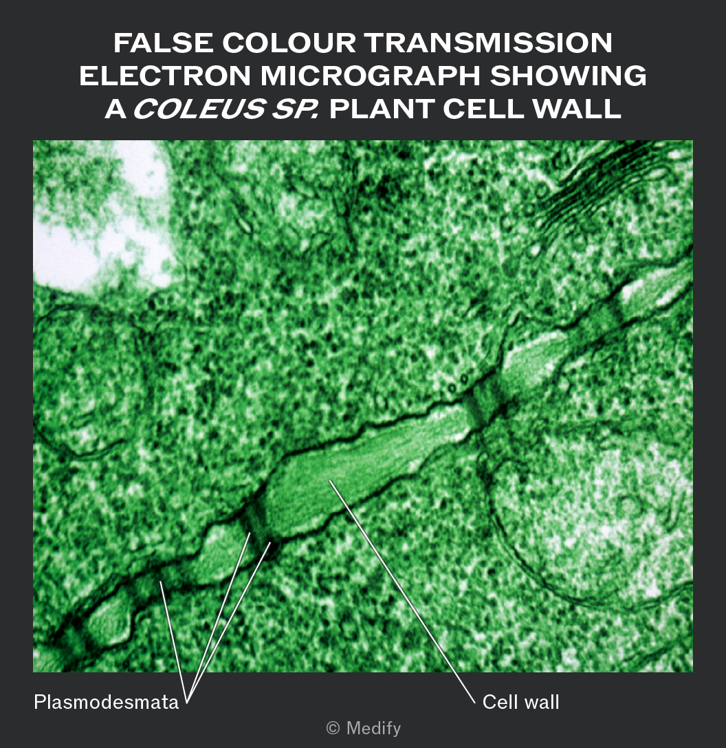
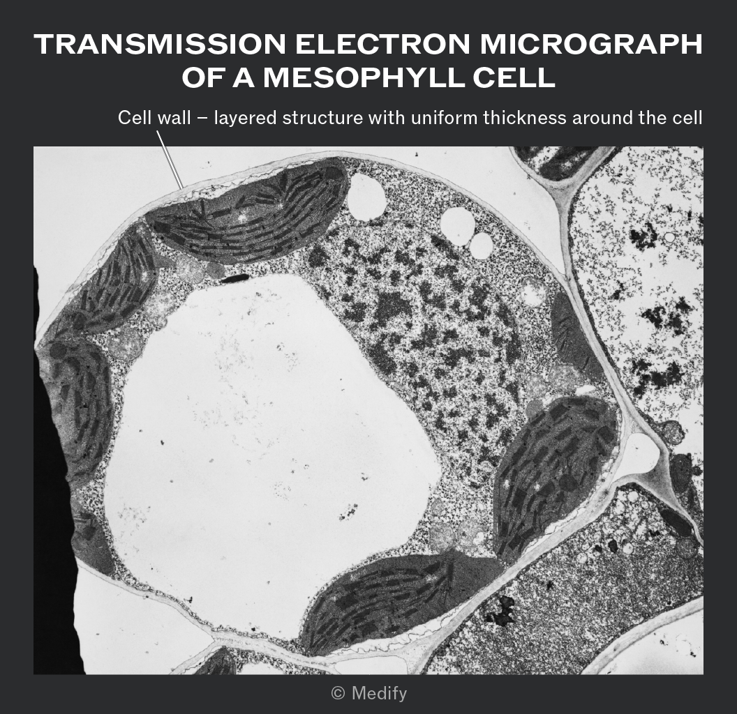
The image is a false color transmission electron micrograph showing a Coleus sp. plant cell wall. The micrograph is predominantly green with various textures indicating different cellular structures. Two labels are present: 'Plasmodesmata' points to several small, dark, thread-like structures within the cell wall, and 'Cell wall' points to a thicker, lighter green structure running horizontally across the image. The text at the top reads 'FALSE COLOUR TRANSMISSION ELECTRON MICROGRAPH SHOWING A COLEUS SP. PLANT CELL WALL'.Transmission electron micrograph of a mesophyll cell. The image shows a detailed cross-section of a mesophyll cell with various internal structures visible. A label points to the 'Cell wall' and describes it as a 'layered structure with uniform thickness around the cell'. The image includes dense regions and organelles within the cell, such as chloroplasts with visible grana stacks, and other cellular components. The micrograph is black and white, highlighting different textures and densities within the cell. © Medify is noted at the bottom.


14
New cards

flagella structure and anatomy

Flagella are;

  1. long,

  2. whip-like extensions of cells

  3. formed from microtubules.

  4. They can facilitate cell motility by propelling cells through their environment. Sperm cells and some bacterial cells are examples of cells that have flagella.
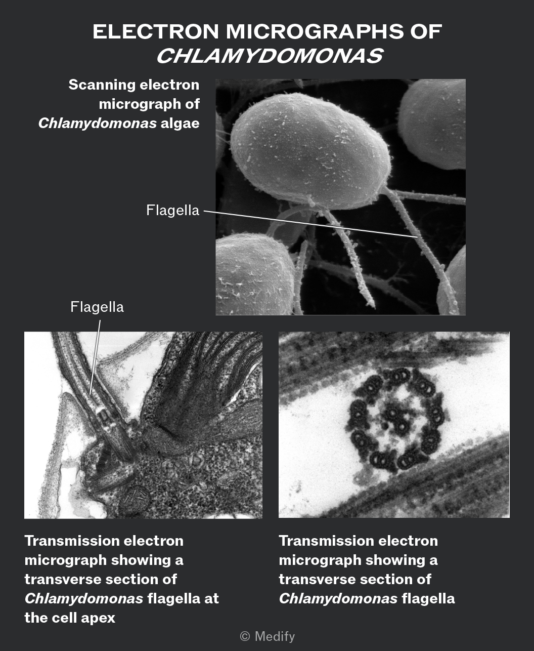

The image consists of three electron micrographs of Chlamydomonas with text labels. At the top, the title reads 'ELECTRON MICROGRAPHS OF CHLAMYDOMONAS'. Below, the leftmost image is labeled 'Scanning electron micrograph of Chlamydomonas algae' with an arrow pointing to a structure labeled 'Flagella'. The middle image is labeled 'Transmission electron micrograph showing a transverse section of Chlamydomonas flagella at the cell apex' with an arrow pointing to a structure labeled 'Flagella'. The rightmost image is labeled 'Transmission electron micrograph showing a transverse section of Chlamydomonas flagella'.


15
New cards

anatomy and structure of cilia

Cilia are

  1. short,

  2. hair-like extensions of cells,

  3. composed of microtubules

  4. and shorter than flagella.

  5. Large groups of cilia can beat in coordinated waves

  6. to move cells or organisms (e.g., Paramecium) or to movesubstances across cell surfaces (e.g., mucus in the respiratory tract).
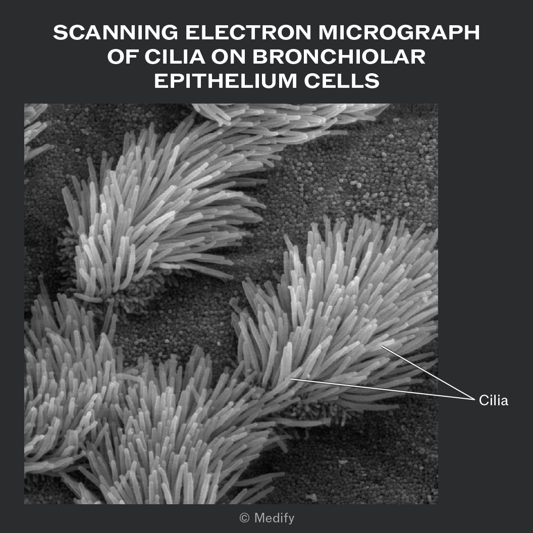

A scanning electron micrograph showing dense clusters of cilia on bronchiolar epithelium cells. The top of the image contains the text: 'SCANNING ELECTRON MICROGRAPH OF CILIA ON BRONCHIOLAR EPITHELIUM CELLS'. Several arrow annotations point to the cilia, with the label 'Cilia'. The cilia are depicted as numerous, elongated, hair-like structures protruding from the surface of the cells. The background shows a textured surface of the epithelium cells.


16
New cards

In eukaryotic cells, protein production and secretion require the coordinated action of several organelles:


  • mRNA is transcribed from DNA in the nucleus.

  • It travels to ribosomes on the rough ER, where the protein is synthesised, powered by ATP generated in the mitochondria.

  • The protein moves through the rough ER, folding into its 3D shape.

  • They are packaged into transport vesicles that bud off and are carried to the Golgi apparatus via the cytoskeleton.

  • Within the Golgi, proteins are modified (e.g., the addition of carbohydrate to form glycoproteins), then pinched off in secretory vesicles.

  • These vesicles fuse with the cell-surface membrane, releasing the protein (such as extracellular enzymes) by exocytosis.


17
New cards

what is the cytoskeleton function

The cytoskeleton

  1. helps maintain cell shape,

  2. provides structural support,

  3. facilitates intracellulart ransport,

  4. and enables cell movement. I

  5. forms the structural basis of cilia and flagella, allowing their movement.
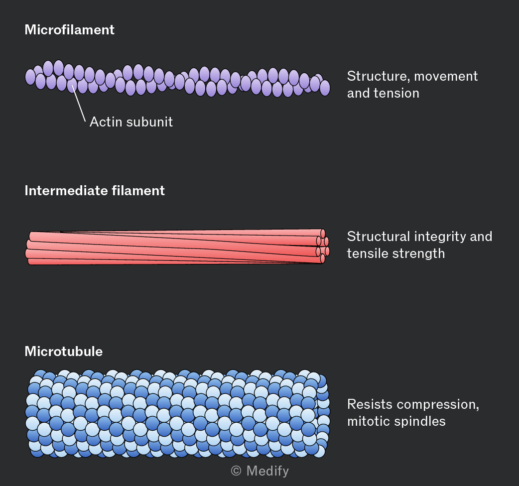
The image shows three types of cytoskeletal filaments with labels. At the top is a purple microfilament composed of oval-shaped actin subunits, labeled 'Actin subunit'. The label 'Structure, movement and tension' is to the right. In the middle is a pink intermediate filament consisting of cylindrical strands, labeled 'Structural integrity and tensile strength' on the right. At the bottom is a blue microtubule made of spherical subunits, labeled 'Resists compression, mitotic spindles' on the right. Each filament type is labeled: 'Microfilament', 'Intermediate filament', and 'Microtubule', respectively.


18
New cards

what are microfilaments and function

Microfilaments are

  1. thin protein filaments of the cytoskeleton made from actin.

  2. They are involved in maintaining and changing cell shape

  3. and play important roles in cell movement,;

  4. muscle cell contraction, endocytosis, exocytosis, phagocytosis and cytokinesis.


19
New cards

microtubules anatomy function

Microtubules are

  1. hollow protein filaments of the cytoskeleton

  2. made from tubulin.

  3. They providemechanical strength, helping maintain cell shape and resist compression.

  4. Microtubules also act as tracks

  5. for the movement of vesicles, organelles and molecules within the cell and help position organelles in the cytoplasm.

  6. They are involved in spindle fibre formation during mitosis and meiosis.


<p><strong>Microtubules</strong> are </p><ol><li><p><strong>hollow</strong> <strong>protein</strong> <strong>filaments</strong> of the cytoskeleton </p></li><li><p>made from <strong>tubulin</strong>.</p></li><li><p> They providemechanical <strong>strength</strong>, helping maintain cell shape and resist compression.</p></li><li><p>Microtubules also act as <strong>tracks</strong> </p></li><li><p>for the <strong>movement</strong> of vesicles, organelles and molecules within the cell and help position organelles in the cytoplasm.</p></li><li><p> They are involved in <strong>spindle</strong> <strong>fibre</strong> formation during mitosis and meiosis.</p></li></ol><p></p>
20
New cards

what does prokaryote cells encounter, how do they differ form eukaryotes

Prokaryotes are unicellular organisms that include bacteria and archaea. They differ from eukaryotes in several ways:

  • They lack membrane–bound organelles, including a nucleus.

  • Their ribosomes are smaller.70s vs 80s

  • They have a cell wall made of peptidoglycan.not chitin of cellulose

  • Some possess flagella, which are structured differently from those of eukaryotes.
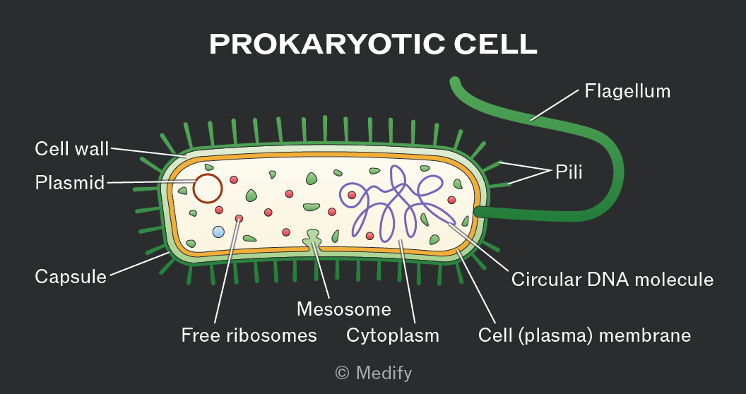
Diagram labeled 'PROKARYOTIC CELL' showing a cross-section of a prokaryotic cell. Outer structures include a capsule, cell wall, and cell (plasma) membrane. A flagellum is attached to the cell exterior. Several pili are protruding from the cell surface. Inside, a circular DNA molecule is visible. There are free ribosomes shown as small dots, and plasmids represented as small circles. The cytoplasm fills the interior, with a mesosome depicted as a folded membrane. The diagram includes annotations pointing to each structure.


21
New cards

explain the genetic material in prokaryotes

Prokaryotic DNA consists of a single circular DNA molecule that is supercoiled, making it more compact.

It is found free in the cytoplasm and is not associated with histone proteins.

Prokaryotes may also contain small additional circular DNA molecules called plasmids.

22
New cards
23
New cards
24
New cards
25
New cards
26
New cards
27
New cards
28
New cards
29
New cards
30
New cards