MLS 4510 - EXAM 2
 Call Kai
 Learn
 Practice Test
 Spaced Repetition
 Match
 Flashcards
 Knowt Play
1/283

There's no tags or description
Looks like no tags are added yet.
284 Terms
What is the approximate volume of spinal fluid in an adult?
90–150 mL
Normal adult CSF may have 0–5 white blood cells (WBCs)/µL. Which of the following cell types accounts for 40–80% of these WBCs?
lymphocytes
Which of the following sites is used most often for CSF collection?
L3-L4
Cell counts on CSF specimens should be performed within what time frame following the collection of the CSF sample?
within one hour
Which of the following are characteristics of normal CSF?
colorless
Which of the following situations suggest a traumatic tap occurred?
blot clots are present
Xanthochromia in a premature infant may be due to an immature blood-brain barrier
true
Clear cerebrospinal fluid samples should be examined and counted undiluted
true
A spinal fluid that is slightly hazy is briefly examined microscopically. The medical laboratory scientist performing the count decides to make a 1:10 dilution using 30 µL of the sample. What volume of diluent should be used?
270 uL
In a moderately bloody specimen, 250 cells are counted in 10 squares. The dilution is 1:100. What is the count/µL?
2.5 × 10⁴
A 1:10 dilution is made on a CSF sample. Five squares on each side of the hemocytometer are counted for a total of 10 squares, and a total of 150 cells are recorded. What is the count per microliter (µL)?
1500
A cell count is ordered on a CSF sample that is bloody. Which of the following procedures would improve count accuracy?
Use Spinal Diluting Fluid, a mixture of acetic acid and crystal violet
Cytocentrifugation is the recommended method for preparing a cerebrospinal fluid (CSF) sample for examination of cellular morphology
true
hemosiderin deposits
Previous subarachnoid hemorrhage (SAH)
neutrophilic pleocytosis
Bacterial meningitis
nucleated RBCs (nRBCs)
Bone marrow contamination of the CSF
lymphocytic pleocytosis
Viral meningitis
The image on the right is a Wright-Giemsa stained CSF smear (1000×). What structures are indicated by the arrows?

bacteria
presumptive malignant cells

lymphocytes

choroidal cells

monomacrophages

A medical laboratory technician decides to make a 1:20 dilution of cerebrospinal fluid (CSF) after briefly evaluating a portion of the sample microscopically. After making the dilution and charging the chambers, the number of cells that are observed in each of the large squares of the hemocytometer is >100. What should the technician do to obtain the most accurate count?
make a larger dilution
A sample of cerebrospinal fluid is diluted 1:100; the standard 9 squares of a hemocytometer are counted on each side for a total of 18 large squares.
Side 1: 186 nucleated cells counted
Side 2: 184 nucleated cells counted
Total nucleated cells = 370
Using the standard hemocytometer formula shown on the right, what is the nucleated cell count per microliter (µL)?
2.06 × 10⁴
Which of the following is not a function of CSF?
Regulate body metabolism
How many white blood cells (WBCs) would be considered normal for adult cerebrospinal fluid?
0–5 WBCs/µL
Upon centrifugation, a cerebrospinal fluid (CSF) sample supernatant exhibited xanthochromia. The image is a Wright-Giemsa-stained smear that was made from that CSF sample. What condition is probably related to these macroscopic and microscopic findings?

Previous subarachnoid hemorrhage
Increased numbers of the cells indicated by the arrows are associated with which of these conditions?

viral meningitis
These 3 tubes of cerebrospinal fluid (CSF) are delivered to the laboratory for analysis. The tube labeled #1 was the first tube collected. Which one of the tubes should be used by the hematology department for cell count and differential?

tube #3
The image is a Wright-Giemsa-stained smear (1000×) of cerebrospinal fluid (CSF). What is the identification of the cells that are indicated by the arrows?

Presumptive malignant cells
What does lymphocyte pleocytosis refer to?
An increased number of lymphocytes in a CSF when compared to a normal sample
These tubes of cerebrospinal fluid (CSF) arrive in the laboratory for evaluation. The tubes were numbered in the order in which they were obtained, with #1 being the first tube collected and #3 being the last tube collected. What may be indicated by the macroscopic appearance of the CSF?

traumatic tap
When preparing a slide for morphologic examination, what is the reason for adding a drop of albumin to the cerebrospinal fluid (CSF) sample prior to cytocentrifugation?
Preserve the integrity of the cells
Where does most cerebrospinal fluid (CSF) originate?
Choroid plexus
The image is a Wright-Giemsa-stained smear (1000×) of cerebrospinal fluid (CSF). The arrows point to cells that may indicate which of the following conditions?

Leukemia with CNS involvement
A cerebrospinal fluid (CSF) sample for hematologic evaluation should be tested within how long of collection?
1 hour
What term is used to describe the color in these tubes of cerebrospinal fluid (CSF)?

xanthochromia
Nucleated blood cells will always have the same appearance on a cytocentrifuged smear as they do on a peripheral blood smear
false
The image on the right is a representative field from a cytospin preparation of a CSF from a patient with a viral infection. Identify the cell indicated by the arrow.

atypical (reactive) lymphocyte
Macrophages are the phagocytes that originate from lymphocytes
false
Match each cell type listed in the drop-down box to the letter that corresponds to its identification in the image on the right.

Cell B = lymphocyte
Cell C = neutrophil
Cell D/D1 = macrophage
What is the identification of this cellular clump found in CSF? Note the presence of many similar-appearing nuclei without distinct lines of demarcation between cells.

Ependymal clump
cerebrospinal fluid (CSF)
Ependymal cells

serous fluid
Mesothelial cells

joint fluid
Synovial lining cells

bronchoalveolar lavage (BAL)
Bronchial lining cells

The elements indicated by the arrows were observed on a Wright-stained smear of a synovial fluid. What is the likely identification of the elements, and what would you do to confirm the finding?

Bacteria; perform a Gram stain
Prominent vacuolation involving the cytoplasm of abnormal lymphoblast-like cells seen in a body fluid preparation is a distinctive feature of Burkitt Lymphoma
true
malignant cells
High nuclear/cytoplasmic (N/C) ratio, irregular nuclear membrane, non-uniform appearance
reactive mesothelial cells
Low N/C ratio, smooth nuclear membrane, uniform appearance
The image on the right is a cytospin preparation of cerebrospinal fluid (CSF). What is the cell type that is indicated by the arrow?

eosinophil
Identify the cells indicated by the arrows in this cerebrospinal fluid (CSF) sample.

blast cells
A patient suffering from typical leukemic symptoms presents to the emergency room. The physician orders a spinal tap after noticing possible central nervous system involvement. What type of cells are indicated by the red arrows?

blasts
What is the predominant cell population seen in this pleural effusion?

eosinophils
The normal serous membranes are lined by which type of cells?
Hint: these cells are commonly shed into pleural, peritoneal, and pericardial fluids.
Mesothelial
A patient suffering from typical leukemic symptoms presents to the emergency room. The physician orders a spinal tap after noticing possible central nervous system involvement. What type of cells are indicated by the blue arrows?

Normal lymphocytes
A patient with suspected intra-abdominal malignancy had a paracentesis performed. The fluid was then sent to the laboratory for examination and fluid differential. Which cells predominate in this cytospin field from the patient's sample?

plasma cells
Identify the cells taken from a pleural effusion.

malignant cells
After suspecting that his patient may have lung disease, the physician sends a bronchoalveolar lavage (BAL) to the laboratory for examination. What is the cell type indicated by the arrows in this cytospin sample?

bronchial lining cells
Identify the binucleate cell indicated by the arrow in the pericardial fluid sample shown here.

mesothelial cell
The image on the right is a cytospin of a pleural fluid. What is the cluster of cells indicated by the arrow?
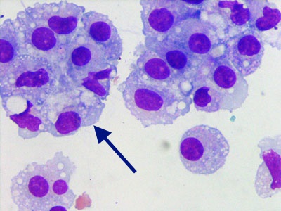
mesothelial cells
What are the cells that are indicated by the arrows in the joint fluid image shown on the right?

Synovial lining cells
A physician ordered a peritoneal lavage on a patient who complained of fever and abdominal pain following an automobile accident. The fluid is analyzed in the laboratory. How should the sample shown in the image to the right be reported?

Neutrophils with intracellular and extracellular bacteria
What is the most likely identification of the clumped cells in this microscopic field?

tumor cells
The image on the right is a cytospin preparation of cerebrospinal fluid viewed at 1000× magnification. What is the identity of the primary blood cell type observed in this microscopic field?

lymphocytes
The membrane that lines the wall of a cavity is the
parietal
Synovial crystals that occur as a result of purine metabolism or chemotherapy for leukemia are:
Monosodium urate
During normal production of serous fluid, the slight excess of fluid is:
Absorbed by the lymphatic system
Synovial fluid for crystal examination should be examined as a/an:
wet preparation
When diluting a synovial fluid WBC count, all of the following are acceptable EXCEPT:
acetic acid
Differentiation between bacterial peritonitis and cirrhosis is done by performing a/an:
absolute neutrophil count
An increase in the amount of serous fluid is called a/an:
effusion
A pleural fluid pH of 6.0 indicates:
esophageal rupture
Another name for a peritoneal effusion is:
ascites
The procedure for collecting synovial fluid is called:
arthrocentesis
The chemical test performed most frequently on synovial fluid is:
glucose
transudate
caused by increased capillary permeability, caused by decreased oncotic pressure, caused by congestive heart failure, clear appearance
exudate
tuberculosis related, caused by increased hydrostatic pressure, malignancy related, endocarditis related
The highest WBC count can be expected to be seen in a patient with:
septic arthritis
All of the following are abnormal when seen in synovial fluid EXCEPT:
Synovial lining cells
Which of the following best represents a hemothorax?
Blood HCT: 30, Fluid HCT: 20
All of the following are characteristics of malignant cells EXCEPT:
Absence of nucleoli
In an examination of synovial fluid under compensated polarized light, rhomboid-shaped crystals are observed. What color would these crystals be when aligned parallel to the slow vibration?
blue
Fluid:serum protein and lactic dehydrogenase ratios are performed on serous fluids:
To classify transudates and exudates
Negative birefringence occurs under red-compensated polarized light when:
Fast light runs against the molecular grain of the crystal
The primary function of synoviocytes is to:
Provide nutrients for the joints
cerebrospinal fluid (CSF)
supplies nutrients to nervous tissue, removes metabolic wastes, maintains intracranial pressure, produces a mechanical barrier for brain and spinal cord
meninges
brain/spinal cord lined by __
dura mater
outer layer, lines skull, vertebral canal
arachnoid
inner membrane, filamentous
pia matter
thin membrane, lines brain and spinal cord surfaces
meninges
made up of dura mater, arachnoid mater, pia mater
CSF formation
in choroid plexuses, 20mL produced an hour in adults, flows through subarachnoid space, a volume of 90-150mL maintained in adults at a given time, circulating fluid reabsorbed back into capillaries at about the same rate it is produced, tight-fitting endothelial cells in choroid plexuses comprise the blood-brain barrier (BBB)
blood-brain barrier (BBB)
prevents passage of many harmful molecules to the brain, also prevents passage of antibodies in medication, selective filtration (does not resemble an ultrafiltrate of plasma)
disruption of BBB
allows WBCs, proteins, and chemicals to enter the CSF (meningitis, multiple sclerosis)
specimen collection of CSF
lumbar puncture (between L3-L4 and L4-L5), careful technique and mindful of intracranial pressure and infection prevention, elevated pressure of CSF requires fluid to be drawn slowly, pressure measured when needle first enters subarachnoid space, specimens collected in 3-4 sterile tubes labelled in the order in which they are drawn, freeze leftover sample and store until no longer needed, CSF testing typically performed STAT (should be appropriately stored if testing is delayed)
CSF tube 1
chemical, serological testing, store frozen
CSF tube 2
microbiology testing, store RT
CSF tube 3
hematology (cell count, differential), store refrigerated (up to 4 hours)
CSF tube 4
may be used for additional microbiology or serology testing