Therapeutic Approaches - Interventions, Effusions and other Therapeutic Considerations
 Call Kai
 Learn
 Practice Test
 Spaced Repetition
 Match
 Flashcards
 Knowt Play
1/9

There's no tags or description
Looks like no tags are added yet.
10 Terms
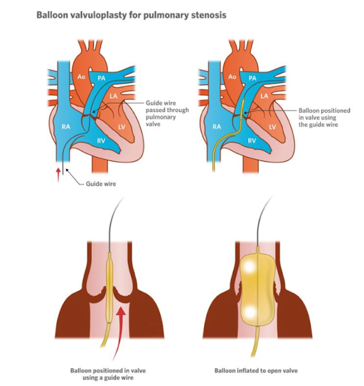
What do treatment options look like for pulmonic stenosis?
• Valvular stenosis can be well ballooned
If the stenosis is caused by a muscular component, it is more difficult to be effective.
Muscular/Subvalvular Stenosis: A balloon cannot stretch or permanently fix abnormal, dynamic heart muscle (myocardium). Inflating a balloon against thick muscle bundles does not permanently relieve the obstruction because the muscle will not tear or remodel the way valve tissue does
Different types of balloons catheters can be used (standard, high pressure, cutting).
Usually placed through the cranial vena cava into right ventricle, then towards the pulmonary artery
Valve implantation is not a standard procedure.
PS with transvalvular pulmonic stent angioplasty
Access: A catheter and guide wire are inserted via a transvenous approach (typically through a vein in the leg or neck) and navigated into the right side of the heart and across the stenotic pulmonic valve.
Stent Deployment: A pre-mounted, balloon-expandable metallic stent is positioned across the narrowed valve/artery region and expanded to prop the pathway open. (Inside the pulmonary artery)
Hemodynamic Result: This permanently widens the restrictive channel, immediately reducing the pressure gradient between the right ventricle and the pulmonary artery.
Surgical repair described with cardiopulmonary bypass.
• Use of atenolol (betablocker) may be combined or used to reduce stress on right side of the heart.
Slows down the HR, slows down the forces of contraction
What do treatment options look like for PDA?
Right to Left shunt - cannot be closed
Due to damage to pulmonary vessels, goes from pulmonary artery to aorta
Shunt is now a safety valve,
Left to Right shunt - can be closed
Lungs being overloaded, aorta diverting to pulmonary artery
• Surgical ligation more invasive.
Complex morphology
Very small animals - too small to place device or morphology of PDA is too complex for placement of device or if there are multiple openings
• Minimally invasive (catheter-based occlusion) - good to go from arterial side, left to right, flow aids you, right side - more vessels (Against flow)
Embolization coil - less in favor now, can compact, dislodge
Duct Occluder (ACDO) - must go from arterial side - directionality of device, difficult to implant in small dogs
Vascular plug - symmetrical - easier to place
Why is closing a right-to-left shunt PDA dangerous?
Loss of Cardiac Output: In right-to-left shunting, pulmonary vascular resistance (PVR) has risen so high that blood in the pulmonary artery is forced backward across the PDA into the aorta. Closing the PDA traps the right ventricle behind a massive, high-pressure wall.
Right Heart Failure: Without the PDA to vent high pressure from the right side of the heart, the right ventricle quickly fails to pump against the stiff lung vasculature, leading to acute, catastrophic right-sided heart failure and death.
Maintenance of Systemic Flow: Though the shunted blood is deoxygenated (causing cyanosis/low oxygen levels), keeping the PDA open allows the right ventricle to maintain baseline systemic blood flow that would otherwise plummet if the channel were plugged.
When are pacemakers indicated?
A pacemaker is a small, battery-powered device placed under the skin in your chest that monitors your heartbeat and sends tiny electrical pulses to keep your heart beating at a normal rate.
• Indicated in Bradyarrhythmias
High-grade second-degree atrioventricular block (I| AVB)
Third degree AVB
Sick Sinus Syndrome
Atrial standstill
• Two types:
Transvenous
Wires (leads) are threaded through a large vein (such as the subclavian or jugular vein) directly into the interior of the heart, where the electrode tips lodge against the heart wall (the endocardium). The main pulse generator box is placed under the skin in the upper chest, just below the collarbone.
Epicardial (cats and very small animals)
Instead of threading wires through the veins, the pacing leads are attached directly to the epicardium (the outer surface of the heart muscle) by a surgeon. The wires are guided out through the chest wall and connected to an external or implanted pulse generator. [1, 2, 3, 4]
• Most often pace only the right ventricle.
Right sided congestive heart failure may cause what consequences?
VIDEO
• Pericardial effusion
Cause (tamponade)
Consequence (usually small amount; cats left sided heart failure possible too)
• Pleural effusion
Cats potentially left and right sided heart failure
• Ascites
What are the main right sided heart problems?
• Pericardial tamponade or constrictive pericarditis
• Right sided cardiomyopathy (or biventricular)
DCM, ARVC/AC
• Tricuspid valve dysplasia or degeneration
• Pulmonary stenosis
• Pulmonary hypertension
What are the main steps and tools needed to treat thoracocentesis?
• Sedation
• Clip and prepare the area
• Ultrasound-guided - to look for pockets of fluid
• Butterfly-needle
Describe how effusion types can be classified?
**Most important modified transudate and chylothorax
Chylothorax - milky color due to triglycerides
Modified Transudate - would not expect to see proteins and high cellularity in pericardial fluid

How does effusion fluid type guide common disease and subsequently further diagnostic testing?

Examples of fluid classification for abdominal effusion includes:
• Transudate
• Modified transudate → Cardiac
• Exudate (inflammatory): Septic & non septic
• Haemorrhage
• Chyle
• Bile
• Urine