Cardiac Brig
 Call Kai
 Learn
 Practice Test
 Spaced Repetition
 Match
 Flashcards
 Knowt Play
1/134

There's no tags or description
Looks like no tags are added yet.
135 Terms
6 main things housed in the mediastinum
heart
great vessels
esophagus
trachea
thoracic duct
lymph nodes
most anterior structure of the heart
right ventricle
Where does the right ventricle inferior lie?
junction of the sternum and xiphoid process
structures at the base of the heart
where the right ventricle joins the pulmonary artery
under sternal angle
what produces the point of maximal impulse?
cardiac apex
external jugular vein location
over the SCM
internal jugular vein location
under the SCM
best place to hear the aortic valve
Right 2nd intercostal space
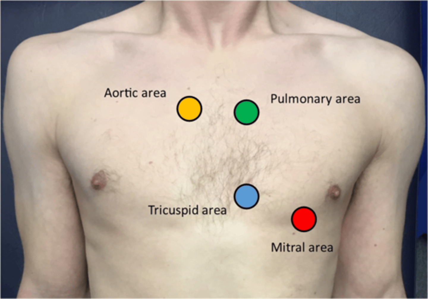
best place to hear the pulmonic valve
Left 2nd intercostal space
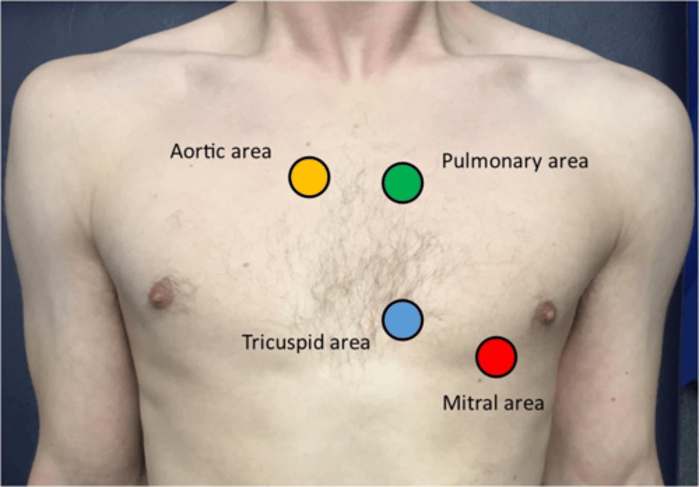
best place to hear the tricuspid valve
Lower left sternal border
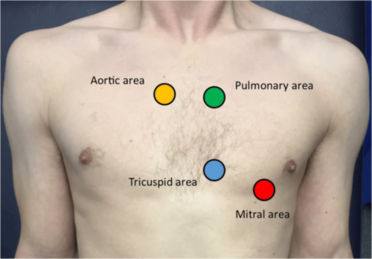
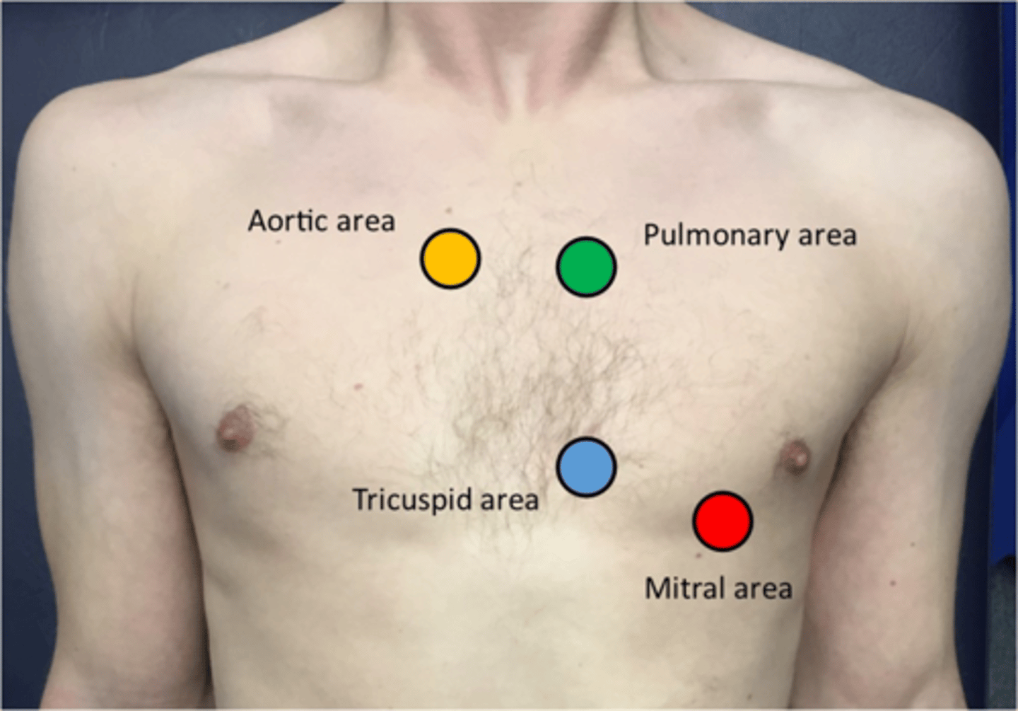
best place to hear the mitral valve
apex
5th intercostal space on the midclavicular line
4 pulse points on a baby
radial
brachial
apical
femoral
Systole
S1/Lub
-semilunar valves open
-AV valves close
-Ventricular contraction
Diastole
S2/Dub
-Semilunar valves close
-AV valves open
-Ventricles relax
best way to evaluate the heart's electrical system
EKG
cardiac output
how much blood each ventricle ejects in one minute
cardiac output formula
CO = HR x SV
stroke volume
amount of blood ejected with each heart beat
ejection fraction
percent of ventricular volume ejected at each heart beat
normal ejection fraction
60%
preload
volume of blood/stretch in a ventricle before it contracts
myocardial contractility
the ability of the heart muscle to shorten
afterload
the degree of vascular resistance
when does pulse wave occur
shortly after ventricular contraction
what can impact pulse wave
cardiac output and afterload
pulse pressure
difference between systolic and diastolic
what impacts BP
cardiac output, afterload, blood volume
3 things we are looking for with cardiac history and exam
1. is there good blood supply to the heart?
2. is the electrical system of the heart working?
3. is the heart adequately pumping to all organs?
-valves functioning properly?
Cardiac history ROS
CAD
HTN
Rheumatic fever
H/O cardiac testing (why, when, where?)
Heart murmur
Chest pain
Palpitations
SOB
Swelling/edema
Fainting/Syncope
3 types of SOB
Dyspnea: abnormal breathing
Orthopnea: hard to breathe when laying down
Paroxysmal nocturnal dyspnea: difficulty breathing at sleep
Typical MI presentation and associated symptoms
-Exertional pain
-Pressure/discomfort in the chest, shoulder, back, neck, or arm
Atypical MI symptoms
-cramping or grinding pain
-tooth or jaw pain
-SOB
-fatigue
-N+V w/o pain
What to ask if patient states chest pain
-OPQRST
-Baseline activity - has it changed?
-Associated symptoms (other organ systems like pulm, GI, MSK, anxiety)
Pericarditis
Retrosternal, knife-like pain
Worse when laying down or deep inspiration
Dissecting aortic aneurysm
Tearing or ripping sensation
-Radiates to the back between shoulder blades
-Rapid decline and LOC
pulmonary embolus
Sharp and worse on inspiration
-SOB
-Hemoptysis
-LOC
better ways to ask if a patient is having palpations
heart racing
fluttering
skipping a beat
irregular
If patient says they have palpitations, what do you ask
-can they replicate it?
-slow or irregular?
-how many episodes? frequency? timing?
-abrupt or gradual onset/offset?
Is SVT abrupt or gradual onset
abrupt
cause of paroxysmal nocturnal dyspnea
fluid accumulation during the day
How much fluid accumulates before pitting edema?
5L
Questions to ask to evaluate any swelling or edema
Rings tight?
Eyelids puffy in morning?
Clothing tight in short pd?
Legs or feet tight in shoes?
Gained 1-2lbs in a day
Questions to ask w/ fainting and syncope
Near-syncopal or syncope?
How long was LOC?
Warning signs?
Position when waking up?
Did they fall/injury?
How did they feel after?
What does it mean if a patient feels crappy post-syncope
usually vasovagal
what to think with abrupt onset/offset of syncope
cardiac in nature, specifically ventricular
3 steps to cardiac examination
1. JVP
2. Carotid pulse
3. Heart exam
-inspection
-palpation
-auscultation
What is JVP a reflection of
Right atrial pressure which correlates to central venous pressure and right ventricle end-diastolic pressure
what can cause jugular venous distension
fluid overload
what are jugular vein pulsations
pressure changes in the cardiac cycle cause JVPs
what is the best vein to measure JVP
right external jugular vein
what do the level of pulsations in the neck correlate to
fluid status
patient positions for JVP
-laying at 30 with head supported
-head turned slightly to left
-ideally tangenital lighting
steps to measure JVP
1. look between SCM attachments for pulsations of R internal jugular
2. find the point of highest pulsation and put an index card across it
3. identify the sternal angle and place a ruler perpendicular to it
4. measure where they cross over
5. add 5cm
normal JVP
6-8cm
>8cm JVP
Hypervolemia
-acute/chronic HF
-tricuspid stenosis
-pulmonary HTN
-SVC obstruction
-cardiac tamponade
-constrictive pericarditis
<5cm JVP
hypovolemia
COPD impact on JVP
elevated on expiration, vein collapse on inspiration
carotid inspection
inspect for visible pulsations then auscultate
auscultating carotids
1. have pt hold breath while listening
2. normal finding: pulse
3. abnormal finding: bruit
Bruit
Murmur-like sound due to turbulent flow (carotid stenosis)
-best heard w/ diaphragm
-most often atherosclerotic stenosis and can lead to stroke
-heart murmur can radiate to carotid
Palpation of the carotid
-amplitude (strength)
-contour of pulse wave
-variations in amplitude
-timing of pulse related to S1 and S2, should follow S1
-thrills (abn vibrations - think fistula)
reporting amplitude of carotid
strong
bounding
thready
weak
reporting contour of pulse wave of carotid
Speed of upstroke, summit, and downstroke
-N: brisk
-Abn: varied peak, double systolic peak (bisferiens), bigeminal, rapid rise or collapse
Pulsus alternans
regular pulse rhythm but force of pulse alternates
-severe L ventricular dysfunction (myocontractility problem)
-on BP: alternating loud and soft korotkoff sounds OR sounds double as pressure declines
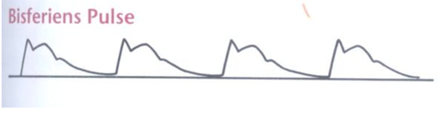
Bisferiens pulse
Increased pressure with double pulse peak (feels like 2 consecutive pulses)
-aortic disease (specifically aortic regurg and hypertrophic obstructive cardiomyopathy)
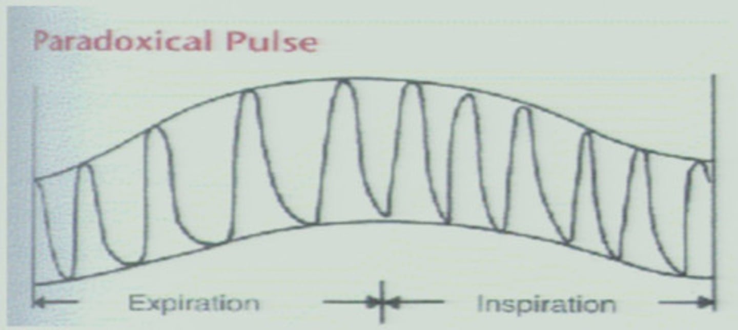
Paradoxical pulse
You can feel pulse w/ expiration but not inspiration
-large drop in systolic during inspiration w/ low pulse amplitude
-pericardial tamponade, COPD or asthma, constrictive pericarditis
Pericardial tamponade
fluid in the pericardium that exerts pressure on the heart and hampers its ability to contract normally
Bigeminal pulse
Extra, premature contraction between each normal beat
- regular beats = normal strength
- extra beats = dec strength
cause of bigeminal pulse
electrical abnormality/arrhythmia cause this if impacting ventricles
Hyperkinetic/bounding pulse
Hits fingers very hard due to high pulse amplitude
-rapid upstroke and rapid descent
Physiologic causes of bounding pulse
fever, anemia, systolic HTN, cirrhosis
-aortic or mitral regurg, ductus arteriosus
physiologic causes of bounding pulse
stress, exercise, pregnancy
PMI (apical impulse)
locates the left border of the heart
Is normal to see a small area on a thin person
Location of the PMI (apical impulse)
4th or 5th intercostal space @ midclavicular line
aligned w/ nipple under breast
normal size of PMI
1-2.5 cm
cause of an abnormal PMI
-Lateral displacement = heart enlargement
-Double pulse = hypertrophic cardiomyopathy
-Epigastric and subxiphoid movement = RV hypertrophy or AAA
palpation of right ventricle
L sternal border
3-5th intercostal space
palpation of LV/apex
L 5th intercostal space
midclavicular line
abnormal PMI palpation findings
Heaves
Thrills
Heaves PMI palpation
sustained impulse from an enlarged ventricle
is best felt with palm or pads of fingers
thrills PMI palpation
buzzing or vibration due to turbulent blood flow
is best felt with ball of hand
when will you see a displaced apical impulse
Pregnancy
HF
Cardiomyopathy
Ischemic heart disease (from MI damage at apex)
Thoracic deformaties
when may you see a double apical impulse
hypertrophic cardiomyopathy
what is diaphragm best used for
high pitched sounds
-S1, S2
-aortic and mitral regurg and pericardial friction
what is bell best used for
low pitched sounds w/ light pressure
-S3 and S4 in mitral stenosis
-used at apex and medially along tricuspid area
what does cardiac auscultation screen for
valvular heart disease
Valvular heart disease
changes in normal blood flow in the heart creating altered or additional sounds which are murmurs
stiffened valves
altered sounds when opening and closing = snaps or clicks
S2 splitting
Delay of pulmonary/aortic valve closure
- low intrathoracic pressure
- normal w/ inspiration
where do you expect to hear S2 splitting best?
Aortic and pulmonic areas
what to note w/ murmurs
Timing
Shape
Location
Radiation
Intensity
Pitch
Quality
Variation w/ respiration and special maneuvers
S3
Occurs very close to S2
-low pitch, use bell
-diastole
-rapid filling of ventricle
-apex
-"ken-tuc-ky"
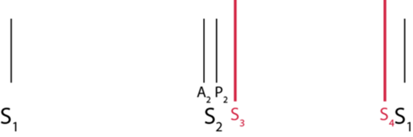
what can S3 be a sign of
heart failure
Normal in athletes, young kids, pregnancy
What is likely if the sound heard after S2 is stronger under the breast
S3
what is likely if the sound heard after S2 is stronger at the 2nd ICS
S2 splitting
S4
Extra atrial contraction
Low pitches, use bell
-late diastole
-best in apex
-atrial contraction to push blood into stiff ventricle
-"te-nuh-see" (S4-S1-S2)
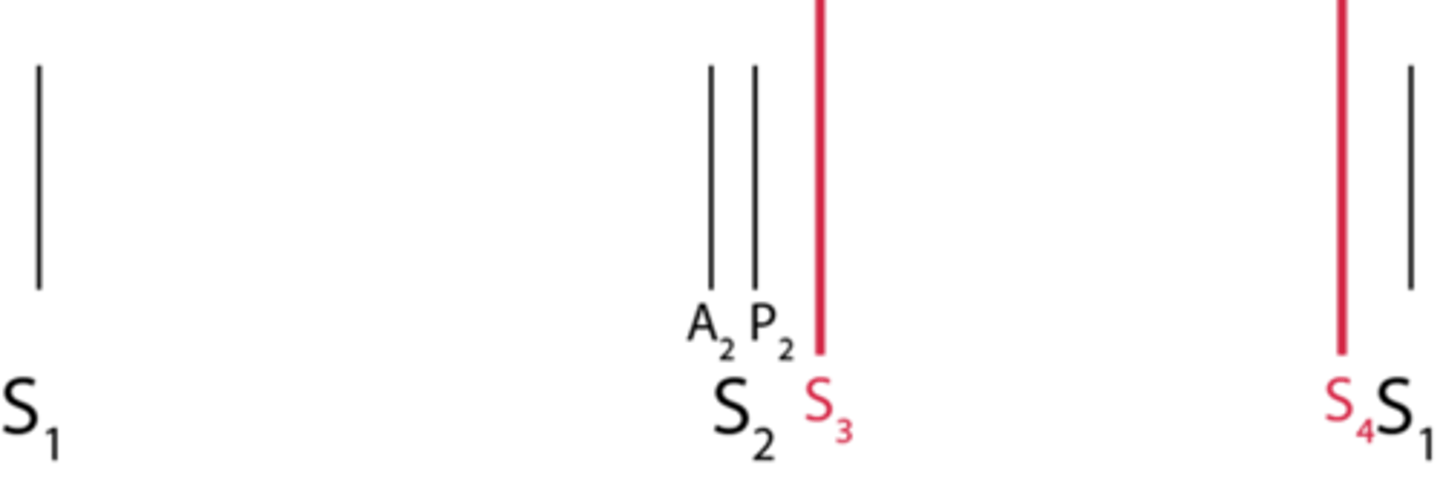
causes of S4
normal in athletes
HOCM or fibrosis
what sided heart issues are most clinically relavent
left
mitral or aortic valve
2 most common causes of heart murmurs
1. narrowing of a heart valve
2. regurg blood flow over a heart valve
-pressure is too high due to outflow obstruction
-damaged valve cant do its job
what to write for timing of a murmur
Note systolic or diastolic
early-
late-
mid-
pan or holo-
pre-