3 FINAL: 11 05 PNS & Reflexes - PHYL 301
 Call Kai
 Learn
 Practice Test
 Spaced Repetition
 Match
 Flashcards
 Knowt Play
1/46

There's no tags or description
Looks like no tags are added yet.
47 Terms
What vertebrae makes up the brachial plexus?
Ventral rami (roots) of C5 to T1 spinal nerves
Brachial Plexus
Roots (C5 to T1)
Trunks (superior, middle, and inferior trunk)
Divisions (anterior and posterior divisions)
Cords (posterior, lateral, and medial cords)
Spinal nerve branches
Brachial Plexus Major Branches
Radial nerve
Ulnar nerve
Median nerve
Musculocutaneous nerve
Axillary nerve
Erb Palsy
Caused by damage to C5–C6 roots of brachial plexus
Frequently due to obstructed labor
Atrophy of affected arm
Atrophy
Decrease in size or wasting of a tissue
Neurogenic Atrophy
Muscle atrophy due to peripheral nerve damage

Median nerve
Innervation
Forearm flexors
Pronator teres & quadratus
Digital flexors
Digits 1 to 3
Thumb, Pointer, Middle
Inner ½ of digit 4
Half of Ring Finger
Carpal Tunnel Syndrome - Causes & Symptoms
Many causes, all involve median nerve
Paresthesia: numbness, tingling, or pain
Affects thumb and fingers
Freq. loss of grip
Innervation of wrist
Carpal Tunnel Syndrome - Diagnosis
Nerve conduction test
Tinel sign
Phalen maneuver
Carpal Tunnel Syndrome - Treatment
transverse carpal ligament = flexor retinaculum
Ligament over median nerve
May cut ligament to give median nerve more room
Wrist splint: Keep wrist in neutral postion to reduce pressure on median nerve
Which spinal nerve levels contribute to the lumbar plexus?
Branches from L1 to L4 spinal nerve roots (some fibers from T12)
L4 & L5 fuse to form lumbosacral trunk
Lumbosacral trunk
L4 & L5 spinal nerve roots

Lumbar Plexus
Major branches:
liohypogastric nerve: innervates external & internal obliques & transverse abdominis
Ilio-inguinal nerve: innervates external & internal obliques & transverse abdominis
Lateral femoral cutaneous nerve: carries sensations from skin of anterior, lateral, & posterior thigh
Genitofemoral nerve: sensory information from skin of anteromedial surface of thigh and external genitals
Femoral nerve: innervates quadriceps, sartorius, pectineus, & iliopsoas
Obturator nerve: innervates gracilis, obturator externus, & adductor magnus
Liohypogastric nerve - Lumbar plexus
Innervates external & internal obliques & transverse abdominis
Ilio-inguinal nerve - Lumbar plexus
innervates external & internal obliques & transverse abdominis
Lateral femoral cutaneous nerve - Lumbar plexus
carries sensations from skin of anterior, lateral, & posterior thigh
Genitofemoral nerve - Lumbar plexus
sensory information from skin of anteromedial surface of thigh and external genitals
Femoral nerve - Lumbar plexus
innervates quadriceps, sartorius, pectineus, & iliopsoas
Obturator nerve
innervates gracilis, obturator externus, & adductor magnus
What spinal nerve roots form the sacral plexus?
Formed from spinal nerve roots L4 to S4
Sacral Plexus
Major branches:
Superior gluteal nerve: innervates gluteus minimus, gluteus medius, & tensor fasciae latae
Inferior gluteal nerve: innervates gluteus maximus
Sciatic nerve: innervates semimembranosus, semitendinosus, & adductor magnus
Posterior femoral cutaneous nerve: sensory from perineum, posterior thigh & lower leg
Pudendal nerve: innervates genitalia, perineum, external anal sphincter, and urethral sphincter
Superior gluteal nerve - Sacral Plexus
innervates gluteus minimus, gluteus medius, & tensor fasciae latae
Inferior gluteal nerve - Sacral Plexus
innervates gluteus maximus
Sciatic nerve - Sacral Plexus
innervates semimembranosus, semitendinosus, & adductor magnus
Posterior femoral cutaneous nerve - Sacral Plexus
sensory from perineum, posterior thigh & lower leg
Pudendal nerve - Sacral Plexus
innervates genitalia, perineum, external anal sphincter, and urethral sphincter
Sciatica
radiating pain and/or numbness from spine to the back of the leg
Usually unilateral
Commonly caused by sciatic nerve compression
Herniated disc common cause
Spinal compression of the nerve •
Piriformis syndrome (related, but not the same as sciatica
Sciatica – Foot Drop
Caused by compression/lesions of the peroneal/fibular nerve
Peroneal n. innervates anterior & lateral compartment of the leg
Sciatica – Treatment
Non-steroidal anti-inflammatory drugs (NSAIDs)
Corticosteroid injection
Muscle relaxants
Physical therapy – strengthening core muscles
Chiropractic alignment?*
Reflex
Quick, almost immediate involuntary motor response
Reflex arc Definition
Path from sensory stimulus to motor response
Begins at sensory receptor and ends at peripheral effector

Reflex arc Pathway
Activation of a sensory receptor
Relay of information to the CNS
Information processing
Activation of a motor neuron
Response by the effector
Example of parallel processing: girl’s right foot steps in class
Parallel processing: spinal cord coordaites both responses at same time
Right leg — ipsilateral withdrawal reflex. She steps on glass with her right foot. The right leg flexes to lift that foot away from the glass.
Left leg — contralateral crossed-extension reflex. Interneurons in the spinal cord relay the signal to the opposite side. The left leg extends to support her weight and keep her from falling.
Ipsilateral
Same Side
Contralateal
Opposite side
Different Reflex Classifications
Development: Innate or acquired reflex
Response: Somatic, visceral, or autonomic reflex
Circuit complexity: Monosynaptic or polysynaptic
Processing site: Spinal, cranial (cerebral), or enteric reflex
Innate Reflexes
Instinctive reflex that doesn’t require learning
Withdrawal reflex
Crossed-extensor reflex
Patellar reflex
Plantar (Babinski) reflex
Learned Reflexes
Reflex acquired through experience and repetition
Swallowing, micturition, defecation
Athletic & rehearsed movements
Transcribing and typing
Braking at red lights
Positive Babinski Sign
Poke feet → Toes go up
Normal in infants
Bad for adults
Negative Babinski sign
Poke feet → Toes curl down
Normal in adults

Monosynaptic Reflex
Involves a single segment of the spinal cord
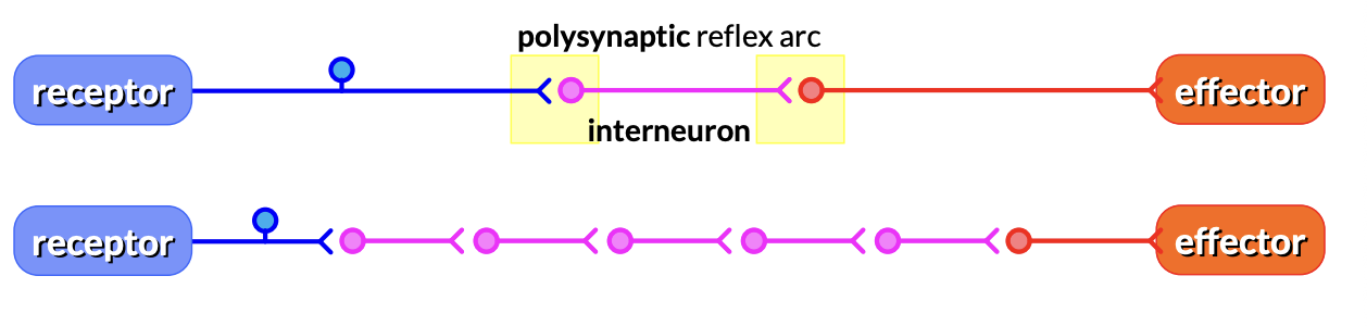
Polysynaptic Reflex
Integrates motor output from several spinal segments
Stretch Reflex
Examples: patellar reflex, postural reflexes
Stretch reflex arc:
Stimulus stretches muscle spindle fibers
Activates a sensory neuron
Information is processed in the spinal cord
Motor neurons are activated
Effector muscle(s) contract(s)
Baroreceptor Reflex
Baroreceptors
Regulate cardiovascular center activity in medulla oblongata
Monitors internal pressure; blood pressure
Detect pressure changes via stretch & mechanoreceptors
Locations: • Carotid sinus • Aortic arch • Cardiac atria
more representative, if blood pressure drop, baroreceptor fires less, increases sympathetic blood pressure will increase
Sympathetic System
Fight or Flight
Parasympathic System
Rest and digest