Histology Lab Midterm
 Call Kai
 Learn
 Practice Test
 Spaced Repetition
 Match
 Flashcards
 Knowt Play
1/78

Earn XP
79 Terms
a dense collagenous tissue surrounding each fascicle
perimysium

a dense collagenous sheath that binds the fascicles into a single nucleus
epimysium

Occupying the spaces between individual muscle fibres (barely visible)
endomysium
Where can you find Type I collagen fibers
bones
What are type I collagen fibers made up of
bundles of fibers that roughly run parallel to each other and have transverse bands
Where can you find type II collagen fibers
cartilage
What are Type II collagen fibers made up of
fibers that are organized in random directions
What forms the basal lamina
Type IV collagen fibers
Name layers of epidermis from deep to superficial
Stratum Basale
Stratum Spinosum
Stratum Granulosum
Stratum Lucidum
Statum Corneum
Which layer is found in thick skin
stratum lucidum
What cells are found in stratum basal
basal keratinocytes, cuboidal stem cells and precursors
What cells are found in spinosum, granulosum, lucidum
keratinocytes, stratified squamous epithelial cells
what cells produce pigment
melanocytes
why is the stratum spinosum “spiky”
from desmosomes that hold cells tight
Single row of cells attached to the dermis
stratum basal
which layer receives adequate nourishment, but as cells are pushed higher, away from source of nutrition, they gradually die. Layer is Spike
statum spinosum
which layer has abundant granules in cells
stratum granulosum
Very thin translucent band of flattened dead keratinocytes. Found in thick skin
stratum lucidum
What is the outermost layer. These are dead cells. Constantly rubbing off and being replaced by deeper cells
Stratum Corneum
What does adipose tissue look like under a microscope
Matrix is sparse, but resembles areolar matrix. Closely packed adipocytes

utermost layer of the skin
composed primarily of stratified squamous epithelial cells called keratinocytes
Epidermis

composed of mostly dense connective tissue
Fibers: collagenic and elastic fibers
Cells: fibroblasts, adipocytes; various phagocytes
Dermis

deepest layer of the skin, located beneath the epidermis and the dermis
hypodermis

What section is this
oblique

what section is this
Cross

What Section is this
longitudinal

What tissue is this
simple cuboidal

What tissue is this
Simple squamous

What tissue is this
Simple columnar

What tissue is this
stratified squamous

What tissue is this
stratified cuboidal

what tissue is this
stratified columnar

What tissue is this
reticular
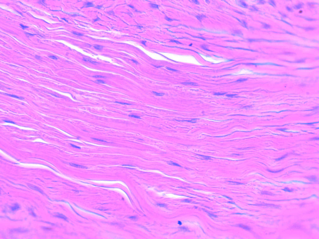
What tissue is this
dense regular

what tissue is this
dense irregular

What tissue is this
elastic

What type of bone is this
spongy bone

What type of bone is this
compact bone
What does spongy bone contain
trabeculae and bone marrow
Where is spongy bone found
epiphysis
What does compact bone contain
osteons, ECM, blood vessels, and nerves
Where is compact bone found
diaphysis

What kind of cartilage is this
elastic

What kind of cartilage is this
fibrocartilage

What kind of cartilage is this
hyaline
What is a mature cartilage cell
chondrocytes

Where are chondrocytes found
in lacunae

What tissue is this
Adipose

What is the arrow pointing to on the myofibril
M Line

What is the arrow pointing to on the myofibril
I Band

What is the arrow pointing to on the myofibril
Thick Filaments (myosin)

What is the arrow pointing to on the myofibril
Thin Filaments (actin)

What is the arrow pointing to on the myofibril
A Band

What is the arrow pointing to on the myofibril
Z Disc/Z Line

What is the arrow pointing to on the myofibril
H Band
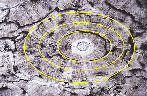
What is outlined on the bone
Lamella/Concentric Circles

What is the arrow pointing to on the bone
Central Canal/Haversian Canal

What is the outlined on the bone
Lacuna

What is the arrow pointing to on the bone (small lines)
Canaliculi

What is the arrow pointing to on the bone
Osteon

What is the arrow pointing to on the bone
Periosteum
Where can you find an osteocyte in bone tissue
Within the lacuna

What is #1 pointing to on the bone
endosteum

What is #9 pointing to on the myofibril
sarcomere

What is #2 pointing to on the muscle
epimysium

What is #3 pointing to muscle
perimysium

What is #4 pointing to on the muscle
endomysium

What is #5 pointing to on the muscle
Myofibril

What is the small blue sheath around on the muscle
fascicle
specialized intercellular connection that directly links the cytoplasm of adjacent cells
Gap Junction

What is #1 pointing to on the synapse
postsynaptic membrane

What is #2 pointing to on the synapse
synaptic cleft

What is #3 pointing to on the synapse
neurotransmitter

What is #4 pointing to on the synapse
synaptic vesicle

What is #5 pointing to on the synapse
presynaptic neuron
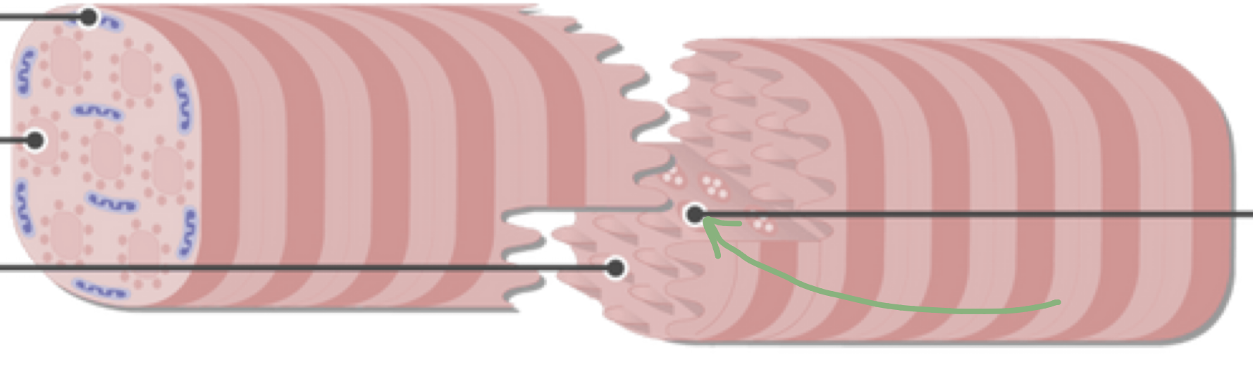
What is the green arrow pointing to cardiac muscle
Gap Junction

What are the black arrows pointing to on the cardiac muscle
intercalated disc
essential chemical messenger (neurotransmitter) that transmits signals across the gap between a nerve cell and another cell
ACH (acetylcholine)

What tissue is this
pseudostratified columnar