Abdomen Anatomy + Positioning & Technical Factors
 Call Kai
 Learn
 Practice Test
 Spaced Repetition
 Match
 Flashcards
 Knowt Play
1/34

There's no tags or description
Looks like no tags are added yet.
35 Terms
AP Abdomen (KUB)
demonstrates borders of psoas major muscles
right under diaphragm
next to spine
stretches out to the top of the femur
Digestive Tract Accessory Organs
not very visible on X-ray
liver
gallbladder
pancreas
spleen
Stomach & Parts of the Small Intestine
Stomach
has rugae or folds
Small Intestine
duodenal bulb
duodenum - shortest and widest
jejunum (2/5 of SI)
ileum (3/5 of SI)
ileocecal valve
Large Intestine
ileocecal valve
cecum
appendix (vermiform)
ascending colon
right colic (hepatic) flexure
transverse colon
left colic (splenic) flexure
descending colon
sigmoid colon
rectum
anus

Order All 11 Parts of the Large Intenstine
ileocecal valve
cecum
appendix (vermiform)
ascending colon
right colic (hepatic) flexure
transverse colon
left colic (splenic) flexure
descending colon
sigmoid colon
rectum
anus
Urinary System Organs
will need contrast to see in an intravenous urogram (IVU)
kidneys
ureter
bladder
urethra
suprarenal/adrenal glands

Label the 9 Parts of This Cross Section
mesentery (double fold)
small bowel
peritoneal cavity
parietal peritoneum
visceral peritoneum
retroperitoneal
right & left kidneys
inferior vena cava
aorta

Retroperitoneal Organs
Nine Organs
adrenal glands
kidneys
pancreas
duodenum
ascending colon
inferior vena cava
aorta
descending colon
ureter
(upper rectum)
Infraperitoneal Organs
Three Organs
bladder
lower rectum
male reproductive (closed sac) or female reproductive (open sac) organs
Intraperitoneal Organs
Eight Organs
liver
gallbladder
spleen
stomach
jejunum
ileum
cecum
transverse colon and sigmoid colon
Abdominal Quadrants & Organs in Each
RUQ
liver
galbladder
right colic (hepatic) flexure
duodenum
head of pancreas
right kidney
right supradrenal gland
LUQ
spleen
stomach
left colic (splenic) flexure
tail of pancreas
left kidney
left supradrenal gland
RLQ
ascending colon
appendix
cecum
2/3 of ileum
ileocecal valve
LLQ
descending colon
sigmoid colon
2/3 of jejunum
Abdominal Regions
right hypochondriac
epigastric
left hypochondriac
right lateral (lumbar)
umbilical
left lateral (lumbar)
right inguinal (iliac)
pubic (hypogastric)
left inguinal (iliac)
Planes Dividing the Abdominal Regions
right lateral plane
left lateral plane
transpyloric plane
transtubercular plane

Label the Abdominal Regions & Planes
right hypochondriac
epigastric
left hypochondriac
right lateral (lumbar)
umbilical
left lateral (lumbar)
right inguinal (iliac)
pubic (hypogastric)
left inguinal (iliac)
right lateral plane
left lateral plane
transpyloric plane
transtubercular plane
Topographic Landmarks
felt by palpation
mid & upper abdomen landmarks
xiphoid process (T9-T10)
inferior costal margin (L2-L3)
iliac crest (L4-L5)
lower abdomen and pelvic landmarks
ASIS (anteriorsuperior iliac spine)
greater trochanter
symphysis pubis
ischial tuberosity

Label Everything Besides the Middle One
ASIS (anteriorsuperior iliac spine)
greater trochanter
symphysis pubis
ischial tuberosity
Positioning of Abdomen
KUB
Acute Abdomen Series
AP Supine Abdomen
AP Erect Abdomen
PA Erect Chest
Decubitus Positions
Lateral Decubitus
Dorsal Decubitus
Routine & Special Abdomen Positionings
Routine
AP supine abdomen (KUB)
Special
PA prone
Lateral decubitus (AP) abdomen
AP erect abdomen
Dorsal decubitus (lateral) abdomen
Patient Preparation
all clothing removed
opaque objects removed
hospital gown worn
pillow for head, clean linen on table
cover patient for warmth & modesty
Positioning Considerations
exposure on expiration
IR markers
collimation
Positioning Factors
kVp (70-85)
short exposure time
adequate mAs

What is this projection?
AP Supine Abdomen
AP Supine Abdomen
CR center to iliac crest
Evaluation Criteria
symphysis pubis visible (just clip the top)
kidneys & lower liver margin included
no rotation
no motion
exposures factors optimal

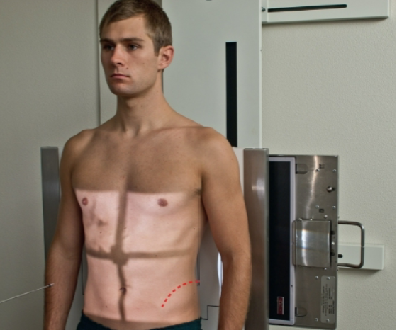
What is this projection?
AP Erect Abdomen
AP Erect Abdomen
CR 2 inches (5 cm) above iliac crest
top of IR in the axilla region
patient erect for at least 5 mins.
Evaluation Criteria
diaphragm included (unless PA chest taken)
no rotation
no motion
exposures factors optimal


What projection is this?
PA Prone Abdomen
PA Prone Abdomen
CR & IR to iliac crest
abdominal tissue will be more compressed than on AP projection
Evaluation Criteria
symphysis pubis visible
kidneys & lower liver included
no rotation
no motion
exposures factors optimal


What position is this?
Left Lateral Decubitus Position
Left Lateral Decubitus Position
CR 2 inches (5 cm) above iliac crest @ midsagittal plane
patient lateral recumbent on left because air collects between liver & diaphragm
gastric bubble down
left for 5 mins. for air-fluid levels
include upside marking
Evaluation Criteria
diaphragm demonstrated
both sides of body included
no rotation (of shoulders & pelvis)
no motion
exposures factors optimal


What is this position?
Dorsal Decubitus Right Lateral Position
Dorsal Decubitus Right Lateral Position
CR 2 inches (5 cm) above iliac crest @ midcoronal plane
patient supine
BARELY USED
Evaluation Criteria
diaphragm demonstrated
no rotation
no motion
exposure factors optimal


What is this projection?
Right Lateral Abdomen Position
Right Lateral Abdomen Position
CR perpendicular to table & centered at iliac crest
patient lateral & recumbent with no tilt
Evaluation Criteria
diaphragm included
no rotation
no motion
exposure factors optimal

Clinical Indications for Acute Abdomen Series
ileus (parastalsis stops)
ascites (accumulation of fluid in peritoneal cavity)
perforated hollow (bowel) viscus
intra-abdominal mass (neoplasms)
post-op (abdominal surgery)
Acute Abdomen Series 3-Way & 2-Way
3-Way
AP supine
AP erect
PA chest
2-Way
AP Supine
AP erect or left lateral decubitus
must include diaphragm