Muscle sensing and motor units
 Call Kai
 Learn
 Practice Test
 Spaced Repetition
 Match
 Flashcards
 Knowt Play
1/35

There's no tags or description
Looks like no tags are added yet.
36 Terms
What is the sensorimotor loop?
The idea that motor control is a layered loop: the brain sends commands down to the muscles, and sensors send feedback back up so movement can be continuously corrected
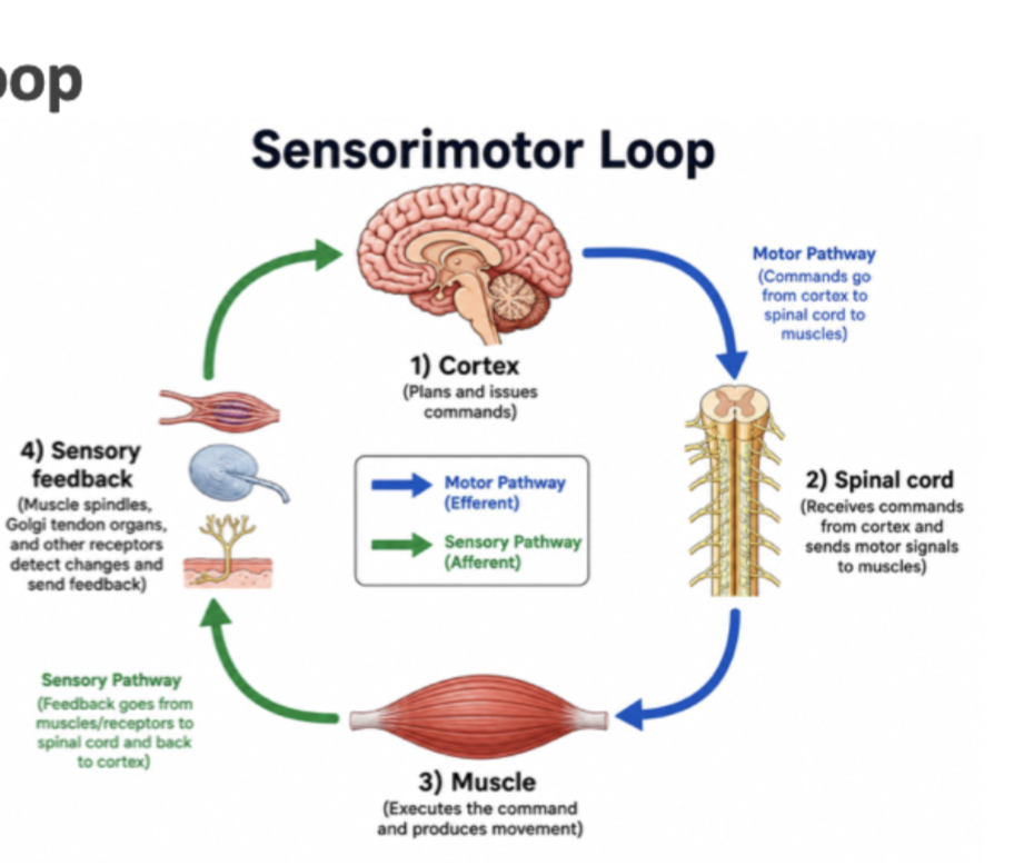
4 parts of the sensorimotor loop
Cortex: plans and commands the movement
Spinal cord: relays commands to muscle and runs fast automatic reflexes
Muscle: generates force and moves the body
Sensors: detect how stretched the muscle is (length) and how hard it’s pulling (tension), and send this info back up to the spinal cord and brain so they can fix the movement if it’s off
How does a command get from the brain to the muscle?
Through the motor (efferent, meaning “carrying away” from the brain) pathway: the cortex sends a signal to the spinal cord, where motor neurons innervate (connect to and activate) the muscles, and once muscles get this input, they contract
How does feedback get from the muscle back to the brain, and why does it matter?
Through the sensory (afferent, meaning “carrying toward” the brain) pathway: sensors in the muscles detect changes and send feedback into the spinal cord, then the cortex; this constant feedback lets us adjust our movements.
What is a skeletal muscle made of?
A bundle of muscle fibres: long, contractile cells (cells that can contract)
joint

DONT NEED TO KNOW THIS: A joint is where two bones meet and can move relative to each other (e.g. the elbow, where the upper arm bone meets the forearm bones).
How does a muscle move a joint?
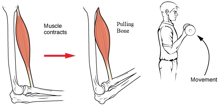
When the muscle fibres contract they shorten, pulling on the tendon, which pulls on the bone, so that bone moves at the joint (e.g. the biceps shortens → pulls the forearm up → the elbow bends)
Why do muscles work in opposing pairs?
Muscles can only PULL, not push, because they can only get shorter (relaxed = long, contracted = short); so one muscle pulls the joint one way and its partner pulls it back (e.g. biceps vs triceps)
Alpha (lower) motor neuron
Neurons with cell body in the ventral (front) spinal cord whose axon runs out to muscle and gives it input, and their axons can be very long (e.g. reaching the toes)

axons travel together in the motor nerve
What is a motor unit?
One motor neuron + all the muscle fibres it controls.
Fire the neuron, and every one of those fibres contracts together (like a puppeteer holding one string tied to many hands: a single pull moves them all at once).
How does motor unit size relate to control vs force?
Small units (a few fibres) allow fine control (e.g. eye muscles); large units (many fibres) produce big force (e.g. biceps).
fine control
Fine control is making small, precise movements (e.g. eyes, fingers). Small motor units have few fibres, so recruiting one more adds only a little force, which lets the brain adjust force in small steps.
lots of motor units (manu fibres) produce big force (biceps)
Motor neuron pool
The whole set of motor neurons that supply a single muscle (e.g. all the motor neurons that project to the gastrocnemius, a calf muscle)
Where are the cell bodies of a motor neuron pool located?
Clustered together in a column running lengthwise (up and down) in the spinal cord, spanning several spinal cord segments


How does the spinal cord turn a muscle up or down?
By changing how many neurons in the pool it activates. Each neuron controls its own group of fibres (its motor unit). Activating fewer neurons means fewer fibres contract, so the force is weaker. Activating more neurons means more fibres contract, so the force is stronger
What are the two ways the spinal cord controls how hard a muscle pulls? (or the 2 knobs that control how hard a muscle pull)
recruitment
rate coding
Recruitment
Activating more motor neurons, so more muscle fibres contract. The cord adds small (weak) units first, then larger ones, so force builds smoothly from a feather-light touch to a heavy lift.
What is rate coding?
Firing each motor neuron faster. More action potentials per second means each unit’s fibres produce more force.
add more players (recruitment) and ask each to play louder (rate coding).
Two kinds of motor neuron activity
Phasic (brief, vigorous bursts of action potentials for fast, dynamic movements)
Tonic (steady, sustained low-level firing to hold a position)

phasic left and tonic right.
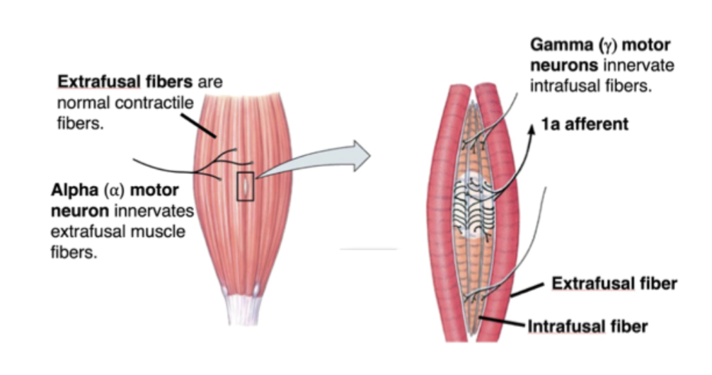
Extrafusal fibre
The ordinary muscle fibres that generate force

Intrafusal fibre
Tiny fibres inside the muscle spindle; sensors, not force producers

Alpha (α) motor neuron
Drives the extrafusal fibres → movement
Gamma (γ) motor neuron
Drives the intrafusal fibres → keeps the spindle sensitive
dont memorize just explanation: The spindle can only sense stretch when it’s pulled tight. When the muscle contracts and shortens, the spindle goes loose. Gamma motor neurons tighten it back up by contracting its intrafusal fibres, so it can keep sensing changes in muscle length
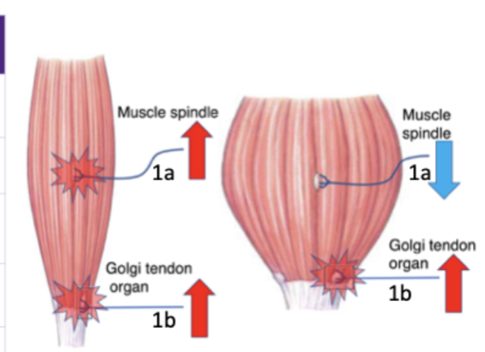
1a afferent
Sensory fibre from the spindle → reports muscle stretch/length

1b afferent
Sensory fibre from the tendon organ → reports muscle tension/force
What is a muscle spindle, and where is it found?
A small sensory organ (stretch detector) lying among the muscle fibres, inside the muscle. It’s made of intrafusal fibres, which are thinner and shorter than regular muscle fibres. Its job is to detect stretch of the muscle.
How does a muscle spindle detect stretch
1a afferent fibres (mechanoreceptors, meaning sensors that respond to physical deformation) wrap around the intrafusal fibres. When the muscle is stretched, the spindle stretches too, which stretches the 1a ending and opens its ion channels. The 1a afferent fires action potentials into the dorsal (back) part of the spinal cord, reporting LENGTH and how fast it is changing.

purple shape is the muscle spindle
Which motor neurons drive the extrafusal vs intrafusal fibres?
Alpha motor neurons drive the extrafusal fibres (the normal contractile fibres that give you the contraction).
Gamma motor neurons, which also sit in the spinal cord, drive the spindle’s own intrafusal fibres
How do you relax a muscle?
Remove the excitation going to it (the alpha motor neurons stop sending signals)
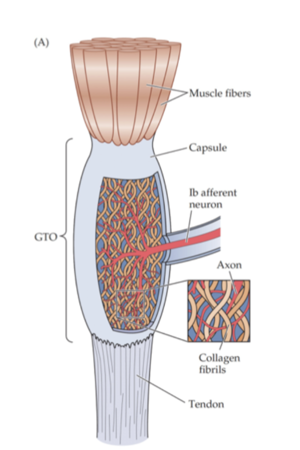
Golgi tendon organ (GTO)
It sits in the tendon, in line with the muscle’s pull. It detects the strain on the muscle, meaning how tightly the muscle is contracting (tension/force)
How does the GTO detect tension?
It contains mechanoreceptors (1b afferent endings) woven between the collagen fibrils of the tendon. When the muscle pulls, the tendon is strained, ion channels open, and the 1b afferent fires in proportion to TENSION, meaning how hard the muscle is actually pulling, whether from stretch or contraction
Why do we have both muscle spindles and Golgi tendon organs?
They sense different things:
the spindle watches length (1a afferent),
the tendon organ watches force (1b afferent)
Spindle vs golgi tendon organ

Where is each sensor positioned relative to the muscle fibres?
The spindle (and its 1a afferent) sits parallel to the muscle fibres (side by side with them).
The GTO sits in series, within the tendon (in line with the muscle, end to end, like links in a chain)
Alpha–gamma coactivation
One command activates both α (shortens the extrafusal fibres) and γ (tightens the intrafusal fibres) at the same time, so the spindle stays taut and keeps sensing length
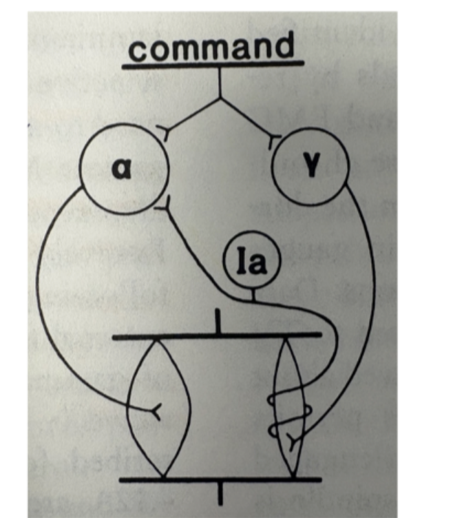
Like reeling in a fishing line as you walk toward the water: keep it taut and you can still feel a bite (you want some tension on the line so you’ll feel if the fish is biting)
What problem does alpha–gamma coactivation solve?
If only alpha fired, the muscle would shorten and the spindle inside it would become loose (no longer pulled tight). A loose spindle can’t sense stretch, so it would stop reporting muscle length. The brain and spinal cord would then lose track of how long the muscle is in the middle of the movement