Lecture 1 - Proteins
 Call Kai
 Learn
Learn Practice Test
 Spaced Repetition
 Match
Match Flashcards
 Knowt Play
1/89

There's no tags or description
Looks like no tags are added yet.
Name | Mastery | Learn | Test | Matching | Spaced | Call with Kai | Chat |
|---|
No analytics yet
Send a link to your students to track their progress
90 Terms
What are some functions of proteins?
Promote chemical reactions
Plasma membrane proteins
Signaling b/w cells and within cells
Structural proteins
Motorptoeins
Antibodies
Hormones
What constitutes most of a cell’s dry mass?
Proteins
What are proteins composed of?
Amino Acids
What does an amino acid consist of?
An amino group, carboxyl group, hydrogen and side chain (R) attached to the alpha carbon

Which aa are polar?
Aspartic acid, Glutamic acid, Arginine, Lysine, Histidine, Asparagine, Glutamine, Serine, Threonine, Tyrosine
Which aa are nonpolar?
Serine (Ser / S)
Threonine (Thr / T)
Cysteine (Cys / C)
Asparagine (Asn / N)
Glutamine (Gln / Q)
Tyrosine (Tyr / Y)
How is a peptide bond formed?
alpha-carboxyl group of one amino acid is joined to the alpha-amino group of another amino acid
What is lost when a peptide bond is formed?
Water
What is a steric (spatial) effect?
An effect in which the shape of a molecule influences its reactions
What is the arrangement of peptide bond?
Planar and does not permit rotation
Why is the peptide bond rigid?
Bond between the carbonyl carbon and nitrogen atom of the peptide bond has partical double-bond character
Where are polypeptides made?
In the ribosomes
What is the beginning of a polypeptide chain?
The N (amino) terminal end
What type of bonds are involved in protein folding?
Weak noncovalent bonds
What is protein conformation?
The three dimentional structure of a protein
What three types of noncovalent bonds are involved in protein folding?
Hydrogen bonds, Ionic bonds, Van der Waals attractions
What are hydrogen bonds?
Electropostive hydrogen atom is shared between two electronegative atoms (Nitrogen, Oxygen, Flourine)
What is an ionic bond?
Electrostatic interaction between oppositely charged atoms
What are Van der Waals attractions?
Short-distance interactions between the fluctuating electrical charges of the electron clouds around two atoms
How do hydrophobic nonpolar side chains behave in an aqueous environment?
They tend to be forced together in order to minimize disruption of the hydrogen-bonded network of surrounding water molecules
Where do nonpolar (hydrophobic) side chains of aa’s tend to do?
Cluster in the interior of proteins
Where do polar (hydrophilic) side chains of aa’s tend to do?
Tend to be on the outside of proteins and h-bonded to other polar aa’s or the polypeptide backbone
What is formed when nonpolar amino acid side chains are buried on the inside of a folded protein?
A tightly packed hydrophobic core hidden from water
What does lysozyme do?
Breaks down peptidoglycan
What are disulfide bonds (S-S bonds)?
Covalent links formed between the sulfur atoms in the side chains of cysteines
Between which structures can disulfide bonds form?
Between cysteines in the same protein or between different polypeptide chains in a multisubunit protein
What are the disulfide bonds function?
Stabilize protein structure (folding or mediate attachment of protein subunits)
What are intrachain disulfide bonds?
Joining two parts of the same polypeptide
What interchain disulfide bonds?
Joining two different polypeptide chains
Where is the formation of disulfide bonds catalyzed?
Endoplasmic reticulum
Why do disulfide bonds fail to form in cytosol?
High concentration of reducing agents that convert S-S bond to -SH groups
Why can disulfide bonds have a major stabilizing effect on protein structure, even though they're just one type of bond?
Covalent bonds need much more energy to break than noncovalent bonds → strong "staples."
Where are disulfide bonds often found?
Extraceullar proteins
What is the first level of protein structure?
Primary—the amino acid sequence of a protein determines its conformation (3-D structure)
What is the second level of protein structure?
Secondary—the folding of polypeptide chains into ordered structures maintained by repetitive hydrogen bonding
What are the most common types of secondary-level proteins?
Alpha helix and beta pleated sheets
What is the third level of protein structure?
Teritary— the full 3-dimensional organization of a polypeptide chain
What is the fourth level of protein structure?
Quaternary—the complete 3-dimensional structure of multi-subunit protein complexes (only applies to multi-subunit protein)
What does protein function depend on?
Proper conformation
Why is it theoretically possible to have 20ⁿ different proteins of n amino acids long?
Because each of the 20 amino acids can occur at any position in the chain → 20 choices per position × n positions = 20ⁿ possible proteins.
What defines secondary protein structure (α-helix & β-pleated sheet)?
Regular, repeating conformations from H-bonding between amino & carbonyl groups of the polypeptide backbone — NOT side chains.

/What does an alpha helix consist of
Single polypeptide chain that twists around on itself to form a rigid cylinder

Where do H-bonds on alpha helix occur?
Occur between every fourth peptide bond

What does the H-bond link?
Carbonyl group (C=O) of one peptide bond to N-H of another

What are the key features of an α-helix?
Turn every 3.6 aa; H bonds parallel to helix.
What is the length of each turn of an alpha helix?
3.6 residues (pitch 0.54 nm)
How do α-helices allow cell membrane proteins (transporters, receptors) to cross the lipid bilayer?
Nonpolar side chains face out toward lipids; hydrophilic backbone H-bonds inward → stable α-helix in membrane.
What is a coiled-coil and how does it form?
2–3 α-helices wrap together, nonpolar side chains face inward → stable rod. E.g., α-keratin, myosin.

In a coiled-coil, how are amino acid side chains arranged along a single α-helix?
Sevenfold "abcdefg" repeat; hydrophobic "a" & "d" cluster (every four apart) → stripe on helix surface.

How do two α-helices arrange themselves in a coiled-coil?
Hydrophobic stripes face inward (each other); hydrophilic side chains face outward (water).
What factors destabilize an α-helix?
Like charges repel (Asp/Glu or His/Lys/Arg)
Bulky β-carbon R groups crowd the helix (Ile, Thr, Val)
Why is proline incompatible with the α-helix?
Proline = helix breaker:
Restricted rotation (cyclic structure)
No H bonding (α-amino group can't participate)
In which protein was the β-pleated sheet first identified?
Fibroin — the major constituent of silk.
What do β-pleated sheets and α-helices have in common?
Both involve H-bonding between carbonyl and amino groups of peptide bonds → form a regular, repeating conformation.
How do H-bond directions differ between β-sheets and α-helices?
β-sheet: H bonds are perpendicular to the direction of the protein chain
α-helix: H bonds are parallel to the helix axis
What chain arrangements are possible in β-pleated sheets (unlike α-helices)?
H bonds can form:
Intrachain — between different parts of a single chain doubled back on itself
Interchain — between different chains
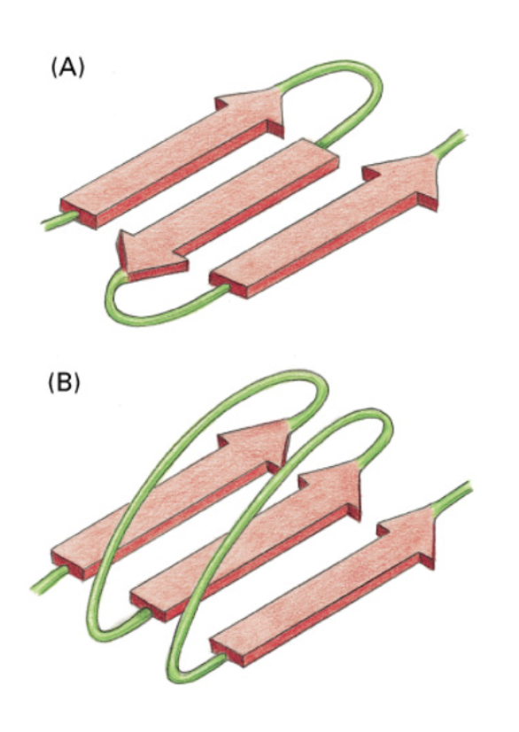
What are the two types of β-pleated sheets?
Parallel = same direction (N→C aligned)
Antiparallel = opposite directions (N/C flipped)

What does “pleated” mean in β-pleated sheets?
Refers to the zigzag nature of the structure

What are the three representations of antiparallel β-sheets?
Left: All atoms of backbone shown with H bonds
Middle: Only atoms of backbone shown
Right: Shorthand ribbon drawings
What are the two types of β-sheet structure, and where are they found?
Antiparallel & parallel → both in protein core; both rigid (H-bonded).
Besides the α-helix, what other helical structures can disrupt it?
Short stretches of other helices characterized by different numbers of amino acids per turn (e.g., 2, 3, 4.4), held together by H bonds.
What are reverse turns in secondary structure?
Regions where the chain changes direction or folds back.
Which amino acids are typically found in reverse turns, and why?
Glycine — small R group prevents aa crowding (steric reasons)
Proline — cyclic structure facilitates turn structure
What are motifs?
Repeated combinations of supersecondary structure within a protein (usually short repetitive units)
What is a domain or module?
A specific region of protein that can fold independently of the rest of the protein into a discrete stable structure and which has its own function
What is the difference between a motif and domain?
A protein domain can fold and function independently, while a protein motif cannot fold on its own and relies on the rest of the protein structure for stability
Is there a sharp distinction between domain and motif?
No clear-cut boundaries
How long are domains?
40-350 aa’s long
What are the modular units from which many larger proteins are construcuted?
Domians (modules)
How many domains may a small protein contain?
A single domain
How many domains may a larger protein contain?
Several dozen domains
In larger proteins with many domains, how are the domains usually connected?
By relatively short unstructured lengths of polypeptide chain
What is the relationship between protein size and number of domains?
Small proteins → may contain a single domain; larger proteins → may contain several dozen domains.
True or false: Domains are the modular units from which many larger proteins are constructed.
True
What are multidomain proteins?
Believed to have originated when the DNA sequence encoding each domain accidentally became joined, creating a new gene
What is domain shuffling?
The rearrangement/recombination of domains between proteins, creating new protein architectures (illustrated by EGF and protease domains)
Through mutation, where do novel binding surfaces often appear?
At the junction (or borders) of different domains
What can be created through mutations at domain junctions without affecting domain structure?
New binding sites for small molecules
True or false: New binding sites created at domain junctions by mutation disrupt the domain structure.
False — they are created without affecting the domain structure

Give an example of a protein formed from four domains
Src protein kinase
What is the function of Src protein kinase?
It is a cytoplasmic kinase that adds phosphate groups to the tyrosines of other proteins
What are the four domains of Src protein kinase, and how are they categorized?
Two regulatory domains (SH2 and SH3 — Src homology regions) and two catalytic domains
What does "SH2" and "SH3" stand for in Src protein kinase?
Src Homology region 2 and Src Homology region 3
In Src protein kinase, where is the ATP substrate located?
At the interface of the two kinase (catalytic) domains

How many fibronectin type 3 domains make up the extracellular matrix molecule fibronectin?
Four
What type of protein is fibronectin, and what kind of domains does it contain?
An extracellular matrix molecule composed of fibronectin type 3 domains
What does the EGF domain example illustrate about protein evolution?
Domains can be shuffled between different proteins, so the same domain (e.g., EGF) appears in combination with different domains in different proteins
What is a protease?
a type of protein enzyme that breaks down dietary proteins into smaller pieces called peptides or single amino acids
What is the key takeaway from comparing chymotrypsin with other proteases?
Proteins with related functions can differ in their domain composition — some are specialized and contain EGF and/or other domains in addition to protease domains