Week 6: nutrition, skin, assessment
 Call Kai
 Learn
 Practice Test
 Spaced Repetition
 Match
 Flashcards
 Knowt Play
1/127

There's no tags or description
Looks like no tags are added yet.
128 Terms
nutritional status
reflects the balance between nutrient intake and metabolic requirements
optimal nutritional status
undernutrition
overnutrition
optimal nutritional status
Achieved when sufficient nutrients are consumed to support daily body needs and increased metabolic demands
undernutrition
Occurs when nutritional reserves are depleted or nutrient intake is inadequate
Vulnerable Populations: Infants, children, pregnant women, recent immigrants, low-income individuals, hospitalized patients, and older adults.
Clinical Risks: Impaired growth/development, lowered infection resistance, delayed wound healing, longer hospital stays, and higher health care costs.
overnutrition
Caused by the consumption of nutrients (especially calories, sodium, and fat) in excess of body needs
Clinical Risks: Increases risk for obesity, type 2 diabetes, cardiovascular disease, osteoarthritis, sleep apnea, chronic kidney disease, gallstones, and acid reflux
obesity is multifactorial
culture and nutrition
Occupation, socioeconomic status, gender, religion, health awareness all impact eating customs
Religions use food as symbols in celebrations & rituals and also have restrictions:
Hinduism: lacto-vegetariansim favored
Islam & Orthodox Judaism: no pork or pork products, meat not slaughtered according to ritual
types of nutritional assessment
helps nurses determine whether a patient is getting enough nutrients, such as protein, vitamins, minerals, and calories, to stay healthy
malnutrition universal screening tool (MUST)
Mini nutritional assessment (MNA)
comprehensive nutritional assessment
24 hour diet recall
food frequency questionnaire
food diaries
mobile apps
malnutrition universal screening tool
Identifies patients who are at risk for malnutrition.
MUST evaluates three main factors:
Body Mass Index (BMI): Is the patient underweight?
Unintentional weight loss: Has the patient lost weight without trying during the past 3–6 months?
Acute illness: Is the patient seriously ill and likely to have no nutritional intake for more than 5 days?
malnutrition
occurs when the body does not receive the right amount or balance of nutrients. This can include undernutrition or overnutrition.
score for MUST test
0 | Low risk |
1 | Medium risk |
2 or more | High risk |
mini nutritional assessment (MNA)
Evaluates nutritional status and malnutrition risk, especially in older adults (65 years and older).
The MNA looks at factors such as:
Recent weight loss
Appetite and food intake
Mobility (ability to move around)
Recent illness or psychological stress
Cognitive problems, such as dementia
BMI or calf circumference
comprehensive nutritional assessment
Provides a detailed evaluation of a patient's overall nutritional health ( a complete picture of nutritional health)
This assessment is more thorough than MUST or the MNA.
It includes:
Medical history: Does the patient have conditions affecting nutrition?
Dietary history: What does the patient normally eat?
Physical examination: Are there signs of muscle loss, fat loss, or nutrient deficiencies?
Body measurements: Weight, height, BMI, and weight changes
Laboratory tests: Blood tests that may help identify nutritional problems when interpreted with other findings
24 hour diet recall
Determines what a patient ate and drank during the previous 24 hours.
The nurse asks the patient to remember everything they consumed yesterday.
This helps the nurse evaluate the patient's recent calorie and nutrient intake.
food frequency questionnaire
Determines how often a patient eats certain foods over a longer period, such as the past month or year.
The nurse might ask:
How often do you eat vegetables?
How many times per week do you eat fast food?
How often do you drink soda?
How frequently do you eat fruits?
This helps the nurse identify long-term dietary patterns.
food diaries
Tracks everything a patient eats and drinks over several days.
Unlike a 24-hour recall, the patient records their food intake as they eat throughout the day.
A food diary may include:
Type of food eaten
Amount consumed
Time of eating
Beverages consumed
Snacks
mobile apps
Uses technology to help patients track their nutritional intake (digital food diary)
Patients may use apps to monitor:
Calories consumed
Protein, carbohydrate, and fat intake
Water consumption
Meals and snacks
Dietary patterns
The nurse can review this information to help identify nutritional concerns.
subjective assessment
Eating patterns
Usual weight
Changes in appetite, taste, smell, chewing, swallowing
Nausea, vomiting, diarrhea, constipation
Food allergies or intolerances
Medications and/or nutritional supplements
Patient centered care
Meal preparation
Transportation/access to grocery store, income for food
Environment during mealtimes
nutrition for infants
Breastfed or bottle fed
Timing and method of weaning
Introducing solid foods
nutrition for children
Special likes or dislikes
How much do they eat
teaching about aspiration
general appearance
The nurse looks at the patient's overall physical appearance to identify signs of poor nutrition
inspecting for signs of cachexia
looking at anthropometric measures
cachexia
is a condition characterized by severe loss of muscle mass, often accompanied by loss of body fat and weight. (disease related muscle loss)
It is commonly associated with serious chronic illnesses, such as cancer, heart failure, or COPD.

signs of cachexia
Extremely thin appearance
Prominent bones (ribs, collarbones, shoulders)
Significant muscle wasting
Loss of body fat
Weakness and reduced strength
anthropometric measures
are measurements of the human body used to evaluate nutritional status and health risks
weight
BMI
waist circumference
weight
The nurse measures the patient's body weight and monitors changes over time.
Why is this important?
Unintentional weight loss may indicate malnutrition or illness.
Excessive weight gain may indicate increased body fat or fluid retention.
Sudden weight changes may indicate fluid imbalance.
body mass index (BMI)
is a measurement that compares a person's weight to their height, is only a screening tool. It does not directly measure body fat or determine nutritional health by itself
Below 18.5 - Underweight
18.5–24.9 - Healthy weight
25.0–29.9 - Overweight
30.0 or higher - Obesity
below 18.5
underweight BMI
18.5 - 24.9
healthy weight BMI
25.0 - 29.9
overweight BMI
30.0 or higher
obesity
waist circumference
Waist circumference measures the distance around a patient's abdomen.
Why is this important?
Excess fat around the abdomen is associated with an increased risk of:
Heart disease
Type 2 diabetes
High blood pressure
Metabolic syndrome
Larger waist circumference = Greater risk of certain metabolic and cardiovascular diseases
women
waist circumference = 35 inches
anything above this level is increased risk
men
waist circumference = 40 inches
anything above this level is increased risk
objective nutritional assessment
Look at the patient's general appearance for signs of muscle or fat loss.
Measure the patient's weight and calculate BMI.
Measure waist circumference when appropriate to evaluate abdominal fat-related health risks.
clinical signs of malnutrition
Skin: dry, flaking, scaling, petechiae or bruising,
Hair: dull, dry, or sparse
Eyes: plaques, dryness, pale or red conjunctivae
Lips: cheilosis, angular stomatitis
Tongue: glossitis, pale
Gums: bleeding
Nails, brittle, ridged, splinter hemorrhages
Musculoskeletal: pain in legs, osteomalacia,
rickets, joint pain
Neurological: hyporeflexia, disorientation or irritability
skin
dry, flaking, scaling, petechiae or bruising
hair
dull, dry, or sparse
Eyes
plaques, dryness, pale or red conjunctivae
Lips
cheilosis: Red, swollen cracks or sores at the corners of the mouth
angular stomatitis: Red, smooth, beefy-appearing tongue (often due to vitamin B complex deficiency
vitamin b complex deficiency
can cause red, smooth, beefy appearing tongue (angular stomatitis)
vitamin C deficiency
can cause bleeding gums
gums
bleeding
Nails
brittle, ridged, splinter hemorrhages
Musculoskeletal
pain in legs, joint pain
osteomalacia (soft bones)
rickets (bowing of leg bones in children due to vitamin D / calcium deficiency)
vitamin D / calcium deficiency
can cause rickets - bowing of leg bones in children
neurological
hyporeflexia - exaggerated or overactive reflexes
disorientation or irritability
melanin and skin tone
Melanin protects skin from UV rays
Incidence of skin cancer differs with skin tone
Differences in hair with ancestral descent
skin subjective assessment
History of skin diseases
Changes in pigmentation
Change in mole
Excessive dryness or moisture: seborrhea, xerosis
Pruritis
Excessive bruising
Rash or lesions
Medications
Hair loss: alopecia, hirsutism
Changes in nails
Environmental or occupational hazards
skin objective assessment
nurses observe the patient's normal skin pigmentation and look for unusual color changes
general pigmentation
vitiligo
pallor, cyanosis, erythema
jaundice
note any tattoos - Nurses should inspect tattooed areas for irritation, infection, or other abnormalities, documenting tattoos can help establish a baseline of the patient's skin appearance.
general pigmentation
pigmentation refers to the natural color of a person's skin.
Skin color is mainly determined by melanin, a pigment produced by cells in the skin.
During an assessment, the nurse looks for:
Changes in the patient's normal skin color
Unusually pale, blue, red, or yellow areas
Uneven pigmentation
Areas where skin color has been lost
vitiligo
is a condition in which the skin loses its natural pigment, causing lighter or white patches.
This happens because melanocytes (cells that produce melanin) are destroyed, usually through an autoimmune process.
White or lighter patches on the skin
Irregularly shaped areas of pigment loss
Can occur anywhere on the body
pallor
pale skin
It can occur when there is decreased blood flow to the skin or reduced hemoglobin.
cyanosis
occurs when the skin or mucous membranes develop a bluish or grayish appearance due to increased deoxygenated hemoglobin in the blood
erythema
means redness of the skin caused by increased blood flow near the skin's surface
Possible causes:
Inflammation
Infection
Allergic reactions
Sunburn
Pressure-related skin injury
jaundice
yellowing of the skin, yellowing of the sclera occurs when too much bilirubin builds up in the blood
Bilirubin is a yellow substance produced when the body breaks down old red blood cells. The liver normally processes bilirubin so it can be eliminated.
objective assessment pt 2
Moisture
Texture
Edema
Mobility & turgor - skins elasticity, how quickly skin returns tot its normal position after being gently pinched and released
lesions
Vascularity & bruising
Petechiae
Ecchymosis/Contusion/Hematoma
Cherry (senile) angiomas
petechiae
are tiny, pinpoint red, purple, or brown spots caused by bleeding from small blood vessels under the skin.
they do not blanch / turn pale when pressed
ecchymosis
A larger flat area of discoloration caused by bleeding under the skin
contusion
A bruise caused by an injury or trauma
hematoma
A localized collection of blood outside blood vessels, sometimes forming a swollen lump
cherry (senile) angiomas
are small, usually harmless red or purple growths formed by clusters of tiny blood vessels.
They become more common with age.
Small, round spots
Bright red to dark purple
Usually smooth and slightly raised
Common on the trunk, arms, and shoulders
lesions
any traumatic or pathologic changes in skin structure
can develop because of:
Injury or trauma
Infections
Allergic reactions
Skin diseases
Inflammation
types: primary and secondary
primary lesions
Traumatic or pathological changes on previously healthy, intact skin, represent the initial skin change
Examples include:
Macule: Flat spot with a different color, such as a freckle.
Papule: Small, raised bump, such as a pimple.
Vesicle: Small, fluid-filled blister.
Pustule: Raised lesion containing pus.
secondary lesions
develop when a primary lesion changes over time, such as from scratching, infection, or healing, the original lesion has changed
Examples include:
Crust: Dried blood or fluid on the skin (scab).
Scale: Flaking skin.
Ulcer: Open sore with loss of skin tissue.
Scar: Tissue that forms after an injury heals.
Excoriation: Scratch or abrasion caused by rubbing or scratching.
note for assessing lesions
Color
Elevation
Pattern or shape
Size
Location & distribution
Exudate (if any) - fluid or drainage coming from a lesion
lesion patterns
describes the shape or arrangement of skin lesions
annular
confluent
discrete
gyrate
grouped
linear
annular
ring shaped or circular
Lesion forms a ring.
The center may appear clearer than the edges.
Example: Ringworm (tinea corporis

confluent
lesions merge together
Several individual lesions join to form a larger area.
The borders between lesions become less distinct.
Example: Some widespread inflammatory or drug-related rashes.

discrete
separate, individual lesions
Lesions are not connected.
Each lesion has its own distinct borders.
Example: Several separate insect bites.
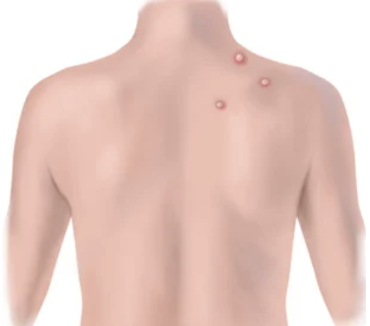
grouped
Lesions clustered closely together
Several small lesions appear in one area.
They remain distinguishable rather than merging into one large lesion.
Example: Herpes simplex blisters.

linear
Lesions arranged in a line
Lesions follow a straight or nearly straight path.
Example: Scratches or poison ivy contact dermatitis

gyrate
curved or winding lesions

primary skin lesions
macule / patch
papule
plaque
nodule
wheal / urticaria
vesicles / bulla
cyst
pustule
macule
flat, color changed spot eg. freckle (non palpable)

patch
flat macule larger than 1 cm (eg. vitiligo) (non palpable)
papule
solid, elevated lesion less than 1 cm (elevated mole, wart)
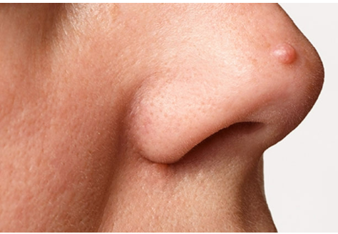
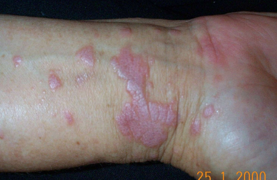
plaque
disk shaped, elevated lesion wider than 1 cm
nodule
solid, hard, or soft elevation extending deeper into the dermis, greater than 1 cm

tumor
Larger nodule, greater than 2-3 cm, firm or soft, deep into dermis.
wheal
Superficial, raised, transient, erythematous lesion with irregular borders caused by fluid in tissues (e.g., mosquito bite, allergic reaction)

urticaria (hives)
Confluent eruption of multiple wheals causing intense itching.
vesicles
Elevated cavity containing clear fluid, up to 1 cm (e.g., herpes zoster/shingles, varicella/chickenpox); fluid filled
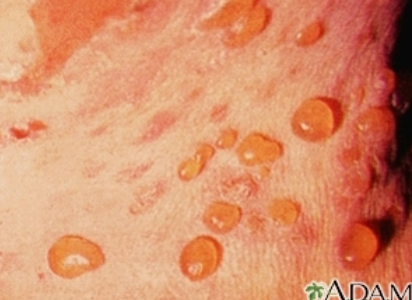
bulla
Elevated fluid-filled cavity larger than 1 cm (e.g., friction blister, severe burn)
bigger vesicle
pustule
Turbid, pus-filled cavity (e.g., acne, impetigo)

cyst
Encapsulated fluid-filled cavity in dermis or subcutaneous layer

secondary skin lesions
crust
scale
fissure
erosion
ulcer
excoriation
scar
atrophic scar
keloid
lichenification
crust
Thickened, dried-out exudate left when vesicles/pustules burst or dry up (e.g., impetigo)

scale
Compact, dried flakes of skin shedding excess dead keratin cells (e.g., psoriasis, eczema)

fissure
Linear crack with abrupt edges extending into the dermis (e.g., cheilosis, athlete's foot).

erosion
Scooped-out, shallow depression; superficial loss of epidermis (moist, no bleeding, heals without scar)
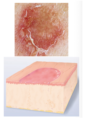
ulcer
Deeper depression extending into dermis, irregular shape, bleeds and leaves a scar (e.g., pressure injury, stasis ulcer)
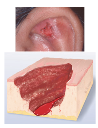
excoriation
Self-inflicted superficial abrasion from intense scratching (e.g., insect bite scratches)

scar
A scar is a mark left on the skin after an injury or wound heals.

atrophic scar
is a scar that appears sunken or depressed below the surrounding skin.

keloid
is an overgrowth of scar tissue that extends beyond the boundaries of the original wound.

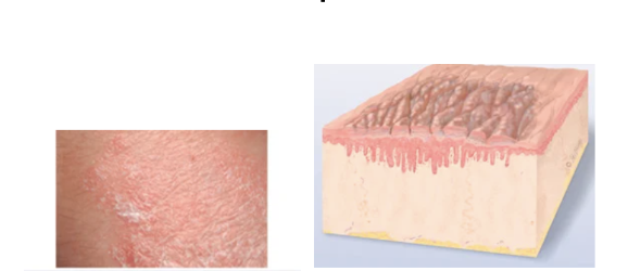
lichenification
Prolonged, intense scratching that thickens skin and produces tightly packed sets of papules (looks like surface of moss or lichen)
moles
Clump of melanocytes (nevus)

danger signs of moles
(ABCDEF) - looking at moles for potential malignant melanoma
Asymmetry - The two halves of the mole do not match.
Border irregularity - Notched, scalloped, ragged, or poorly defined edges.
Color variation - non uniform color; areas of brown, black, tan, blue, red, or white
Diameter - greater than 6 mm
Elevation - Elevation above skin surface.
Evolution - evolution/changes over time in size, shape, color, or symptoms (itching, bleeding)
Funny looking - (Ugly Duckling Sign): A mole that looks noticeably different from all the other moles on the client's body
pressure injuries
(also called a bedsore or pressure ulcer)
occur over bony prominences due to localized pressure, friction, or shear forces
occurs when prolonged pressure, or pressure combined with shear, damages the skin and underlying tissues.
Pressure injuries commonly occur in patients who are bedridden, use wheelchairs, or cannot reposition themselves
(4 stages)
stage 1
non blanchable erythema
The skin is still intact (not broken), but there is persistent discoloration caused by pressure.
Redness does not turn white when pressed (non-blanchable).
May feel warmer, cooler, firmer, or softer than surrounding skin.
In darker skin, discoloration may appear different from red.
stage 2
partial thickness skin loss
The epidermis and part of the dermis are damaged.
Open, shallow wound with a pink or red, moist wound bed.
May appear as an intact or ruptured serum-filled blister.
Fat, muscle, and bone are not visible.
stage
full thickness skin loss
The injury extends through the epidermis and dermis into deeper tissue.
Subcutaneous fat may be visible.
Wound is deeper than Stage 2.
Muscle, tendon, and bone are not exposed.