Chapter 5: Tissues (Epithelial, Connective, Muscular, Nervous)
 Call Kai
 Learn
 Practice Test
 Spaced Repetition
 Match
 Flashcards
 Knowt Play
1/81

Earn XP
Description and Tags
Anatomy & Physiology (McGraw Hill)
82 Terms
tissues
groups of similar cells and extracellular material that perform a common function
4 types: epithelial, connective, muscular, nervous

epithelial tissue
closely packed cells with minimal extracellular matrix
covers body surfaces
line body cavities
forms glands
classified by cell layers: simple, stratified, pseudostratified
classified by cell shape: squamous, cuboidal, columnar, transitional

characteristics of epithelia (6)
cellularity - cells packed tightly
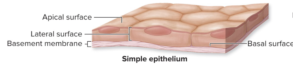
polarity - distinctive cell surfaces (apical, lateral, basal)
attachment to basement membrane (barrier between epithelium and connective tissue)
avascularity - no blood vessels
innervation - supplied with nerves to detect changes in environment)
high regeneration capacity due to wear/tear

function of epithelia
selective permeability
physical protection of internal and external surfaces
absorption (substances must cross to enter blood or lymph)
secretion (glands)
sensory reception
simple epithelium
one cell layer thick
all cells contact base membrane
filtration, absorption, or secretion is primary function
stratified epithelium
two or more layers of epithelia cells
only basal layer in contact with basal membrane
areas subjected to mechanical stress

squamous cells
flat/wide, irregular in shape, nucleus is flat with bulging center (like a fried egg)

cuboidal cell
height and width are similar, nucleus is spherical and central

columnar cell
slender and tall, nucleus is oval and oriented lengthwise in basal region

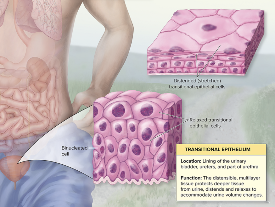
transitional cells
epithelial cells that can change shape, depending on stretch (ex: lining of bladder)
relaxed - basal cells are cuboidal or polyhedral, apical cells are round
stretched - apical cells flattened
binucleated - two nuclei
simple squamous epithelium
single layer of flat cells
thinnest barrier
function - rapid diffusion and filtration, serous membrane secretion
location - alveoli, endothelium (vessels walls), mesothelium/ (serous membranes)

simple cuboidal epithelium
single later of uniformly shaped cells
function - absorption and secretion, secretory tissue of glands and small ducts
location - kidney tubules, ovary surface, exocrine glands

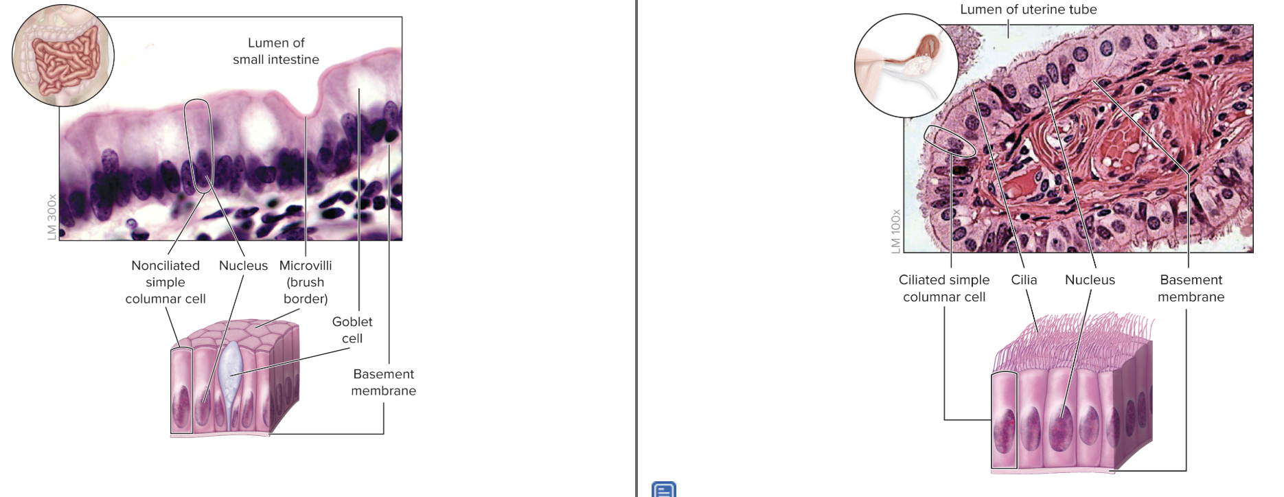
simple columnar epithelium
single layer of tall cells
function - absorption/secretion
nonciliated - may have goblet cells or microvilli “brush border”
location - GI tract
ciliated - cilia move substances across surface
location - fallopian tube (moving oocyte)
pseudostratified epithelium
appears as multiple cell layers, but all cells in direct contact with basement membrane
function - protection and/or secretion
ciliated - goblet cells secretes mucin, cilia moves/traps particles on apical surface
location - reparatory passageways
nonciliated - lack goblet cells and cilia, protection
location - male urethra and epididymis only

stratified squamous epithelium
apical cells are squamous, basal cells are cuboidal
basal stem cells continuously divide to replace lost cells at surface
function - protect underlying tissues from abrasion/friction
keratinized - superficial layers of dead cells contain keratin and lack nuclei
location - skin, hair
nonkeratinized - all cells alive, kept moist with secretions
location - esophagus, vagina

stratified cuboidal epithelium
apical cells are cuboidal
function - protection, secretion
location - exocrine gland ducts

stratified columnar epithelium
apical cells are columnar
function - protection, secretion
location (rare) - large ducts of salivary glands, conjunctiva, male urethra membrane

glands (overview)
cells or multicellular organs composed of predominately epithelial tissue
secrete substances for use within body or elimination from body
two types - endocrine, exocrine
endocrine glands
lack ducts
function - secrete hormones into blood
location - thyroid, adrenal
exocrine glands (overview)
discharge secretions directly onto epithelial surface via ducts
cell classification - unicellular or multicellular
anatomic classification - simple or compound; tubular, acinar, or tubuloacinar
secretory classification - merocine, apocrine, holocrine
unicellular exocrine gland
goblet cells
do not contain a duct
located close to epithelium surface
location - simple columnar, pseudostratified columnar

multicellular exocrine gland
ancini (“grape”) - clusters of cells that produce secretion
ducts - epithelium-lined tube where secretions are discharged
surrounded by fibrous capsule
septa (extensions of capsule) partition gland into lobes

simple exocrine glands
single, unbranched duct

compound exocrine glands
branched ducts

merocine glands
exocrine
function - package secretions into vesicles, released by exocytosis
location - salivary, sweat, lacrimal grands

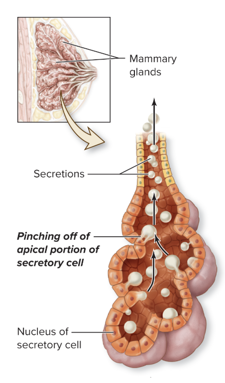
apocrine glands
exocrine
function - produce their secretions when cell’s apical surface pinches off, releasing cytoplasmic content
cell must repair itself to regenerate secretion
location - mammary glands
holocrine glands
exocrine
function - cell accumulates secretion and then disintegrates to release secretion and cell fragment
ruptured cells continuously displaced by epithelial cells
location - sebaceous glands

connective tissue (overview)
most diverse and abundant tissue, derived from mesenchyme
function - binds, supports, protects, storage, transport, immune response
location - tendons, ligaments, adipose, cartilage, bone, blood
3 components - cells, protein fiber + ground substance (extracellular matrix)
3 classifications - connective tissue proper, supporting connective tissue, fluid connective tissue

resident cells
cells permanently housed within connective tissue
Fibroblasts
Adipocytes
Mesenchymal
Fixed macrophages
Mast cells

wandering cells
cells that move continuously move throughout connective tissue
part of immune system
help repair damaged extracellular matrix
protect body against harmful agents
fibroblasts
most abundant resident cell in connective tissue
structure - flat cells with tapered ends
function - produce protein fibers and ground substance components of extracellular matrix

adipocytes
fat cell, resident cell in connective tissue
structure - cell full of lipid droplets, nucleus pushed to edge of cell
function - insulate, protects, stores energy

mesenchymal cells
embryonic stem cell, resident cell in connective tissue
structure - stellate or spindle shaped
function - divide to repair damaged tissue

fixed macrophages
resident cell in connective tissue, derived from monocytes (type of leukocyte)
structure - large, irregular shaped
function - engulf damaged cells or pathogens via phagocytosis, release chemicals to stimulate immune system/attract wandering cells

mast cells
resident connective tissue cell
migrates from bone marrow
structure - leukocytes
function - immune response, releases pro inflammatory molecules
protein fibers in connective tissue (overview)
strengthen and support tissue
3 types - collagen, reticular, and elastic fibers

collagen fiber
connective tissue protein fiber
structure - unbranched, fibrous
function - strong, resistant to stretching
location - tendons, ligaments (25% of body’s protein)

reticular fiber
connective tissue protein fiber
structure - interwoven 3D framework, thinner than collagen
same protein subunits as collagen, arranged differently
function - tough and flexible
location - stroma of organs
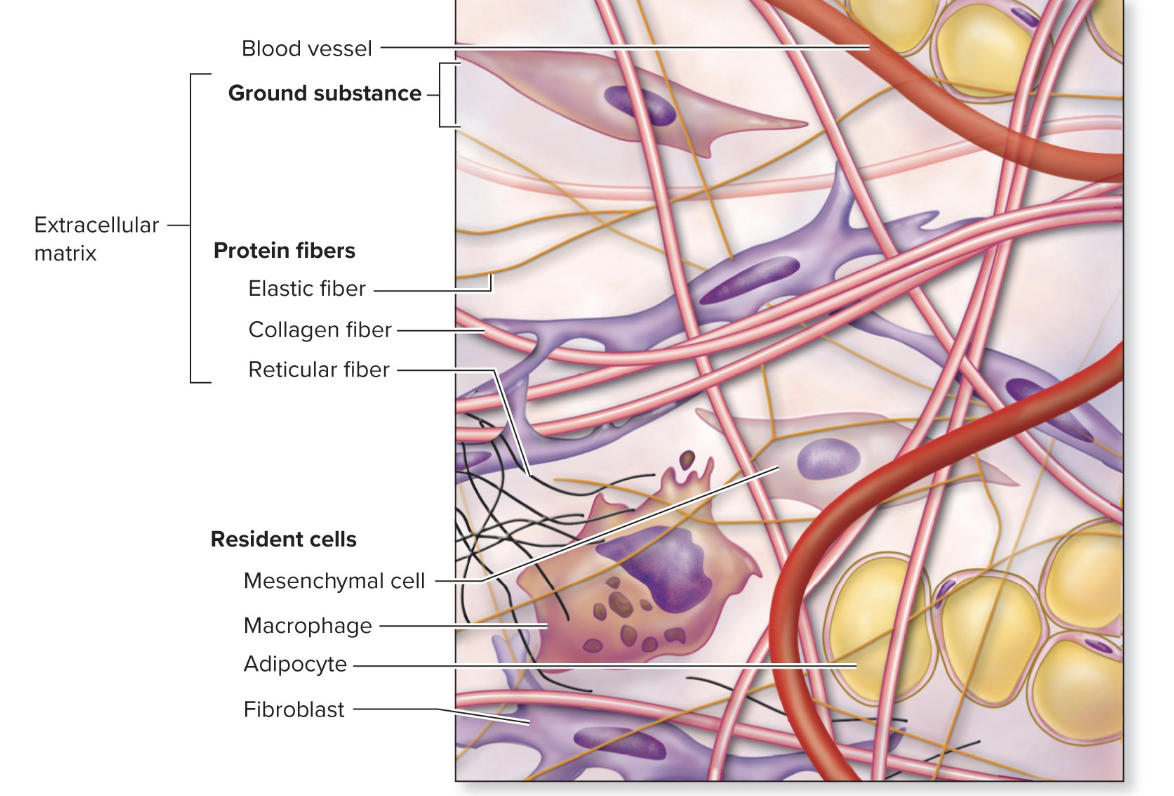
elastic fiber
connective tissue protein fiber
structure - branch and rejoin (wavy appearance), contain elastin protein
function - stretch and recoil easily
location - skin, walls of arteries

ground substance
molecular material produced by connective tissue cells
structure - viscous (blood), semisolid (cartilage), solid (bone)
GAGs pull water to change viscosity
function - houses connective tissue cells and protein fibers
glycoproteins act as glue to bond connective tissue cells and protein fibers to ground substance

mesenchyme connective tissue
embryonic connective tissue
first tissue to emerge from developing embryo
structure
cells - mesenchymal cells
protein fiber - immature protein fibers
ground substance - gel-like
location - origin tissue for all other connective tissues (proper, supporting, fluid) act as stem cells in adult connective tissue

mucous connective tissue
embryonic connective tissue
structure
cells - mesenchymal cells
protein fibers - more immature protein fibers
ground substance - viscous
location - umbilical cord only

connective tissue proper
classification of connective tissue
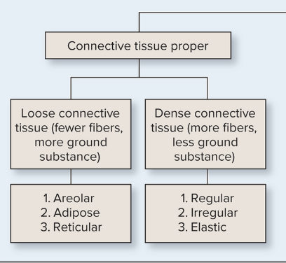
subclassifications based on proportions of cells, fibers, and ground substance
Loose Connective Tissue - fewer cells and fibers
areolar, adipose, reticular
Dense Connective Tissue - more fibers and less ground substance
regular, irregular, elastic
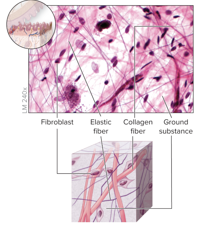
areolar connective tissue
loose connective tissue (connective tissue proper)
highly vascularized
structure
cells - mostly fibroblasts
protein fiber - unconfined organization of collagen and some elastin
ground substance - abundant and viscous
function - binds skin and epithelia to deeper tissues, protects/surrounds internal organs and vessels
location - skin (papillary + subcutaneous layers), organs
adipose connective tissue
loose connective tissue (connective tissue proper)
commonly referred to as “fat”
highly vascularized
structure
cells - adipocytes
protein fiber - sparse collagen and elastin hold cells together
ground substance - minimal
function - generates heat, insulation, protection, energy storage
brown adipose - newborns
white adipose - adults
location - subcutaneous layer, surrounds some organs

reticular connective tissue
loose connective tissue (connective tissue proper)
structure
cells- leukocytes, some fibroblasts
protein fiber - reticular fibers
ground substance - viscous
function - stroma (supportive framework) in lymphatic organs
location - spleen, lymph nodes, red bone marrow

dense regular connective tissue
dense, connective tissue proper
low blood supply, slower healing
structure
cells - few fibroblasts
protein fiber - abundant, tightly packed and stacked collagen
ground substance - limited
function - withstands stress
location - tendons, ligaments

dense irregular connective tissue
dense, connective tissue proper
highly vascularized
structure
cells - more fibroblasts than dense regular
protein fibers - bundles/clumps of projecting collagen fibers
ground substance - more than dense regular
function - resists stress
location - reticular layer of dermis, epimysium covering skeletal muscle, epineurium covering nerves, periosteum covering bone, perichondrium covering cartilage, capsules around organs

elastic connective tissue
dense, connective tissue proper
structure
cells - fibroblasts
protein fibers - branching, densely packed elastic fibers
ground substance - present between protein fibers
function - stretch and recoil
flocation - artery walls, trachea, vocal cords

cartilage
supporting connective tissue
structure
cells - chondrocytes
protein fibers - collagen and elastic
ground substance - semisolid
function - support, withstand compression
avascular - chondrocytes secrete chemical that inhibit blood vessel growth
slow healing
3 types: hyaline, fibrocartilage, elastic

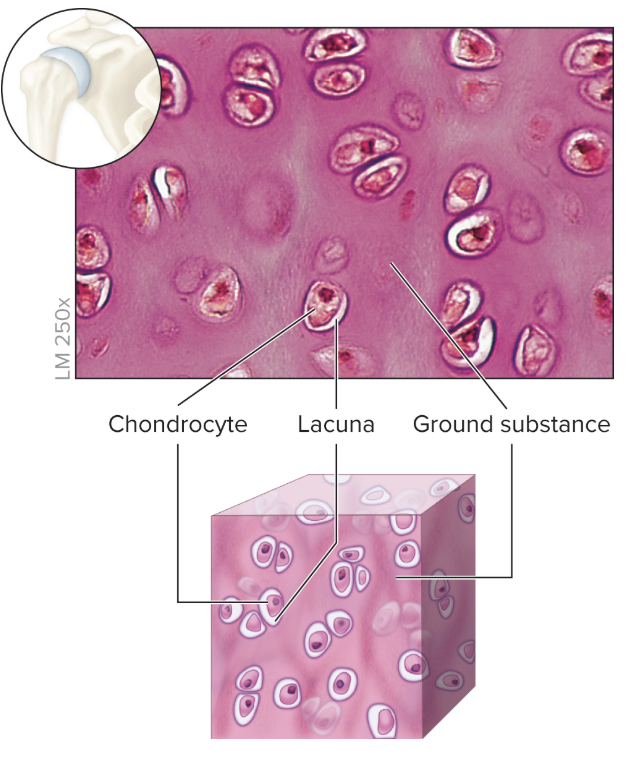
chondrocytes
mature cartilage cell
encased in lacunae (small space in extracellular matrix)

hyaline cartilage
supporting connective tissue
structure
cells - irregularly arranged chondrocytes
protein fibers - collagen (not visible)
ground substance - glycoproteins, proteoglycans, water (makes it compressible)
function - support
location - tip of nose, fetal skeleton, ribs, larynx, epiphyseal and articular ends of long bones
fibrocartilage
supporting connective tissue
structure
cells - large chondrocytes
protein fibers - numerous, coarse, irregular bundles between cells
ground substance - sparse
function - resists compression, shock absorber
location - intervertebral discs, menisci of knee

elastic cartilage
supporting connective tissue
structure
cells - closely packed chondrocytes
protein fibers - numerous, densely packed elastic fibers in weblike mesh
ground substance - sparse
function - flexible and maintains shapes
location - external ear, epiglottis of larynx

bone
supporting connective tissue
more solid, less flexible than cartilage
extensively vascularized
structure
cells - osteocytes
protein - collagen fibers (osteoid)
ground substance - crystal deposits
function- movement, support, protection, stores mineral, stores hematopoietic cells (red blood cell production)
2 types - compact, spongy
compact bone
supporting connective tissue
“dense”; “cortical”
form osteons - rings of lamellae (bone connective tissue)
lamellae encircle central canal that houses blood vessels and nerves

spongy bone
supporting connective tissue
“cancellous”; “trabecular”
contains latticework of bone connective tissue
strong, lightweight
located within interior of bone

blood
fluid connective tissue
structure
cells - erythrocytes, leukocytes, platelets (cellular fragments)
protein fibers - fibrin (helps form sticky mesh for clotting)
ground substance - plasma (water proteins, solutes)
function - transportation of materials, immune response
location - blood vessels, heart

lymph
fluid derived from blood plasma (water, protein, solutes)
no cellular component or fragments
skeletal muscle
“voluntary”, “striated”
structure
multinucleated cells
long and cylindrical, parallel and unbranched
function - movement of skeleton, thermoregulation
location - attached to bones

cardiac muscle
structure
cells have 1-2 nuclei
short, striated appearance
connected by intercalated discs
function
involuntary movement of heart initiated by pacemaker cells
intercalated discs strengthen connection and promote rapid conduction of electrical activity
location - heart
minimal capacity for regeneration

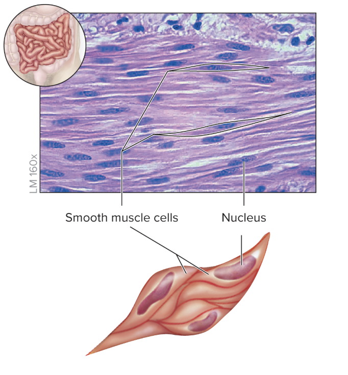
smooth muscle
structure
short fusiform (thick in middle, tapered end) cells
lack striation
function
involuntary contraction movement propels material through organ
controls size of lumen
location - walls of hollow internal organs, GI tract, stomach, airways, urinary tract, blood vessels
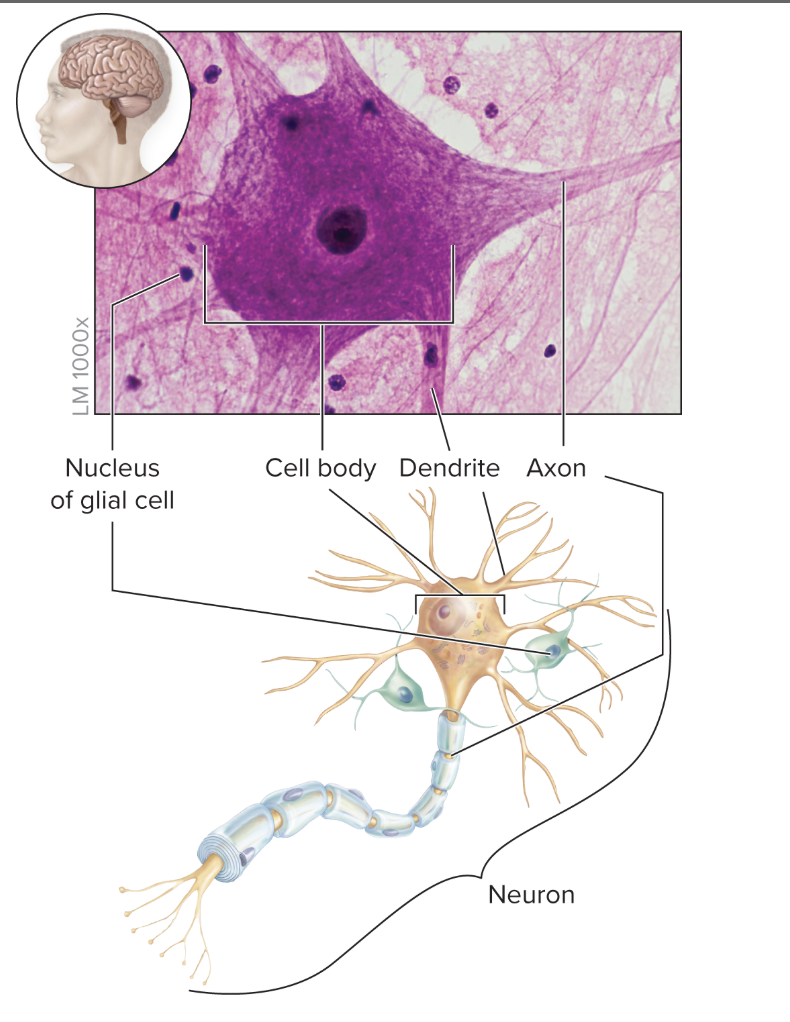
nerve tissue
structure
neurons have cell body, dentrites, axon
glial cells
function
neurons - receive, process, relay nerve impulses
glial cells - protect, nourish, support neurons
location - brain, spinal cord, nerves
body membranes
epithelial layer that is bound to underlying connective tissue
forms internal lining or external covering
4 types: mucous, serous, cutaneous, synovial
mucous membrane
mucosa
structure - epithelium and lamina propria (underlying connective tissue)
function - mucus secretion, absorb material, protection
location - lines passageways that open to external environment

serous membrane
structure
mesothelium (simple squamous epithelium)
thin underlying areolar layer (loose connective tissue)
function - produce thin, watery serous fluid derived from blood plasma
location - external surface of organs
parietal layer: lines body cavity
visceral layer: lines organ surfaces

cutaneous membrane
structure
keratinized stratified epithelium (epidermis)
underlying connective tissue (dermis)
function - protecting organs, prevent water loss
location - skin

synovial membrane
specialized type of connective tissue that secrete synovial fluid
function - reduce friction between bones, distributes nutrients to cartilage and bone
location - joints

zygote
diploid cell formed from sperm fertilizing an oocyte

blastocyst
multicellular structure formed when zygote undergoes multiple cell divisions

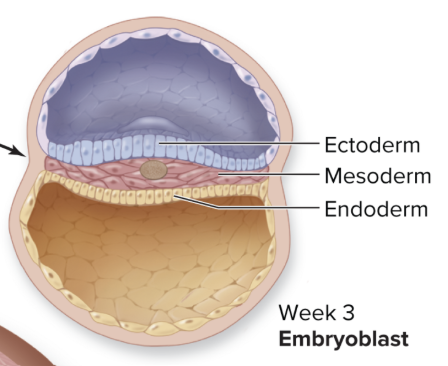
embryoblast
cells that form embryo
differentiate by 2-3 weeks development
develop into 3 primary germ layers: ectoderm, mesoderm, endoderm

ectoderm
germ layer that develops into:
epidermis of skin
epidermal derivatives (hair nails, mammary glands, sweat glands)
nervous tissue and sense organs
pituitary gland
adrenal medulla
teeth enamel
eye lens
mesoderm
germ layer tha develops into:
dermis of skin
epithelial lining of vessels and serous membranes
muscle tissue
connective tissue
adrenal cortex
heart
kidneys and ureters
reproductive organs
spleen

endoderm
germ layer that develops into:
epithelial lining of respiratory, GI, urinary, reproductive tracts
liver, gallbladder
pancreas
thyroid/parathyroid glands
thymus

embryonic stem cells
originate from dividing cells of zygote or embryoblast
totipotent
formed from zygote
give rise to every differentiated cell in organism and placenta
pluripotent
derived from embryoblast
able to mature and specialize into a cell within any body tissue (except placenta)

adult stem cells
undifferentiated stem cells after birth
multipotent - can differentiate into restricted number of some cell types
unipotent - can differentiate into single type of cell

hypertrophy
increase in size of existing cells in a tissue
number of cells remains constant
ex: muscle hypertrophy
hyperplasia
increase in number of cells in a tissue (ex: callus development)
atrophy
shrinkage of tissue by a decrease in either cell size or cell number
metaplasia
mature epithelium changes in response to environment (ex: smoker’s trachea)
dysplasia
abnormal cell development (size, shape, organization); may be precancerous or may revert back to normal
neoplasia
tumor growth from excessive cell proliferation
benign - localized, does not spread
malignant (cancer) - metastasizes/invades other tissues of body