NE203 Lecture 16 - Taste & Olfaction
 Call Kai
 Learn
 Practice Test
 Spaced Repetition
 Match
 Flashcards
 Knowt Play
1/122

There's no tags or description
Looks like no tags are added yet.
123 Terms
Gustation
The sense of taste
5 basic tastes
Sweet, sour, salty, bitter, umami
Other possible tastes
Metallic, carbonated, fatty (depending on context)
Cranial nerves carrying taste to the brain
VII (facial), IX (glossopharyngeal), X (vagus)

Taste pathway to cortex
Taste buds → cranial nerves VII, IX, X → nucleus of the solitary tract (gustatory nucleus) → parvicellular portion of ventral posteromedial nucleus (VPMpc) of thalamus → insular gustatory cortex (and frontal cortex)
Nucleus of the solitary tract
Brainstem gustatory nucleus; first relay for taste information from cranial nerves VII, IX, X
VPMpc
Parvicellular portion of the ventral posteromedial nucleus of the thalamus; thalamic relay for taste
Insular gustatory cortex
Primary cortical area for taste
Taste buds
Sensory end organs for taste
Where taste buds are found
Dorsal tongue, soft palate, larynx, pharynx, and upper esophagus
Taste bud size
Cluster of 40-80 cells

Circumvallate papillae
Taste papillae innervated by cranial nerve IX
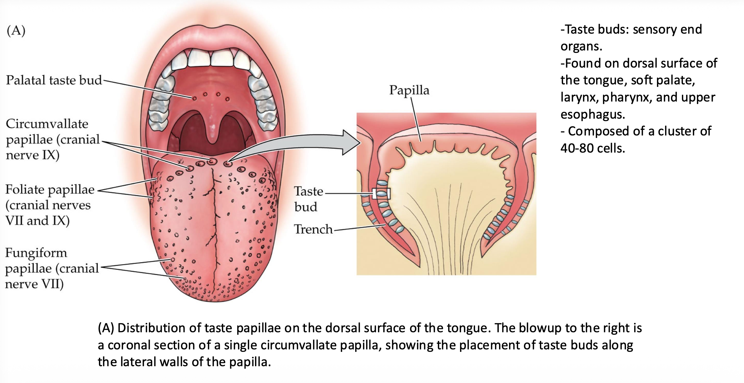
Foliate papillae
Taste papillae innervated by cranial nerves VII and IX

Fungiform papillae
Taste papillae innervated by cranial nerve VII
Palatal taste buds
Taste buds on the palate

Taste pore
Opening formed by the apical tips of taste bud cells where tastants are detected
Tastants
Chemicals that produce the sensation of taste
Microvilli (taste cell)
Finger-like projections at the apical end of taste cells at the taste pore
Basal cell (taste bud)
Cell in the taste bud that gives rise to new taste cells
Trigeminal afferent axons (in taste bud)
Axons near taste buds that carry touch, temperature, and chemesthetic sensations

Type I taste bud cell
Glia-like (astrocyte-like) support cell; performs many of the same functions as astrocytes in the CNS

Type II taste bud cell
Chemoreceptor for sweet, bitter, umami (and salty) tastants; specialized for only 1-2 submodalities; releases ATP through a channel

Type III taste bud cell
Chemoreceptor for sour (and some salty) tastants; releases serotonin at a synapse

Neurotransmitter of Type II cells
ATP, released through a channel (CALHM)

Neurotransmitter of Type III cells
Serotonin
Taste transduction for salty and sour
Ion channels
Taste transduction for sweet, bitter, umami
G-protein-coupled receptors (GPCRs)
Salty receptor
Amiloride-sensitive Na+ channel; Na+ enters and depolarizes the cell
Sour receptor
OTOP1 H+ channel; protons enter, and an inward rectifier K+ channel is blocked, depolarizing the cell
Sweet receptor
TAS1R2 + TAS1R3 heterodimer (GPCR)
Umami receptor
TAS1R1 + TAS1R3 heterodimer (GPCR)
Bitter receptor
TAS2R (GPCR) coupled to gustducin
Gustducin
G-protein coupled to bitter taste receptors (TAS2R)
TAS1R family
GPCR taste receptors for sweet and umami; they are heterodimers
Shared GPCR taste cascade (Type II cells)
Tastant binds TAS1R/TAS2R → G-protein activates PLCβ2 → IP3 → IP3 receptors on ER release Ca2+ → opens TRPM5 and voltage-gated Na+ channels → depolarization → opens CALHM channels → ATP release → ATP binds P2X receptors on gustatory afferent axon → action potentials
PLCβ2
Enzyme in the taste GPCR cascade that produces IP3
IP3 role in taste
Binds IP3 receptors on the ER membrane, releasing Ca2+ into the cytoplasm
TRPM5
Ion channel opened by Ca2+ in Type II cells; contributes to depolarization
CALHM channel
Voltage-gated large-pore Ca2+ homeostasis modulator channel through which ATP is released
P2X receptor
Ionotropic ATP receptor on the gustatory afferent axon that depolarizes it
Ageusia
Loss of taste
Dysgeusia
Altered or distorted taste
SARS-CoV-2 receptor
Binds angiotensin-converting enzyme-2 (ACE2) and is endocytosed
ACE2 expression in smell system
Expressed in olfactory sustentacular cells
COVID-19 and ageusia
About 1/3 of COVID-19 patients report ageusia
Where ACE2 might be in the taste system
On taste bud cells and/or on the surrounding lingual epithelium
Extraoral taste receptors
Taste receptors outside the mouth that act as sentinels, alerting the body to chemicals that must be chemically dealt with or physically removed
Orexigenic
Stimulates appetite
TAS1Rs in the intestine
Detect sugars; sugar absorption and GLP secretion (related to drugs like Zepbound and Ozempic)
TAS2Rs in the sinonasal epithelium
Detect bacterial metabolites; mucociliary clearance, NO secretion, antimicrobial peptides
TAS2Rs in bronchioles (smooth muscle)
Detect irritants; cause bronchodilation
TAS2Rs in the GI tract (SCC)
Detect pathogens and toxins; gut motility, vomiting, diarrhea
TAS1Rs in the stomach
Detect nutrients; orexigenic regulation
FFARs in the intestine
Detect fatty acids; various energy metabolism outcomes
GPR92 in the stomach
Detects protein hydrolysates and other nutrients; orexigenic regulation
Labeled line coding
Taste is mediated by specialized neurons each dedicated to one taste quality; each taste travels from receptor cells to cortex via a dedicated line
Across neuron pattern (ANP) coding
Population/distributed coding; each taste is represented by the combined activity of many neurons, and each neuron can respond to several tastes with excitation or inhibition
Unitary view of taste coding
Both labeled line and ANP coding are valid in different contexts
Labeled line coding may suit
Narrowly tuned neurons; initial acceptance or rejection of a taste; low arousal
ANP coding may suit
Broadly tuned neurons; integration of sensory, metabolic, and cognitive/emotional signals; high arousal
Gustatory cortex temporal coding
Early responses are somatosensory, middle responses are chemosensory (taste identity), and late responses are palatability-related
Flavor
Perception of food and drink that involves all senses: taste, smell, touch/texture, sound, and vision
Orthonasal olfaction
Smelling food while it is still external (through the nostrils)
Retronasal olfaction
Smelling food while it is in the mouth being eaten (odors travel through the retronasal passage)
Odor-taste association
Associating an odor with a taste allows the odor to acquire a gustatory quality
Integration of taste and smell
Gustatory and olfactory pathways share neural pathways and neurons, e.g., thalamic neurons and orbitofrontal cortex
Orbitofrontal cortex (OFC)
Cortical area that integrates taste, smell, and texture
Chemesthesis
Chemical sensations such as burning, cooling, and tingling, mediated by trigeminal afferent endings
Nerve for chemesthesis
Trigeminal nerve (V)
Receptors for chemesthesis
TRPV1-3, TRPA1, TRPM8, acid-sensing ion channels (ASICs), and K+ channels
TRP channels on cold-sensing neurons
Give "cold" sensations to certain chemicals (e.g., TRPM8 for cooling)
Capsaicin and substance P
Capsaicin releases substance P, which can diffuse to nearby gustatory receptors, causing cross-talk between gustatory and trigeminal nerves
Food texture
Stimulates mechanoreceptive trigeminal neurons and adds to the food experience
Sounds and images in taste
Sounds (e.g., a soda "pop") and images add to taste sensation and trigger expectations; visual stimuli cannot be referred to intraoral events
Visual cues and food quality
Brand names or someone else's reaction to food can create inferences about food quality
Gustatory and insular cortices integration
Integrate sounds and images; processing from the amygdala and mediodorsal thalamus
Insular cortex responses
Responds to images of food and facial expressions of disgust
Anticipatory neural responses (2 functions)
1) Prepare the cortex to encode gustatory stimuli 2) Guide food-related actions based on expectations
Olfactory receptor neurons (ORNs)
Neurons in the olfactory epithelium that bind odorants
Olfactory pathway
ORNs → olfactory nerve (CN I) → through cribriform plate → olfactory bulb → olfactory tract → pyriform cortex and other forebrain structures
Cribriform plate
Bone that olfactory axons pass through to reach the olfactory bulb
Olfactory bulb
Where ORN axons first synapse; becomes the olfactory tract
Pyriform cortex
Primary olfactory cortex; produces concentration-invariant odor representations
Olfactory epithelium cell types
Neurons (ORNs), supporting (sustentacular) cells similar to glia, mucus-secreting cells, and basal stem cells
Bowman's gland
Mucus-secreting gland in the lamina propria that sends mucus to the epithelium surface
Lamina propria
Layer under the olfactory epithelium that contains blood vessels, Bowman's glands, ensheathing cells, and macrophages
Olfactory ensheathing cells
Glia that wrap the unmyelinated axons of ORNs and support growth of new axons
Macrophages (olfactory epithelium)
Mediate immune surveillance
Sustentacular cells
Supporting cells in the olfactory epithelium that express ACE2
Zinc nasal sprays
Historically destroyed ORNs, causing irreversible anosmia
COVID-19 and anosmia
Infection destroys sustentacular cells and causes general inflammation in the olfactory epithelium, leading to anosmia
Vomeronasal system
Accessory olfactory system; prominent in carnivores and rodents, less in humans; targets brain regions for homeostasis (hypothalamus)
Vomeronasal organ (VNO)
Receptor cell population in the nasal epithelium that detects pheromones and kairomones
Vomeronasal receptor neurons (VRNs)
Sensory neurons of the VNO; similar in appearance to ORNs
VRNs vs. ORNs
VRN receptors, signal transduction pathways, and ion channels differ from those of ORNs
Pheromones
Chemical signals released by one animal that affect the behavior of the same species
Kairomones / allomones
Chemical signals from other species (e.g., predators) detected by the VNO
Olfaction and innate behaviors
Drives seeking food, mood, arousal, social approach, and avoidance of danger
ORN regeneration
ORNs and VRNs are at high risk from inhaled air and constantly regenerate from basal stem cells; they can find their targets faithfully
Basal cells (olfactory epithelium)
Stem cells that regenerate all cells of the olfactory epithelium within about 21 days after injury