A&P Exam 2
 Call Kai
 Learn
Learn Practice Test
 Spaced Repetition
 Match
Match Flashcards
 Knowt Play
1/96

There's no tags or description
Looks like no tags are added yet.
Name | Mastery | Learn | Test | Matching | Spaced | Call with Kai | Chat |
|---|
No analytics yet
Send a link to your students to track their progress
97 Terms

Fibroblasts
Cells that produce fibers and ground substance that form the matrix of connective tissue
mast cells
Cells that secrete heparin (inhibits blood clotting) and histamine (dilates blood vessels)
collagenous fibers
fibers made of collagen
tough, flexible, resistant to stretching
reticular fibers
thin collagen fibers coated with glycoprotein
form spongelike framework for certain organs
elastic fibers
made of elastin coated with glycoprotein
stretches and recoils

simple squamous epithelium
-allows for rapid diffusion/transport of substances through membrane in alveoli, the inner lining of blood vessels, and Bowman’s capsule of kidneys
-mesothelium of serous membranes that secretes lubricating serous fluid
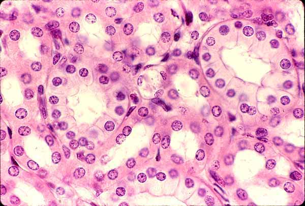
simple cuboidal epithelium
absorption & excretion ; production of mucous coat ; movement of respiratory mucus
bronchioles, most kidney tubules, glands such as mammary, thyroid, salivary, and liver
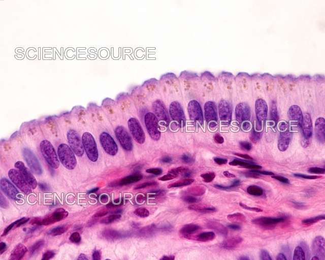
simple columnar epithelium
rapid absorption and secretion of protective mucus
inner lining of stomach, intestines, gallbladder, uterus, and uterine tubes

pseudostratified columnar epithelium
propels and secretes mucus
much of the inner lining of the airway including the nasal cavity, trachea, and bronchi

stratified squamous epithelium
can be keratinized or unkeratinized (mucous membranes)
keratinized: epidermis ; unkeratinized: oral cavity, vagina, esophagus, anal canal
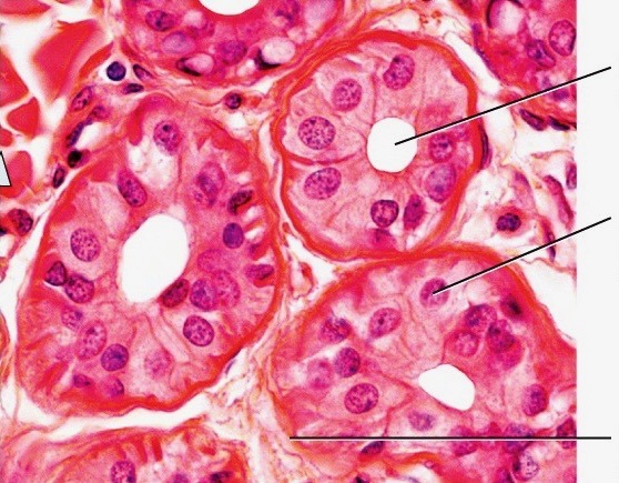
stratified cuboidal epithelium
contributes to sweat secretion, secretes ovarian hormones, produces sperm
sweat gland ducts, egg-producing vesicles of ovaries, sperm-producing ducts of testes
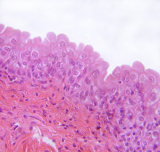
transitional epithelium/urothelium
can expand and retract; protects against osmotic damage
urinary tract: bladder, ureters, urinary ducts, part of kidney, part of urethra

dense connective regular tissue
densely packed collagen fibers and fibroblasts
resists stress, transfers tension, and forms tight bonds
tendons and ligaments
tendon
connects muscle to bone
ligament
connects bone to bone and stabilizes joints
joint
place where two bones meet

dense connective irregular tissue
wavy, nonuniform collagen fibers
imparts durability to tissues and resists stress in unpredictable directions
deep dermis, capsules around viscera, fibrous sheaths around cartilage and bone

areolar tissue
collagenous fibers, elastic fibers, and numerous blood vessels
provides nutrients to overlying epithelia and allows nerves and blood vessels to penetrate into other tissue
surrounds blood vessels and nerves and underlies almost all epithelia

adipose tissue
dominated by adipocytes
provides cushioning, energy storage, and thermal insulation
subcutaneous fat, heart surface, breast

reticular tissue
reticular fibers and lymphocytes
forms supportive sponge-like framework in many lymph tissues (stroma)
lymph nodes, spleen, bone marrow
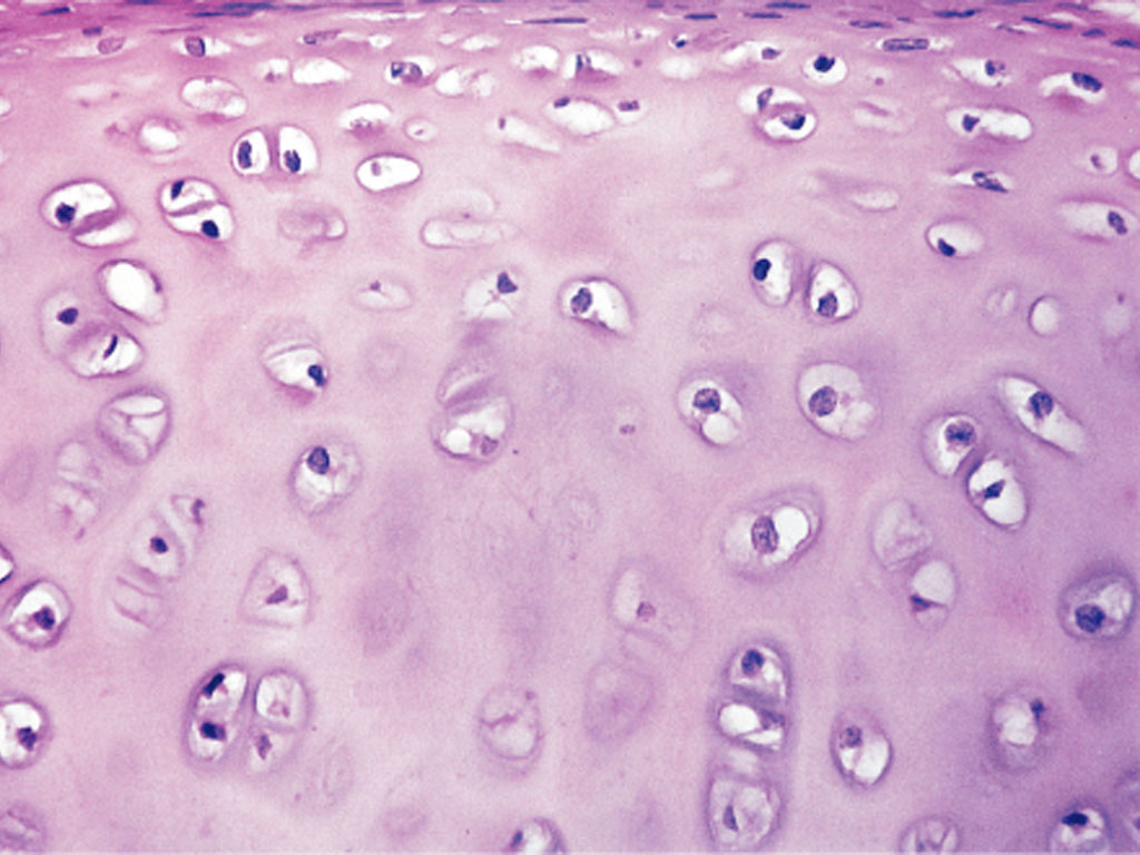
hyaline cartilage
fine, dispersed collagen fibers that are not usually visible
allows smooth movement of joints and growth of long bones
costal cartilage of rib cage, joints at the ends of bones, supportive rings of the trachea and bronchi

fibrocartilage
many collagen fibers
resists compression, absorbs shock in particular joints
pubic symphysis, intervertebral discs, pads of shock-absorbing cartilage

elastic cartilage
elastic fibers
bendy and flexible
outer ear and epiglottis
ectoderm
the outermost primary germ layer
gives rise to the epidermis and nervous system
endoderm
the innermost primary germ layer
gives rise to mucous membranes of digestive and respiratory tracts as well as digestive glands
mesoderm
middle primary germ layer
gives rise to mesenchyme
endosteum
reticular connective tissue that lines internal marrow cavity, the trabeculae of spongy bone, and the canal system of compact bone
marrow/medullary cavity
space in shaft of long bones enclosed by the outer shell of dense bone
contains bone marrow
diaphysis
the shaft of a long bone
epiphysis
the expanded head of long bones that contains spongy bone enclosed by the outer shell of dense bone
articular cartilage
layer of hyaline cartilage at the joint surface where one bone meets another in synovial joints
synovial membrane
lines the inner capsule of a joint and secretes slippery synovial fluid that promotes smooth movement
not true membrane → synoviocytes in place of epithelium
nutrient foramina
minute holes in bones where blood vessels penetrate and enter into perforating canals which travel to central canals
periosteum
tough outer covering of a bone formed by outer collagenous layer and inner osteogenic cell layer
osteogenic cells
stem cells that develop from mesenchyme and produce osteoblasts
in the endosteum and the inner layer of periosteum
osteoblasts
cells that synthesize the organic matter of the bone and promote its mineralization
form rows in endosteum and inner layer of periosteum
also function as endocrine cells
osteocytes
mature osteoblasts that have become embedded in matrix and live for decades
reside in lacunae
also function as endocrine cells
canaliculi
slender channels that connect lacunae which house osteocytes
osteoclasts
bone-dissolving cells on bone surfaces
dissolves hardened matrix
concentric lamellae
layers of matrix that surround a central canal
connected with e/o by canaliculi
osteon
a central canal and its lamellae
perforating canals
transverse passages that connect central canals along their length
lattice of spongy bone
consists of spicules and trabeculae
spongy bone
bone arranged in lattice-like structure, with spaces in between filled with bone marrow, reducing skeletal weight and absorbing shock
trabeculae develop along the bone’s line of stress
red bone marrow
bone marrow that produces blood cells
found in the marrow cavity of nearly every bone in children and in some of the spongy bone in adults
yellow bone marrow
fatty bone marrow
ossification
osteogenesis —> formation of bone
mesenchyme
embryonic loose connective tissue that develops into bones through both intramembranous and endochondral ossification
intramembranous ossification
produces flat bones of skull and parts of clavicle and mandible
bone arising directly from mesenchyme cells surrounding blood vessels
endochondral ossification
produces most bones of the body
bone arising from preexisting model of hyaline cartilage covered with fibrous perichondrium which produces chondrocytes/osteoblasts
epiphyseal plate
thin wall of cartilage that separates the primary and secondary marrow cavities at one or both ends of bone
growth zone for bone elongation through adolescence
metaphysis
transitional zone facing each marrow cavity where cartilage is replaced by bone
mineral deposition
crystallization process in which calcium, phosphate, and other ions are taken from blood plasma and are deposited onto collagen fibers
calcitriol
form of vitamin D produced by skin, liver, and kidneys
secreted into blood stream by kidneys
increases calcium absorption in stomach, increases osteoclast activity, decreases osteoblast activity, and prevents loss of calcium through kidney secretion
calcitonin
released by parafollicular cells of thyroid gland when they detect excess levels of calcium in the blood plasma
downregulates osteoclast activity and upregulates osteoblast activity to increase bone deposition and decrease resorption
parathyroid hormone
hormone released by parathyroid glands in response to low levels of calcium in blood plasma
upregulates osteoclast activity and downregulates osteoblast activity
also prevents loss of calcium in kidney excretion
merocrine/eccrine secretion
secretion through exocytosis
secretory vesicles merge with membrane and release product into duct/lumen
holocrine secretion
secretion by disintegration of cells
final product is substance produced by cell and dead cell fragments
apocrine secretion
secretion by release of vesicles with product into duct
final product contains substance as well as parts of the cell (i.e. plasma membrane)
mucous membranes
found in body cavities that open to outside (oral cavity, anal canal, etc.)
kept moist with secretion
epithelial tissue overlying connective tissue (usually areolar)
serous membrane
surrounds organs in motion, most commonly in closed cavities
double-layered membrane (simple squamous over areolar) with serous fluid in between
found in pleura, pericardium, and peritoneum
cutaneous membrane
the skin!
dry membrane with epithelium (epidermis) and dense irregular connective (dermis)
Lanherhan’s cell
immune system cell that resides in the epidermis with long projections between keratinocytes
mounts an immune system response if detects microbe that breached stratum corneum/granulosum
hirsutism
a condition marked by excessive hairiness in areas that are not usually hairy, most commonly in women and children
papillary layer of dermis
areolar tissue layer directly beneath dermal papillae
reticular layer of dermis
dense irregular tissue beneath papillary layer
hair dermal papilla
bud of vascular connective tissue that provides nutrient to hair
sudoriferous glands
sweat glands of two main types: eccrine and apocrine
eccrine glands
secretes sweat composed mostly of water and salts to cool down body
utilizes merocrine mechanism
apocrine glands
secretes sweat that includes many lipids
ducts directly to hair follicle in places where hair develops during puberty
under the control of sex hormones
utilizes merocrine secretion
sebaceous gland
secretes sebum that ducts onto hair follicle
locks in moisture and contributes to protection of skin
utilizes holocrine secretion
conditions under which bone remodeling occurs
in response to hormonal changes
in response to mechanical stress
turnover is constant
fibrodysplasia ossificans progressiva
genetic condition often triggered by trauma that involves soft tissue inappropriately forming into calcified bone tissue
accessory navicular syndrome
secondary ossification center begins to form own separate bone, commonly in the navicular tarsal bone
acromegaly
condition characterized by the excessive growth of cartilage after the epiphyseal plate has closed in adulthood
osteoporosis
a condition usually caused by the loss of estrogen during menopause
decreased amounts of estrogen results in osteoclast activity outworking osteoblast activity causing the loss of bone, mostly in spongy bone
Paget’s disease
condition marked by excess in deposit of bone causing a deformity in the bone which may lead to decreased function
when bones get thicker/wider, process is more similar to…
…intramembranous ossification
synostosis
bony, immobile joint that forms when a joint ossifies
form by the ossification of either fibrous or cartilaginous joints
includes the ossification of epiphyseal plates
synarthrosis
usually fibrous joint where bones are bound by collagen fibers that penetrate both bones
sutures, gomphoses
amphiarthrosis
usually cartilaginous joint
synchondrosis, symphysis, syndesmosis
synchondrosis
rigid joint in which bones are joined by a hyaline cartilage bridge
includes non-ossified epiphyseal plates
symphysis
joint in which bones are joined by fibrocartilage
diarthrosis/synovial joint
the most mobile joint, contains slippery synovial fluid in joint cavity
articular capsule
connective tissue of the synovial membrane that encloses the joint cavity and retains synovial fluid
outer fibrous capsule is continuous with periosteum of adjoining bones
articular disc
extension of fibrocartilage that forms a pad between two bones across the entire joint cavity
meniscus
extension of fibrocartilage that forms a pad between the femur and tib-fib but does not fully cross the joint cavity, resulting in a left and right pad
bursa
fibrous fluid-filled sacs usually found in places where bones are close to the surface
fibrous joints
joints in which the connecting tissue is dense connective tissue
sutures
joints in which there are very short fibers between the two bones, often in a wavy line to increase articulation surface area
found in the bones of the skull
syndesmosis
a joint where dense regular connective tissue keeps the radius/ulna and tib/fib oriented towards each other
gomphosis
found only between a tooth and its socket
periodontal ligament fibers hold tooth firmly in place
intracapsular ligaments
ligaments found within a joint capsule
generally take a long time to heal due to lack of blood vessels in area
ex: ACL and PCL
anterior cruciate ligament (ACL)
intracapsular ligament found in the joint capsule between the femur and the tibia
osteoarthritis
condition involving the degradation of a joint cavity, resulting in joints that can be bone-on-bone
generally found in bigger joints that undergo high mechanical stress
rheumatoid arthritis
autoimmune disease that often affects distal joints such as in the fingers
WBCs break down synoviocytes, causing swelling and joint deformity