CH 4 The three-dimensional structure of proteins
 Call Kai
 Learn
 Practice Test
 Spaced Repetition
 Match
 Flashcards
 Knowt Play
1/96

There's no tags or description
Looks like no tags are added yet.
97 Terms
native confirmation
three-dimensional shapes of proteins with biological activity
primary structure
the order in which the amino acids in a protein are linked by peptide bonds
1-dimensional
secondary structure
arrangement in space of the atoms in the peptide backbone (C=O and N-H) using H-bonds
conformation of the backbone constrains the possible arrangement of the side chains in tertiary structure
a-helix and beta-pleated sheet
confirmation of the R group/side chains is not part of secondary structure
tertiary structure
includes the 3-dimensional arrangement of all the atoms in the protein
includes R group/side chain
include prosthetic groups
include noncovalent interactions (hydrophobic, electrostatic(saltbridge), h-bonding), ionic, disulfide bridges

domains/super-secondary structure
specific clusters of secondary structural motifs in proteins
combinations of alpha and beta strands produce these
prosthetic group
portions of proteins that do not consist of amino acids
Ex) heme group in myoglobin and hemoglobin
subunits
multiple polypeptide chains that make up a larger molecule/ complete protein
quaternary structure
arrangement of subunits with respect to one another mediated by noncovalent interactions (h- bonding, electrostatic, hydrophobic)
2 to 4 separate chains
sickle cell is caused by a change in 1 amino acid residue, which causes a normal healthy RBC to become sickle shaped and not bind O2 efficiently
give an example how primary structure effects protein function

bond between a-carbon and carbonyl carbon
bond between a-carbon and amino nitrogen
what bond rotations in amino acids allow for secondary structures

a-helix
stabilized by h-bonds parallel to the helix axis
from N-terminus the C-O group of each AA is h-bonded to the N-H group of the AA sequence 4 AA residues away in the sequence
3.6 residues per turn
pitch (the linear distance between corresponding points on successive turns)= 5.4A or 5.4×10^-8cm
confirmation allowed linear arrangement of atoms involved in h-bonds, straight= maximum bond strength
what makes a-helix stable
proline
electrostatic repulsion: due to proximity of charged R groups
steric repulsion: due to proximity of several bulky side chains(all chains outside helix and crowding can occur if bonded with two atoms other than hydrogen)
name some things that can disrupt an a-helix

creates a bend in backbone due to cyclic structure
rotation around bond between a-carbon and N is restricted
prolines alpha-amino group cannot participate in H bonding
why does proline disrupt an a-helix
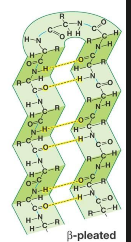
beta-pleated sheets
protein backbone is almost fully extended with h-bonding between adjacent structures (antiparallel orientation allows for maximum hydrogen bonding because the oxygen, nitrogen, and hydrogen are in a straight line)
h-bonds can form between residues on the same or different chains
h-bonds are perpendicular to the direction of the protein chain
interchain bonds
h-bonds formed between different chains
intrachain bonds
h-bonds formed between residues on the same chain

parallel pleated sheet
peptide chain runs in same direction (aligned in terms of N and C terminus)

antiparallel pleated sheet
peptide chains run in alternating directions
irregular structure
can break up helix
found in short stretches
3 residues per turn and 10 atoms in the ring formed by making the h-bond
what is a 3(10) helix

beta bulge
common non-repetitive irregularity found in antiparallel beta pleated sheets
occurs between 2 normal beta structure h-bonds and involves 2 residues on one strand and on on the other
irregular structure
reverse turn
parts of proteins where the polypeptide folds back on itself
Glycine (less crowding) and proline (cyclic structure) often seen at these


BetaAlphaBeta unit
supersecondary structure
2 parallel beta sheets are connected by a stretch of alpha helix
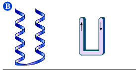
AlphaAlpha unit
supersecondary structure
2 antiparallel alpha helices so that energetically favorable contacts exist between side chains of the helices

Beta-meander
supersecondary structure
antiparallel sheet formed by a series of tight reverse turns connecting stretches of polypeptide chain
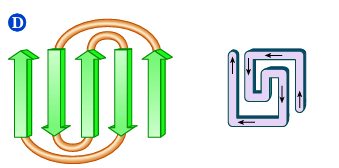
Greek Key
supersecondary structure
polypeptide chain doubles back on itself
motif
term for a repetitive supersecondary structure that can tell us about the folding of a protein but not the function
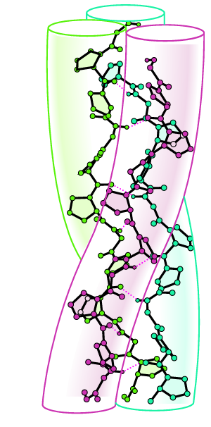
collagen fiber
3 polypeptide chains wrapped around each other in a triple matrix
Left-handed polypeptide helices are twisted together to form a right-handed superhelical structure
every 3rd position has a Gly so that every 3rd residue on each chain is inside the helix
found in connective tissue and bone
Well packed, rigid, triple helix structure responsible for characteristic tensile strength
X-Pro-Gly
X-Hyp-Gly
where X= any amino acid
where Hyp=hydroxyproline (proline with -OH on 4th carbon)
what’s the amino acid sequence for a collage fiber?
less stable
can cause scurvy and bleeding gums etc, asorbic acid (vitamin C) is needed to keep the enzyme active and hydroxylate proline
what happens to collagen without hydroxyproline
fibrous proteins
proteins whose overall shape is that of a long, narrow rod and contain long stretches of regular secondary structure such as coils in keratin and the triple helix of collagen
*not soluble in water
Ex) collagen, fibroin, keratin
Globular proteins
proteins whose overall shape is more or less spherical, helical and pleated sheet sections can be arranged so as to bridge the ends of the sequence close together
*soluble in water
Ex) myoglobin

Covalent
peptide bond between AAs in backbone
Disulfide bonds between cysteine AAs (will restrict folding patterns)
Non-covalent
H-bonding: backbone H bonding and between side chains
hydrophobic: nonpolar residues cluster in the interior of a protein molecule
electrostatic: attraction between oppositely charges groups on surface of a molecule brings groups closer together
complexed: side chains give electrons and support a metal ion or something else (heme group)
name the different forces involved in creating tertiary structure
Lowest
A protein folds into the shape with the ____ possible energy oftentimes stabilized by numerous weak covalent bonds
X-ray crystallography
requires proteins to crystalize, beam exposure to crystal creates a diffraction pattern on a photographic plate or radiation counter
# of electrons in each atom of the molecule scatter the rays and the # of electrons in each atom determine the intensity of the rays (heavier atoms scatter more effectively)
taken at a series of angles
scattered rays lead to constructive and destructive interference giving each protein a unique pattern
can’t use method if protein won’t crystalize
Nuclear magnetic resonance (NMR)
uses proteins in an aqueous solution (most similar to cell environment) to determine 3D structure
Myoglobin
needed for oxygen storage in muscle
globular and composed of 1 polypeptide chain (153 amino acids)
contains 1 heme group
8 a-helical regions and 0 beta-pleated sheets
polar residues on outside and nonpolar found on the inside (except for two polar His residues in the interior)

Fe(II)/Fe2+
protoporyprin IX
6 binding sites
What’s in a heme group

His F8: the N atom of the imidazole side chain occupies 1 binding site of the heme group, perp and opposite form binding site, occupation makes sure that only 1 oxygen can bind
His E7: sterically hinders molecules, decreasing heme affinity for CO2 and bending the O2 ensures that heme will let go
What do the two His residues do for myoglobin

4 Nitrogen from the protoporphyrin
1 nitrogen from the imidazole side chain of the His F8 reside
1 Oxygen molecule
what occupies each of the 6 heme binding sites
hydrophobic interior holds heme group in place and His F8 prevents heme group from becoming free Heme and oxidizing to Fe(III) which will not bind O2
why is the combination of protein and heme needed for O2 storage
denaturation
the unraveling of the 3D structure of a macromolecule caused by the breakdown of noncovalent interactions
heat
extreme pH
detergents
urea and guanidine
beta-Mercaptoethanol
list the ways proteins can be denatured
increase temp, increase molecule vibration, disrupt bond and tertiary structure
how does heat denature proteins
charges on protein missing= loss of electrostatic interactions that would normally stabilize native molecule
how does extreme pH denature proteins
disrupt hydrophobic or electrostatic interactions if detergent is charged (SDS)
how do detergents denature proteins
both form h-bonds with protein that are stronger than native bonds withing the protein itself
how do urea and guanidine denature proteins
reduce disulfide bridges to 2 sulhydryl groups
how does beta-Mercaptoethanol denature proteins
1 subunit =1polypeptide chain, chains can combine to dorm dimers, trimers, and tetramers
What’s a subunit
oligomer
aggregate of several smaller units(monomers)bonding may be covalent or noncovalent
Ex) hemoglobin is an oligomer, while the alpha and beta subunits are still polymers but hemoglobin as a whole being only 4 units is considered one with each subunit being considered a monomer
allosteric
property of multisubunit proteins such that a conformational change in one subunit induces a change in another subunit
hemoglobin
O2 transport to tissues
tetramer, 4 polypeptide chains: a2b2
a: 141 residues
b: 146 residues
4 heme groups
mean the same AA residues are in the same position
what is meant by homologous in regards to the relationship between hemoglobin a and b chains to myoglobin
positive cooperativity
a cooperative effect whereby binding of the first ligand to an enzyme or protein cause the affinity for the next ligan to be higher
hyperbolic
a characteristic of a curve on a graph such that it rises quickly and then levels out
Ex) myoglobin
sigmoidal
referring to an S-shaped curve on a graph, characteristic of cooperative interactions
Ex) hemoglobin
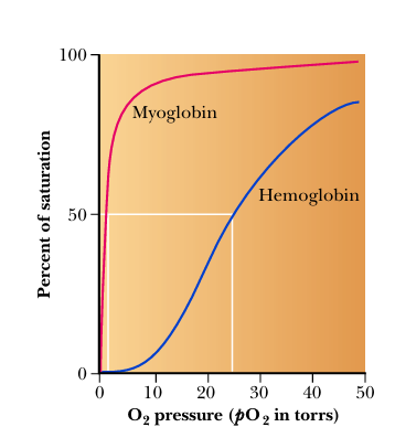
Hemoglobin’s curve being lower that myoglobin’s means at any pressure myoglobin will have a higher saturation percentage than hemoglobin (will always have a higher affinity for O2)
What’s the relationship between Hemo and Myo on binding curve
in lungs O2 pressure=100 torr= 100% oxygen saturation
in capillaries/active muscles= 26 torr= <50% oxygen saturation
positive cooperativity in reverse: if hemo loses 1 O2 it gets easier to lose the rest
How does Hemo’s transport and release of O2 work
binding of O2 and other molecules like CO2 and H+ change the confirmation of the protein which could change function
(O2 brings the b chains closer together)
(H+ and CO2 affect hemo affinity for O2 by decreasing it)
explain the allosteric changes hemo has with O2, CO2, and H+
Cell metabolizes releasing CO2 and H2O
carbonic anhydrase turns it into H2CO3
pKa of H2CO3 is 6.35 while blood is 7.4 so it immediately drops its H becoming H+ +HCO3-
H+ makes the environment more acidic
H+ protonates key amino acids on hemoglobin such as N-terminals of the alpha- chains and His146 of the beta -chains
protonated His is attracted to, and stabilized by, a salt bridge to Asp94. This favors the deoxygenated form of hemoglobin.
hemo releases O2 to the cell or wherever its needed
The oxygenated form of hemoglobin is a stronger acid (has a lower pKa) meaning it wants to give up H more than deoxygenated form
In other words, deoxygenated hemoglobin has a higher affinity for H+ than does the oxygenated form.
explain how H+ affects decreases hemoglobin affinity for O2
cell metabolizes giving off H2O and CO2
carbonic anhydrase combines into H2CO3
pKa of H2CO3 is 6.35 while blood is 7.4 so it immediately drops its H becoming H+ +HCO3-
90% of dissolved CO2 is in this form
mainly transported to the lungs by being dissolved in blood
Hemo binds to O2 at lungs and releases H+
H+ and HCO3- recombine to H2CO3
H2CO3 dissociates to CO2 and H2O
lungs have low CO2 so moves down concentration gradient
CO2 is breathed out
**at high CO2 concentrations hemo binds CO2 as carbamate
alpha amino terminals into anions, which can then interact with the -chain Arg141, also stabilizing the deoxygenated form.
explain how CO2 is transported to the lungs
lowers hemoglobin’s affinity for oxygen, meaning hemoglobin needs a higher pO₂ (more torr) to reach the same saturation.
With BPG: partial pressure at which 50% saturation is at 26 torr
Without BPG: 50% saturation is at 1 torr
No BPG means little O2 would be released from capillaries
how does 2,3-bisphosphylate (BPG) affect oxygen affinity
fetal hemo is a2y2 where y2 doesn’t bind BPG as strongly
one less bond/contact due to Ser instead of His at the salt bridge, neutral instead of + charge)
= fetal hemo has a higher affinity for oxygen
needs to steal from mom so it has to be stronger than mom’s affinity if they were equal strength O2 wouldn’t go anywhere or at least not as effectively
explain how fetal hemo allows the transfer of oxygen from mom to baby
bioinformatics
application of computer methods to processing large amounts of information in biochem
homology
finding known sequences in polymers, finding a similar known sequence in a database and comparing it to the knew protein but must be >25-30% similarity
fold recognition
comparison with well-known folding motifs common to many secondary structures
de novo prediction
bases on 1st principles from chem, biology, and physics confirmed by X-ray crystallography
phospholipids
polar head and nonpolar tail creating a lipid bilayer
liposome
spherical aggregates of lipids arranged so that polar heads face outside and in contact w/ water while nonpolar tails are inside sequestered from water its a spontaneous process (deltaSuniverse>0)
H2O surrounding nonpolar molecules can bind with each other but have fewer possibilities than if they were surrounded by other water molecules which prevents dispersion of energy
each droplet of nonpolar material disrups H2Os ability to h-bond to more H2O
Fewer H2O molecules are inconvenienced by just one big droplet (layers) than a lot of smaller ones (mixed)
why is liposome formation and layer separation a spontaneous process
can form aggregates due to exposed hydrophobic regions that end up on the surface instead of inside which can interact with other hydrophobic regions on other proteins
what happens if proteins don’t fold correctly
prevent misfolding and speed the folding process which prevents aggregates and sometimes require ATP
Ex) Reduce (SH-containing protein disulfide isomerase(PDI) catalyzes the rearrangement of polypeptides non-native disulfide bonds via disulfide interchange reactions to yield native disulfide bonds
what do protein chaperones do
more alpha chain genes than beta (2:1)
means excess of a chain
excess a chains can form aggregates
aggregates=damaged red blood cells
AHSP chaperones a chains to b chains to prevent aggregates and so that there will be enough a chains for b chains
why is the hemoglobin stabilizing protein AHSP important
planar, atoms, plane
the ____ character of the peptide group limits the conformational flexibility of the polypeptide chain and means ___ of the peptide bond are located in a single ____

trans, cis, steric hinderance
peptide groups with few exceptions assume the ___ confirmation in which successive C atoms are on opposite sides of the peptide bond joining them. ___ confirmation can result in _____ in which the atoms would orient closer than their van der waals distance which results in repulsion

psi (Ѱ)
angle between alpha carbon and the carboxy group can rotate
phi (ᵩ)
angle between alpha carbon and amino group
Type I reverse turn
third residue rotated so that the R group (gold) faces outwards and any amino acid can occupy this position
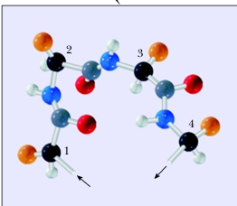
Type II reverse turn
Type I is rotated 180 degrees so that the R group faces inside, the only R group that can fit in this space is the H in Glycine so only Glycine can occupy this 3rd residue position
proline can also occupy the second residue position and help with the turn

Coiled coil
2–7 alpha-helices are twisted together like strands of a rope, facilitating protein-protein interactions.
View down the coil axis shows the alignment of nonpolar residues along one side of each helix. The helices have the pseudo repeating sequence a-b-c-d-e-f-g in which residues a and d are predominately nonpolar

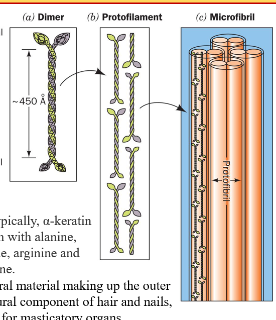
2 keratin polypeptides form a dimeric coiled coil
protofilaments are formed from 2 staggered rows of head-to-tail associated coiled coil
protofilaments dimerize to form a protofibril, four dimers form a microfibril
typically high with Ala, Leu, Arg, and Cys
found in hair, nails, horns, etc
describe alpha- keratin structure
One polypeptide chain folds to form four beta strands which twist to form the distorted beta sheet. Two sheets assemble to form a beta keratin filament
more rigid and water proof
describe beta-keratin structure

irregularities in secondary strucutres
any place where a protein's backbone does not adopt one of the regular repeating patterns of an α-helix or β-sheet.
loops: connect a-helices
turns: allow reverse in direction for alpha or beta sheets (mostly beta)
beta bulge
kink in an a-helices(usually proline)
name some irregularities in secondary structures
certain amino acids tend to exist more in an alpha or beta sheet
what are alpha helix and beta sheet propensities
separated: Nonpolar residues tend to occur in the protein interior and polar residues on the exterior
In an a-helix from sperm whale myoglobin the nonpolar residues are primarily on one side of the helix with polar on the other (A)
An antiparallel beta-sheet from concanavalin A. The protein interior is to the right and has nonpolar amino acids and the exterior to the left with polar AA (B)
where do the locations of polar and nonpolar R groups tend to exist on alpha helix and beta sheet

symmetrical
The majority of soluble and membrane bound proteins in modern cells are _______oligomeric complexes with two or more subunits. Cyclic, dihedral, and tetrahedral
Hydrophobic effect
nonpolar substances minimize their contacts with water
Major determinant of native protein structure
The more positive the value the more hydrophobic are the amino acids located in that region of the protein
A large positive hydropathic index indicated a hydrophobic region of the polypeptide, whereas a large negative value indicates a hydrophilic region
Upper bars denote the proteins interior and lower bars denote the proteins exterior regions as determined by X-ray crystallography
describe hydropathy value

Intrinsically disordered proteins
unstructured in solution but becomes folded structure when interacting with another protein or substance
Ex) CREB
The backbone of the kinase-induced domain (KID) of CREB is drawn as a purple worm. It is unstructured when free in solution but becomes two perpendicular a helices when it forms a complex with the KID- binding domain of another protein
high, high, low, low
folding follows a pathway from ___ energy and ___ entropy to ___ energy and ____ entropy. Driving force of the process is noncovalent interactions (h-bonds) that give off energy= lose energy when folding
hierarchical
Protein folding is a cooperative _______ process in which small elements of structure form coalesce into larger elements which coalesce to yield larger elements (primary—> tertiary)

GroES cap binds to GroEL ring to induce a conformational change in the cis ring that moves the improperly folded protein into the cis cavity to be refolded
catalyzes the hydrolysis of 7 ATPs to 7 ADP and release 7 Ps in order to refold the protein
second molecule of improperly folded protein binds to the trans ring as well as 7 more ATP
cis ring releases GroES cap, ADP, and the properly folded protein
molecule turns 180 degrees such that trans ring becomes the cis ring that is to be capped by GroES and the previous cis ring becomes the new trans ring
describe the GroEL/ES protein chaperone process

Alzheimers
Forms plaques of amyloid deposits of Amyloid betta protein that are surrounded by a halo of neurites (axons and dendrites) from dead and dying neurons
Amyloid-beta accumulates in the brain 10-15 years before tau, a protein that stabilizes microtubules(in cells), aggregates filaments in neurons (tangles) (tau=intracellular neurofibrillary)
Creutzfeldt_Jakob Disease (CJD)
degenerative neurological disorder that is incurable and invariable fatal caused by prions, or misfolded proteins that replicate by converting their properly folded counterparts in their host to the same misfolded structure