VM 602 Neurology
 Call Kai
 Learn
Learn Practice Test
 Spaced Repetition
 Match
Match Flashcards
 Knowt Play
1/106

There's no tags or description
Looks like no tags are added yet.
Name | Mastery | Learn | Test | Matching | Spaced | Call with Kai | Chat |
|---|
No analytics yet
Send a link to your students to track their progress
107 Terms
What are the three parts of a synapse?
1. presynaptic neuron
2. postsynaptic neuron
3. gaps junctions (electrical) or synaptic cleft (chemical)
What is the space between pre and post synaptic neurons called for an ELECTICAL synapse?
gap juction
What is the space between pre and post synaptic neurons called for a CHEMICAL synapse?
synaptic cleft
What are the 3 different anatomical locations of synaptic contacts?
-axodendritic
-axosomatic
-axoaxonic
Where are electrical synapses found?
- smooth muscle
-cardiac muscle
- inhibitory cells
- retina
What is primary function of electrical synapses?
coordinate electrical activity for groups of neurons (ex coordinated contraction of cardiac muscle)
how are neurons connected in electrical synapses?
gap junctions contain connexon proteins that allow ions to flow from cell to cell
Where would chemical synapses be found?
between neurons
neuron-muscle cell
neuron-glandular epithelium
How does an action potential lead to the opening of Ca2+ channels?
Depolarization of the cell leads to the opening of voltage gated Ca2+ channels
How does ionotropic signaling work?
In fast chemical synapses, the NTS bind to ionotropic receptors and directly lead to the opening of Na+ channels and depolarize the postsynaptic cell
How does metabotropic signaling work?
In slow chemical synapses the NTs bind to metabotropic receptors. They bind to a G-coupled protein receptor that indirectly causes the opening of Na+ channels
What do we call a SLOW chemical synapse?
metabotropic signaling
What do we call a FAST chemical synapse?
ionotropic signaling
What is an IPSP?
IPSP is an inhibitory postsynaptic potential - creates transient hyper polarization that prevents an action potential from occurring in the postsynaptic cell
What is an EPSP?
EPSP is an excitatory postsynaptic potential - creates transient depolarization that increases the likelihood of an action potential in the postsynaptic ell?
What kind of synapse can be bidirectional?
electrical synapse (chemical synapse is unidirectional)
what is the resting membrane potential?
the difference in charge between the inside and outside of the cell when it's at rest
What charge is resting membrane potential?
-70 mV
What does the equilibrium potential predict Na+ will do if its channels open?
Na+ will flow into the cell until the cell becomes +71 mV
What does the equilibrium potential predict K+ will do if its channels open?
K+ will flow OUT of the cell until it is around -85 mV. K+ is the only ion that can move in or out of the cell when it is at rest.
What does the equilibrium potential predict Ca2+ will do if its channels open?
Ca2+ will enter in the cell
What does the equilibrium potential predict Cl- will do if its channels open?
Cl- will enter the cell
When the neuron is at rest, what happens to K+?
K+ leaks out through passive potassium channels
When the neuron is at rest, what is happening to Na+ ions?
Most are on the outside of the cell. Na+ on the inside is pumped out through the K/Na+ ion pump.
How does Na+ get into a cell during an action potential?
A stimulus causes some Na+ voltage gated ion channels to be opened. If the stimulus is strong enough and enough Na+ channels are opened, threshold will be reached and an action potential will occur. After threshold, all Na+ channels open and lots of Na+ rush into the cell to depolarize it.
What are the 3 mechanisms that contribute to a negative resting membrane potential?
- Selective permeability where large anion molecules cannot escape the cell
- Concentration gradients: K+ naturally leaks out of the cell down its concentration gradient
- Na/K pump that constantly pumps Na+ out of the cell
What is the absolute refractory period?
the period where no amount of stimuli can create an AP. This occurs immediately after an AP has occurred (cell is still repolarizing).
What is the relative refractory period?
The period when a strong enough stimulus can cause another AP, before the cell has returned to resting potential (usually occurs when cell is in hyperpolarization).
What cells make myelin in the CNS?
oligodenrocytes
What cells make myelin in the PNS?
Schwann Cells
What is saltatory conduction?
The jumping of the nerve impulse from one node of Ranvier to the next. The flow is facilitated by the myelin sheath.
What is a bundle of axons called in the CNS?
tract
What is a bundle of axons called in the PNS?
nerve
What is the function of the CNS?
Integrates sensory information and generates an output
What are afferent nerves?
sensory nerves that bring information from the PNS to the CNS
What are efferent nerves?
motor nerves that bring info from the CNS to the PNS
What is a ganglion?
a group of nerves in the PNS
What is a nucleus in the nervous system?
a group of nerves in the CNS
what is a commissure?
bundle of nerve fibers that cross the midline of the brain
What is white matter?
aggregated fibers (usually myelinated) in the CNS
What is gray matter?
aggregated cell bodies in the CNS
What is the choiroid plexus?
specialized ependymal cells that secrete CSF
what are ependymal cells?
They line the brain ventricles and move the CSF through the central canals
What are the 4 zones of a neuron?
1. integration zone
2. input zone
3. conduction zone
4 output zone
What is the input zone of a neuron?
the soma
What is the integration zone of a neuron?
soma
What is the conduction zone of a neuron?
the axon
What is the output zone of a neuron?
terminal bouton
What is a nerve fiber?
the axon of a neuron
Define a nerve
a group of nerve fibers traveling together.
Where do afferent fibers leave the CNS?
through the dorsal ganglion of the spinal cord
Where do efferent fibers enter the CNS?
through the ventral horn of the spinal cord
What is the general name for supporting cells in the nervous system?
neuroglia
What are the three big differences between neurglia and neurons?
1. neuroglia do not form synapses
2. neuroglia can divide
3. neuroglia do not create action potentials
What are astrocytes?
-support cells of the CNS
-line blood-brain barrier
-recycle neurotransmitters
-maintain homeostasis outside the neurons
-Stimulate growth and maintenance of brain tissue
What are microglia?
- support cell of the CNS
- intrinsic immune system of the CNS
- inflammation response to brain trauma
- remove dead neurons
what are the 4 responses to neural damage?
1. reorganize
2. regenerate
3. survival of the neurons
4. neurogenesis (very rare in mammals)
how does regeneration in the periphery occur?
1. macrophages break down damaged axon
2. Schwann cells guide proximal axon stump toward distal stump physically and by secreting growth factors in the distal stump
3. soma does chromatysis and swells to replenish lost proteins
4. after the stumps connect, the space between the nodes in permanently decreased, so axon is not as fast as it was before.
neurogenic atrophy
damage to muscle immediately after neural injury due to lack of neurotransmitters
why can the CNS not regenerate neurons?
- trauma usuall just kills the cells
- brain damage is seen as inflammation by the immune system and microglia supress regeneration
- overgrowth of glial cells = glial scarring
- glial cells produce growth inhibitors like NOGO
What are the 4 common properties of all muscles?
- contractility
- excitability
- extensibility
- elasticity
muscle fasicle
bundle of muscle fibers (skeletal muscle)

skeletal muscle
striated, voluntary, attached to bone, move bones relative to one another

extrafusal fibers
most of the muscle fibers, provide tensile force on the skeleton
intrafusal fibers
sense muscle stretch and position, run parallel with extrafusal fibers
myofibrils
contain repeating contractile units called sarcomeres

sarcomere
Contractile unit of muscle

myofilaments
make up the sarcomere, myofilaments are made of actin and mysosin = active contractile portion

thick muscle filaments
myosin

thin muscle filaments
actin

sliding filament model of contraction
during contraction the thin filaments slide past the thick ones so that the actin and myosin filaments overlap to a greater degree

sarcolemma
plasma membrane of a muscle fiber, has a -90 mv resting potential
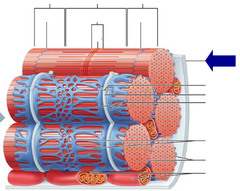
Order of organization of muscle
- fasicle
- muscle fiber
- myofibril
- sarcomere
- myofilaments
transverse tubules
extend from sarcolemma into muscle allow action potentials to get deep into the muscle
sarcoplasmic reticulum
Organelle of the muscle fiber that stores calcium, very important for contraction signals
skeletal muscle innervation pathway
- pyramidal system/ corticospinal tract
- upper motor neurons (conduct voluntary motor signals)
- lower motor neurons (innervate the muscles)
- ACh excites contractions
smooth muscle
involuntary muscle found in internal organs

5 steps of Neurotransmitter production and release
1. synthesis in presynaptic terminal
2. storage
3. release
4. binding to postsynaptic terminal
5. release from receptor
4 main types of neurotransmitters
acetylcholine, amino acids, amines, neuropeptides
synthesis of neurotransmitters
Enzymes to produce the NT are made in cell body and slowly transported down axon. They are stored in the terminal
synthesis of neuropeptides
synthesized on rough ER. Precursor peptide packaged in vesicles then transported quickly down the axon. Mature peptide synthesized by enzymes at the axon terminal
How are neurontransmitters removed from the synapse?
- enzymes degrade them in the synapse
- reuptake by presynaptic transporters
- diffusion and reuptake by glial transporters
Acetylcholine
not an amino acid or derived from one
synthesized from choline and AcetylCoA
Nicotinic (ionotropic) or muscarinic (metabotropic) receptors
nicotinic acetylcholine receptor
found in Neuromuscular Junctions
Found in muscle and the brain
Muscarinic acetylcholine receptor
Found in heart, smooth muscles and brain
Glutamate
excitatory
metabo and ionotropic reception
made from glutamine
Uptake my glial cells and remade into glutamine
GABA
inhibitory - hyperpolarizes postsynaptic cell by opening Cl- channels
made from glutamate
ionotropic
uptake by glia and enzyme degredation
muscle motor unit
single alpha motor neuron + muscle unit of all muscle fibers it innervates for contraction
Alpha (α) motor neurons
innervate the extrafusal muscle fibers that are responsible for generating force
Gamma (γ) motor neurons
innervate contraction of muscle spindle stretch organ intrafusal fibers within skeletal muscle
lower motor neurons
ventral horn motor neurons, innervate skeletal muscles. Gamma and alpha motor neurons are LMNs
upper motor neurons
motor neurons in the central nervous system that control the lower motor neurons in the peripheral nervous system
motor neuron pool
has all the alpha motor neurons that innervate the same muscle.
temporal summation graded contraction
Higher frequency stimulations increase motor unit tension forces
recruiment graded contraction
Stronger stimulations increase number of activated motor units to generate larger muscle forces
muscle twitch
the response of a muscle to a single brief threshold stimulus
3 phases of muscle twitch
1. latent period
2. period of contraction
3. period of relaxation
fused tetanus
when stimulus frequency is so high that no muscle relaxation takes place between stimuli
slow twitch fibers
Small 𝛂-motor neuron • Low input frequency (Hz). Recruited first. Provide sustainable forces (standing walking etc)
Fast twitch Fatigue-resistant fibers
Intermediate 𝛂-motor neuron • Medium input frequency (Hz) recruitment • Medium fibers • Slow peak contraction