Apex TEE
 Call Kai
 Learn
 Practice Test
 Spaced Repetition
 Match
 Flashcards
 Knowt Play
1/42

There's no tags or description
Looks like no tags are added yet.
43 Terms

TEE Component
Top left: Wheel lock
Top right: Control wheels
Middle left: Coaxial cable
Middle lower left: Omniplane button
Lower center-left: Depth markers
Lower right (tip): Piezoelectric crystal (transducer)
Bottom bracket: Shaft


What does the large wheel vs. small wheel control?
Large wheel: Anteflexion and retroflexion (forward/backward).
Small wheel: Lateral movement (left/right).
Memory trick: Large = Forward/Back | Small = Side-to-Side.
TEE—REMEMBER THIS
What to remember
Remove existing OG/NG tubes if possible.
Lubricate the probe with ultrasound gel.
Tip position: Neutral or slightly retroflexed.
Control wheels: UNLOCKED during insertion.
Insertion: Gentle pressure, staying in the midline.
Initial depth: 30–35 cm for a midesophageal view.
Patient position: Neutral neck with anterior displacement of the mandible.
If difficult: Direct visualization using a Macintosh laryngoscope.
Never force the probe. Stop if resistance persists or signs of injury occur.

Where can a TEE probe become lodged during nonmidline insertion?
Pyriform fossa (pyriform sinus).
» Neutral → Unlocked → Midline → 30–35 cm
What 2 things to prevent mucosa injury
30–35 cm and unlocked control wheels to reduce mucosal injury risk.
What forms the heart's base and apex?
Base = both atria (posterior). Apex = LV.
Which aortic structures are clearly visualized by TEE?
Aortic root, ascending aorta, and descending thoracic aorta distal to the left subclavian artery
Why is the aortic arch difficult to visualize on TEE?
The tracheal bifurcation and left mainstem bronchus obstruct portions of the arch.

Identify this TEE view and its primary uses
ME 4-Chamber (ME 4C)
It assess:
1. Ventricular size,
LV/RV wall motion,
MV/TV,
Septal defects,
Pericardial effusion.

Identify the TEE view and its anatomical landmarks.
ME 4C:
RA, RV, LA, LV + mitral and tricuspid valves.

Identify the TEE view and anatomical landmarks.
ME 2C: LA + LV, MV, anterior/inferior LV walls, LAA and coronary sinus.

What does the ME 2C view assess?
LV/LA size, anterior/inferior LV wall motion, LAA thrombus, coronary sinus, MV, LV apex pathology.


Identify the view and its anatomical landmarks.
ME LAX: LA, LV, partial RV, MV, AoV, LVOT, aorta.

What does the ME LAX view assess?
LV size, anteroseptal/inferolateral wall motion, LVOT, septum, MV, aortic root, and LV air.

Quick comparison so far
View | Key recognition | LV walls assessed |
|---|---|---|
ME 4C | 4 chambers, MV + TV | Lateral + septal |
ME 2C | LA + LV, MV | Anterior + inferior |
ME LAX | LA + LV, MV + AoV, aorta | Anteroseptal + inferolateral |
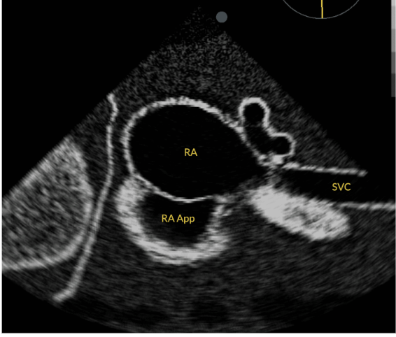
Identify the TEE view and anatomical landmarks.
ME Bicaval: RA, SVC, IVC (when visible), RA appendage.
BI = 2, CAVAL = 2 vena cavae (SVC + IVC).
Exam pearl: When you see the RA with the vena cavae entering it, think ME Bicaval.


What does the ME Bicaval view assess?
RA appendage,
interatrial septum,
IVC cannula,
SVC wires/lines,
intravascular volume status.


Identify the TEE view and anatomical landmarks.
ME AoV SAX:
Circular AoV with 3 cusps (RCC, LCC, NCC), LA, RA, RV.


What does the ME AoV SAX view assess?
LA size,
atrial septal defects,
coronary ostial pathology, and
AoV.

Identify the TEE view and anatomical landmarks
ME RV IF-OF:
RA,
RV,
TV,
AoV,
PA,
LA,
IAS, IAS = Interatrial Septum
PV.
Exam pearl: When you see TV + RV + PA together, think RV Inflow-Outflow.
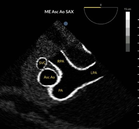

What does the ME RV IF-OF view assess?
IAS, RVOT, PA, TV, pulmonary veins, and RA/LA/RV free-wall motion.


Identify the TEE view and anatomical landmarks.
ME Asc Ao LAX:
Long tubular ascending aorta,
RPA above,
RV and
PA below.


What does the ME Asc Ao LAX view assess?
Ascending aortic pathology
right pulmonary artery pathology, including PE.

Identify the TEE view and anatomical landmarks.
Desc Ao SAX: Circular descending aorta in cross-section, typically at 0°.


What does the Desc Ao SAX view assess?
Descending aortic pathology
left pleural effusion.


Identify the TEE view and anatomical landmarks.
Desc Ao LAX:
Long tubular descending aorta with parallel walls, approximately 90°.


What does the Desc Ao LAX view assess?
Descending aortic pathology
intra-aortic balloon pump (IABP) tip location.


Identify the TEE view and anatomical landmarks.
TG Basal SAX:
Circular LV with MV visible
crescent-shaped RV, RA.


What does the TG Basal SAX view assess?
Basal LV systolic function
RV function
Mitral valve.

Important distinction for your quiz
TG Basal SAX | TG Mid-Papillary SAX |
|---|---|
Mitral valve visible | Papillary muscles visible |
Basal LV level | Mid-LV level |
Assesses basal LV function, RV function, MV | Assesses mid-LV function and wall motion |
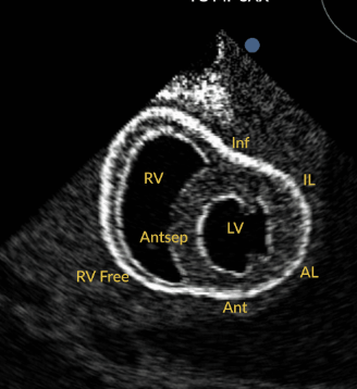
Identify the TEE view and anatomical landmarks.
TG MP SAX:
Circular LV with papillary muscles inside,
crescent-shaped RV.
Exam pearl: If you see a circular LV with papillary muscles inside, identify TG Midpapillary SAX.


What does the TG MP SAX view assess?
LV systolic function, wall motion/ischemia, RV function, LV size/EF, papillary muscles, pericardial effusion.


Identify the hemodynamic condition shown in each.
Small LVEDV → Low preload
Large LVESV → Impaired contractility
Hyperdynamic RV/LV → Low SVR
How does TG Midpapillary SAX distinguish low preload, impaired contractility, and low SVR?
Preload: Filling problem
Contractility: Pumping problem
SVR: Vascular resistance problem

Hemodynamics
Condition | TEE finding | Think |
|---|---|---|
Low preload | ↓ LVEDV | Not enough blood entering |
Impaired contractility | ↑ LVESV | Not enough blood leaving |
Low SVR | Hyperdynamic ventricles | Vasodilation |
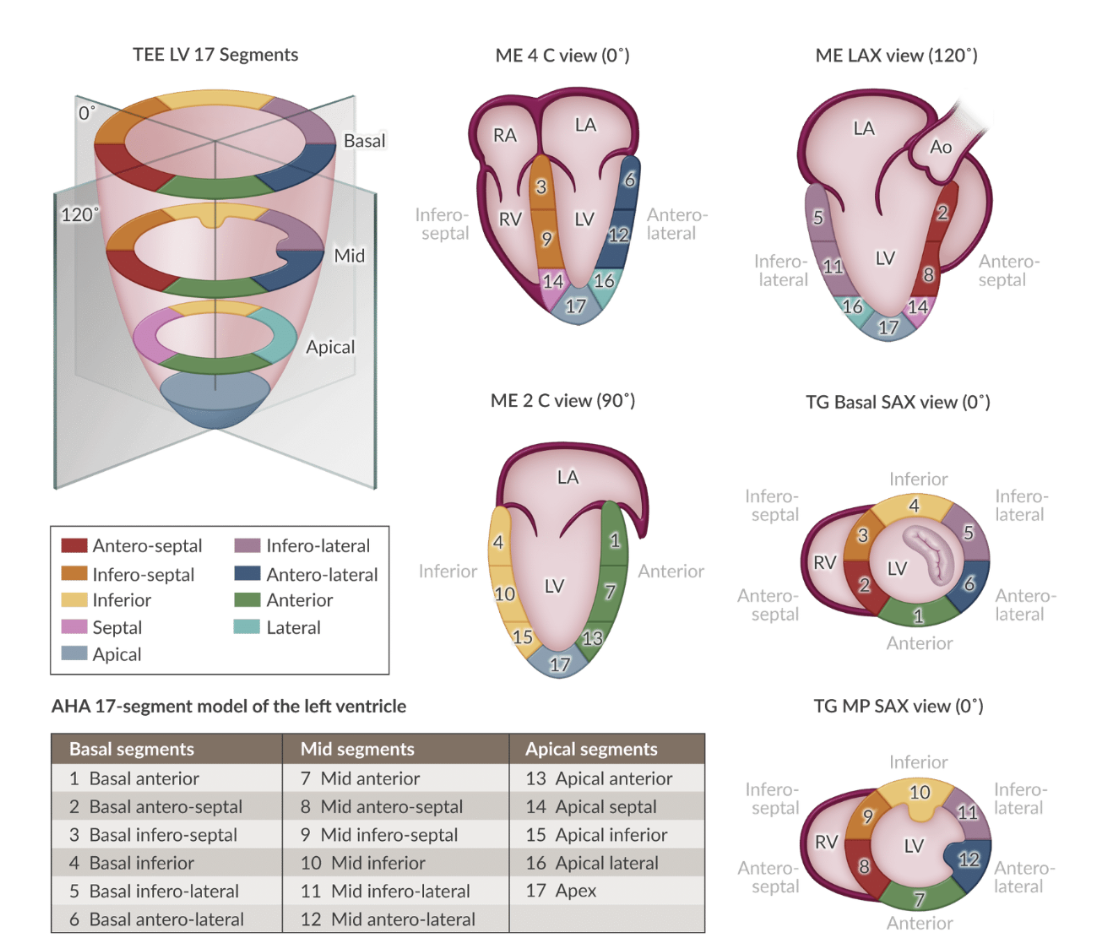
Identify the LV walls visualized in each of the five TEE views.
View | Walls to recognize |
|---|---|
ME 4C | Inferoseptal, anterolateral |
ME 2C | Anterior, inferior |
ME LAX | Anteroseptal, inferolateral |
TG Basal SAX | Six basal LV segments |
TG MP SAX | Six mid-LV segments |

Identify the coronary arteries represented by each color.
coronary territories
Yellow = RCA
Red = LAD
Blue = LCx
The highest-priority associations from this section are:
HOCM: Asymmetric septal hypertrophy → LVOT obstruction → SAM ± MR.
Tamponade: RA collapse in systole, RV collapse in diastole.
RV ischemia: RV dysfunction can accompany LV inferior wall akinesis.
Pulmonary embolism: Acute RH dysfunction and possible leftward septal bulging.
CPR: compression ME4C and for location: ME LAX
What are the major TEE findings and views associated with HOCM?
Asymmetric septal hypertrophy → dynamic LVOT obstruction
Increased blood velocity may cause SAM (Venturi effect) ± mitral regurgitation.
Type 1: basal anterior septal hypertrophy → ME LAX.
Type 2: anterior and inferior septal hypertrophy → ME 4C best shows inferior septum.
What are the characteristic TEE findings and best views for cardiac tamponade?
Pericardial effusion (black/echolucent stripe).
RA systolic collapse + RV diastolic collapse.
Small RV/LV chambers with hyperdynamic systolic motion.
Possible leftward septal bowing.
Best view: RV IF-OF; ME 4C and TG SAX may also suggest tamponade.
“Funky chicken dance” = alternating RA/RV collapse.
What are the causes, TEE findings, and views for RV dysfunction and PE?
Acute causes: PE, RV ischemia/infarction, tamponade.
Global RV dysfunction → increased RV end-systolic volume (TG SAX).
RV ischemia → wall motion abnormalities (RV IF-OF, ME 4C, TG SAX).
Proximal RCA ischemia may also cause LV inferior wall dysfunction.
Best view for pulmonary embolism and what will it look like?
ascending aorta SAX/LAX views.
Acute PE → RH dysfunction ± LEFTWARD interventricular septal bulging.
How is TEE used to assess and optimize CPR?
ME LAX → optimal compression location.
Effective compression → LV emptying, MV closure, AoV opening.
Too cephalad → aortic root compression instead of LV.
ME 4C → assesses RV/LV compression.
6 cm sternal compression = ventricular emptying in adults.
TEE allows real-time monitoring without occupying chest wall space.