PCB3703 - Cardiovascular pt.3 - UCF Ahangari
 Call Kai
 Learn
Learn Practice Test
 Spaced Repetition
 Match
Match Flashcards
 Knowt Play
1/136

There's no tags or description
Looks like no tags are added yet.
Name | Mastery | Learn | Test | Matching | Spaced | Call with Kai | Chat |
|---|
No analytics yet
Send a link to your students to track their progress
137 Terms
Nodal/Conducting cells are a type of ______________ cells.
myocardial
What is contractility of nodal/conducting cells like?
These cells contract very weakly because they contain very few contractile elements (myofibrils).
Nodal/Conducting cells able to _________________ generate action potentials without the help of nervous input like regular neurons
spontaneously
Where is the site of action potentials in the heart? What is this site called?
Although nearly all of the cells in the heart can spontaneously generate action potentials, the sinoatrial node (or SA node) is generally the site of origin. The SA node is located in the upper posterior wall of the right atrium, and it is the first area to spontaneously depolarize, producing an action potential; this is why it is called the pacemaker of the heart.
After an action potential is generated by the SA node, the action potential travels through the _________________ to the ___________________and then to the _____________________ From the Bundle of His, the action potential travels through the ____________________ and then to the _______________________.
After an action potential is generated by the SA node, the action potential travels through the atria to the atrial-ventricular node (AV node) and then to the Bundle of His. From the Bundle of His, the action potential travels through the Purkinje Fibers and then to the ventricular muscle
From the SA node, the action potential spreads throughout the atrial muscle, causing it to _________________.
contract
the atria are electrically ______________ from the ventricles by a fibrous tissue. Therefore, the action potential cannot jump directly down to the ventricles.
isolated
From the AV node, the action potential propagates through the Purkinje Fibers, which rapidly distribute the action potential to the ventricular
muscle which then has ___________________.
contraction
What do chronotropic effect produce?
changes in HR
Differentiate between negative & positive chronotropic effects.
A negative chronotropic effect decreases HR by decreasing the firing rate of the SA node.
-
A positive chronotropic effect increases HR by increasing the firing rate of the SA node
What do dromotropic effect produce? Primarily where?
changes in the conduction velocity, primarily in the AV node
Dromotropic effect DECREASES conduction velocity through the ___________________, slowing/speeding the conduction of action potentials from the ___________ to the __________ and increasing/decreasing the ___________ interval.
Dromotropic effect DECREASES conduction velocity through the AV node, slowing the conduction of action potentials from the atria to the ventricles and increasing the PR interval.
A positive dromotropic effect INCREASES conduction velocity through the ____________, slowing/speeding the conduction of action potentials from the ________ to the ____________ and increasing/decreasing the ___________ interval
A positive dromotropic effect INCREASES conduction velocity through the AV node, speeding the conduction of action potentials from the atria to the ventricles and decreasing the PR interval
Differentiate between a positive and negative inotropic effect.
A negative inotropic decreases force of contraction (contractility), a positive inotropic effect increases force of contraction (contractility)
Which receptor is associated with the heart?
Beta 1
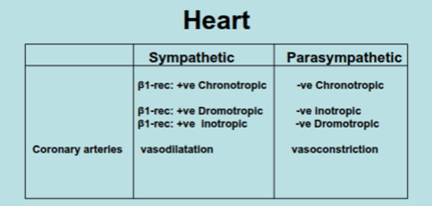
Compare the sympathetic vs. parasympathetic effects of chronotropic, dromotropic, and inotropic in the heart. Also, which one is vasoconstriction/dilation?
At rest, a normal heart beats around ___ to ___ times a minute.
50-99
(usually 72-75)
T/F : arrythmias result from abnormalities in impulse formation or in impulse conduction, Disturbances in the formation of impulses lead to change in the sinus rhythm
true!
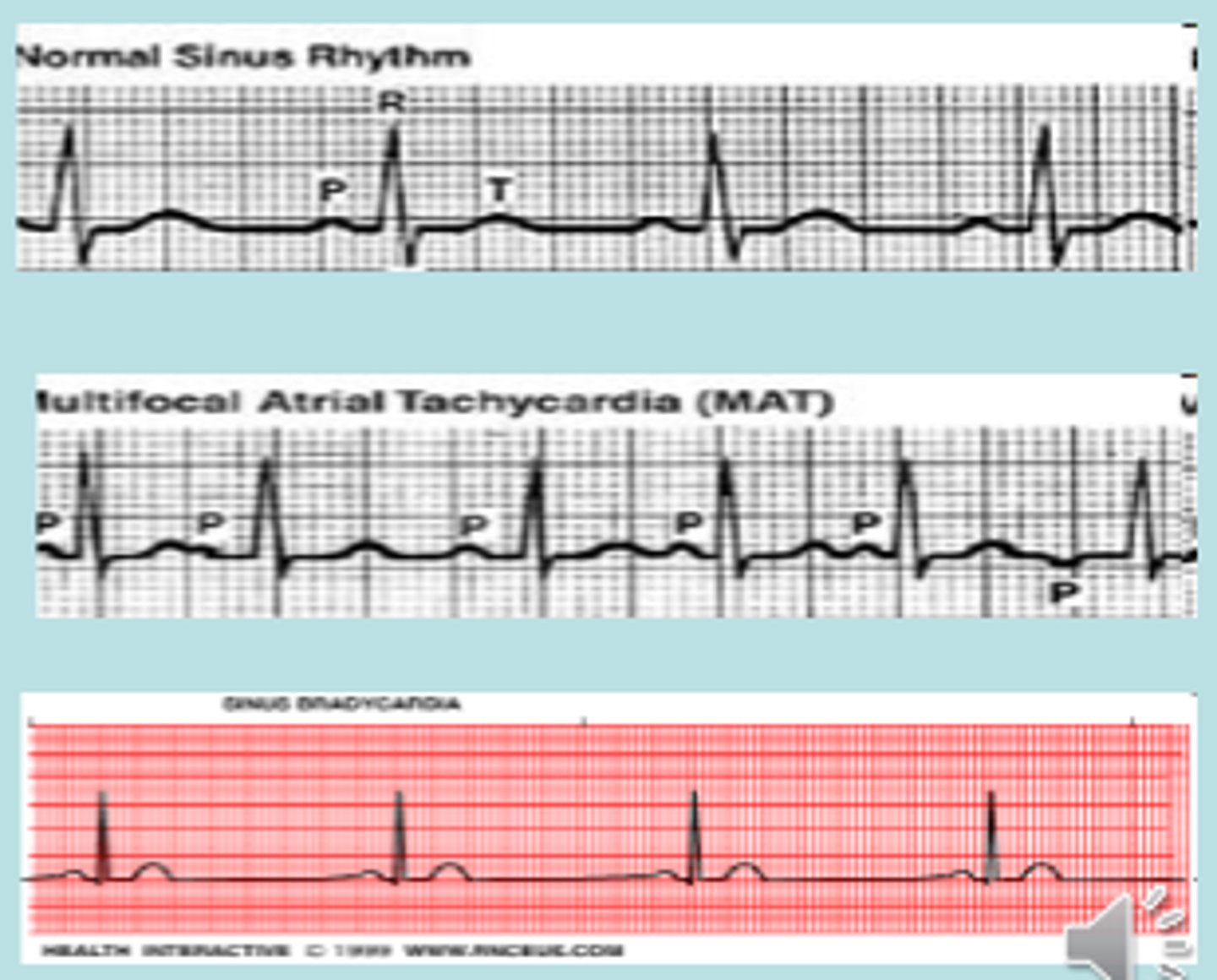
What is sinus tachycardia? During what activities might this occur?
If sinus frequency rises above 100/min (exercise, psychic excitation, fever, rise of 10 beats/min
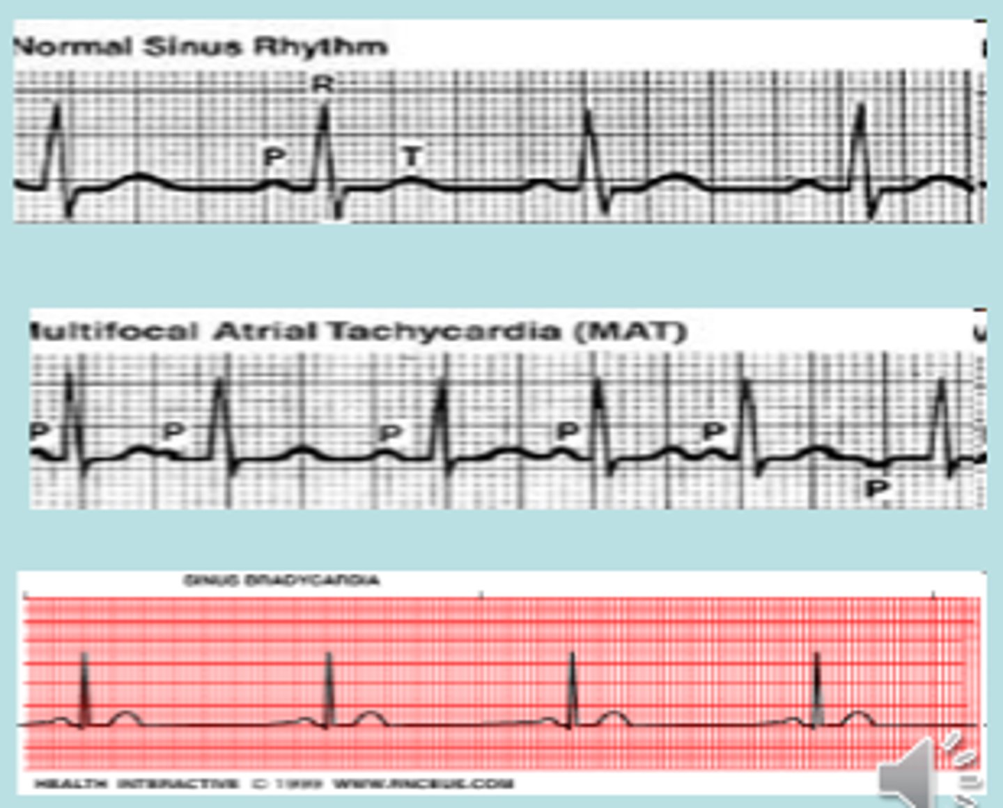
What is sinus bradycardia?
<60 bpm
What are the other words for arrythmia?
extrasystole (ES) or premature contraction
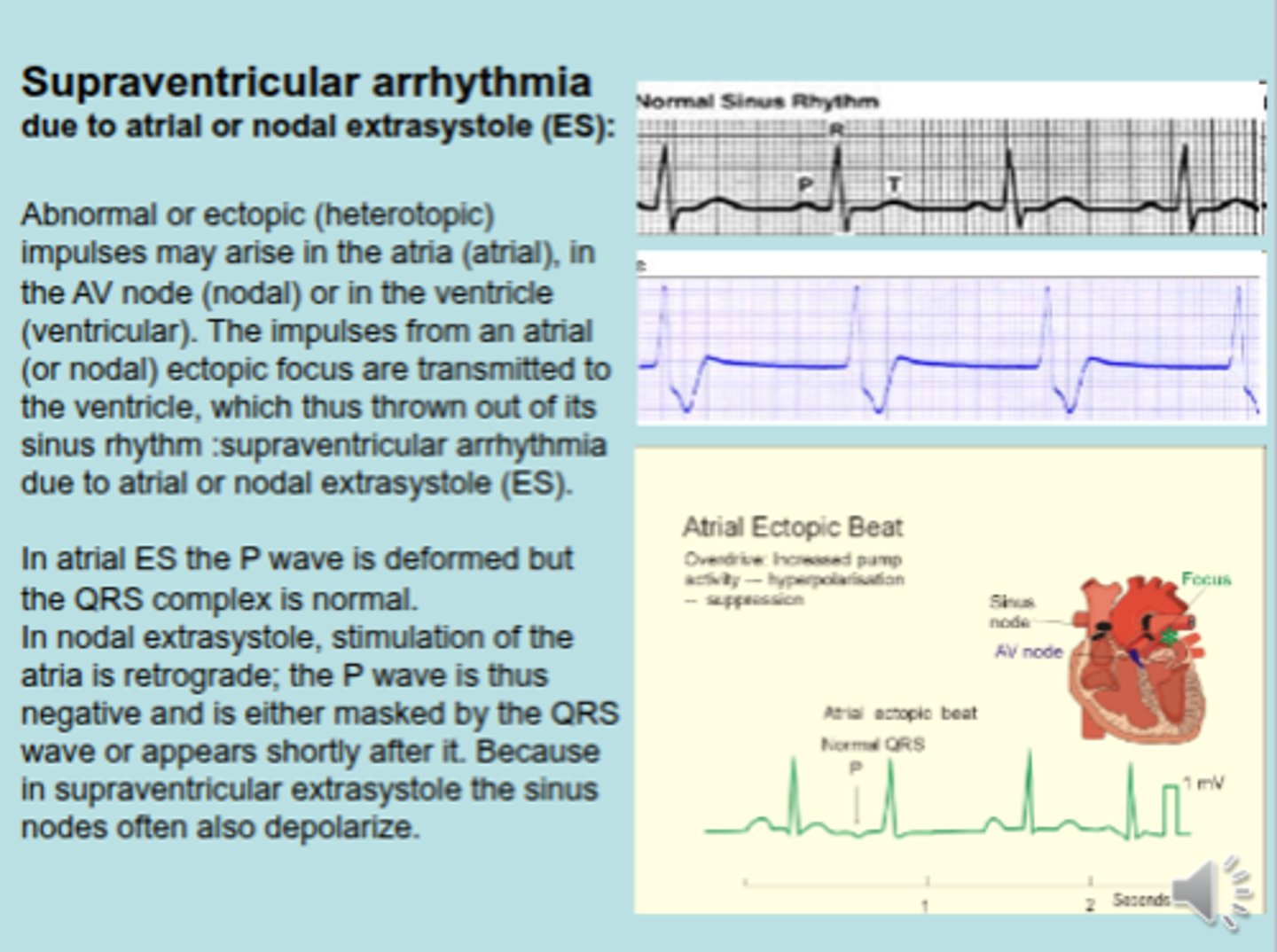
What is a supraventricular arrhythmia?
abnormal contraction of the atrium shows negative P wave, atrium receives abnormal signal from ventricle. That is why it is called atrial ectopic beat
Ventricular premature complexes (VPCs) are ectopic impulses originating from an area distal to the ________________ system.
His Purkinje
What are the most common ventricular arrythmias? What 2 things can cause this?
Ventricular premature complexes (VPCs)
-
could be a result of myocardial infarction or hypercalcemia
What part of the EKG is deformed in Ventricular extrasystole?
QRS complexes
What are the 2 common mechanisms that exist for Ventricular premature complexes (VPCs)?
1. automaticity
2. reentry circuit
What is automaticity for ventricular premature complexes (VPCs)? What could an increased automaticity be caused by?
This is the development of a new site of depolarization in nonnodal ventricular tissue, which can lead to a VPC.
-
Increased automaticity could be due to electrolyte abnormalities or ischemic myocardium
What is reentry circuit for ventricular premature complexes (VPCs)? How does conduction velocity relate to tissue?
Reentry typically occurs when slow-conducting tissue (eg, infarcted myocardium) is present adjacent to normal tissue.
-
The slow-conducting tissue could be due to damaged myocardium, as in the case of a healed MI.
What is ischemia? What can it cause?
deficiency of oxygen
-
premature ventricular contraction
What is digoxin? What does it increase/decrease? What can high doses result in? What can it cause?
medicine given for tachycardia. it decreases HR, but increases strong contractions in the ventricle.
-
High doses causes extra contraction
-
can cause premature ventricular contraction
What is myocarditis? What can it cause
inflammation of myocardium
-
can cause premature ventricular contraction
What is cardiomyopathy? What can it cause?
disease of the myocardium
-
hypertrophic or dilated
-
can cause premature ventricular contraction
What is hypoxia? What can it cause
deficiency of O2 supply
-
can cause premature ventricular contraction
What is hypercapnia? What can it cause?
high CO2 in blood
-
can cause premature ventricular contraction
What can consuming large amounts of smoke, alcohol, drugs like cocaine or weed, and caffeine lead to?
can cause premature ventricular contraction
How can a Mg2+ & K+ deficiency cause premature ventricular contraction?
Mg2+ is a natural Ca2+ channel regulator. deficiency of Mg2+ leads to arrythmia
-
K+ is important because it is important for repolarization. Deficiency of K+ can lead to no relaxation phase.
How can excess Ca2+ cause premature ventricular contraction?
causes extra contraction
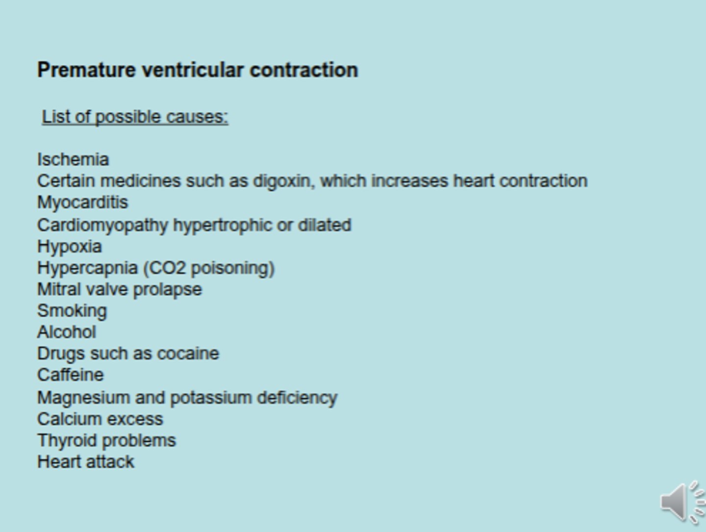
What are the 4 symptoms of arrythmias?
Chest pain
Faint feeling
Fatigue
Hyperventilation (after exercise)
Frequent episodes of continuous PVCs becomes a form of ______________________________, which is a rapid heartbeat, because there is an extra electrical impulse, causing an extra ventricular contraction
ventricular tachycardia (VT)
Ventricular arrythmias/extrasystoles/PVCs can be treated by restoring the balance of ______, ______, or _______ in the body. OR! by _________________ agents.
Ca2+, K+, Mg2+
-
pharmacological
Ventricular arrythmias/extrasystoles/PVCs can be treated either by ___________ agents or _________ agents.
Class I ; Class II
What are class I agents?
Sodium channel blockers .
-
Class I agents are grouped by what effect they have on the Na+ channel, and what effect they have on cardiac action potentials.
-
Lidocaine, Phenytoin
What are class II agents?
Beta blockers.
-
They act by blocking the effects of catecholamines at the β1-adrenergic receptors, thereby decreasing sympathetic activity on the heart. They decrease conduction through the AV node.
-
atenolol, propranolol, metaprolol
What are class III agents?
Block the potassium channels, thereby prolonging repolarization. Since these agents do not affect the sodium channel, conduction velocity is not decreased
-
sotalol
What are Class IV agents?
Calcium channel blockers. They decrease conduction through the AV node, and
shorten phase two (the plateau) of the cardiac action potential. They thus
reduce the contractility of the heart, so may be inappropriate in heart failure.
However, in contrast to beta blockers, they allow the body to retain adrenergic
control of heart rate and contractility
-
Verapamil ; Diltiazem
Which are Class I agents used to treat VPCs?
A. atenolol
B. lidocaine
C. propranolol
D. metaprolol
E. phenytoin
B. ; E.
Which are Class II agents used to treat VPCs?
A. atenolol
B. lidocaine
C. propranolol
D. metaprolol
E. phenytoin
A. ; C. ; D.
Differentiate between Atrial Tachycardia & Ventricular Tachycardia? How many bpm are in both?
Atrial Tachycardia : 100-350 bpm
Ventricular Tachycardia : 120-250 bpm
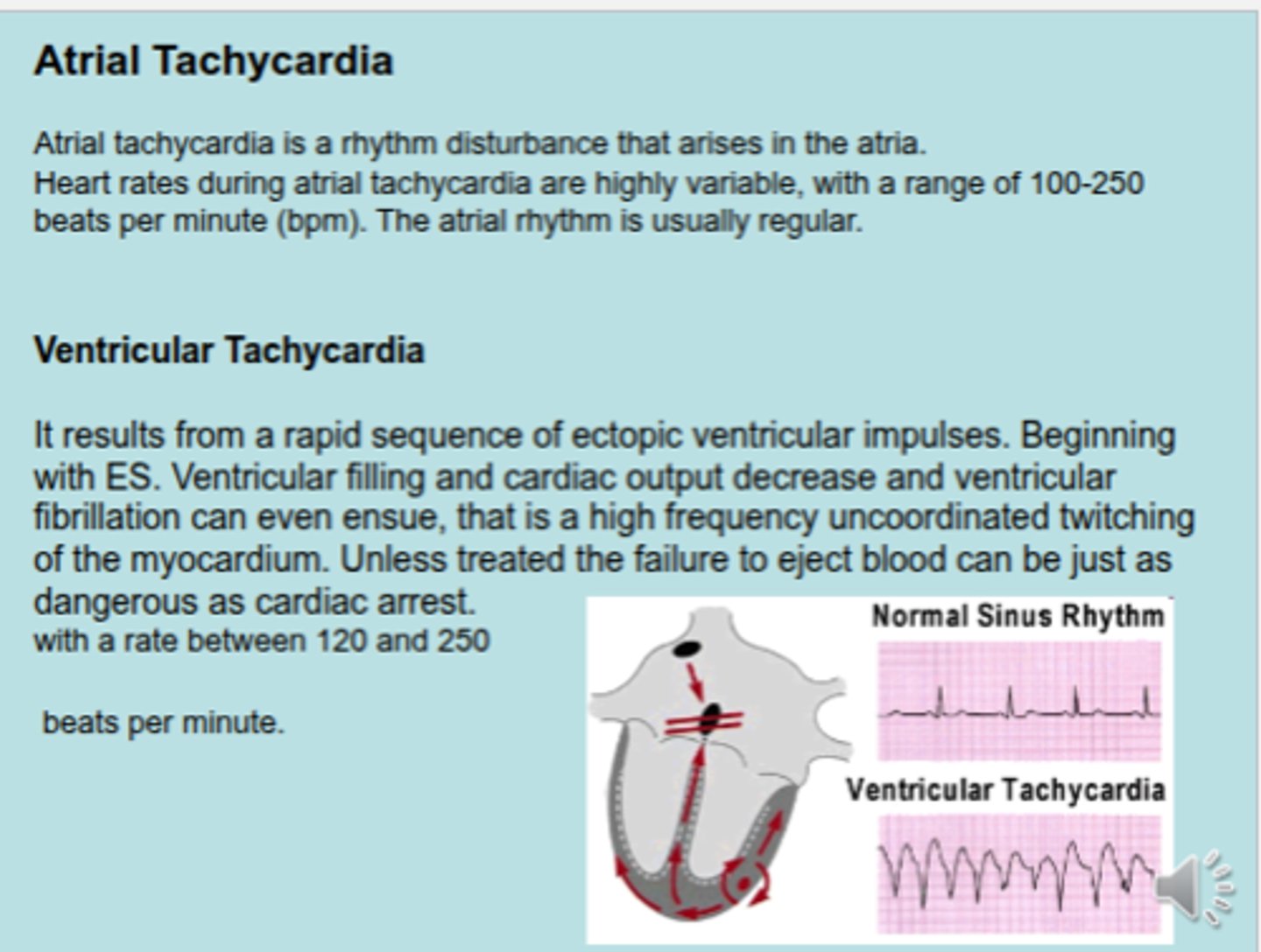
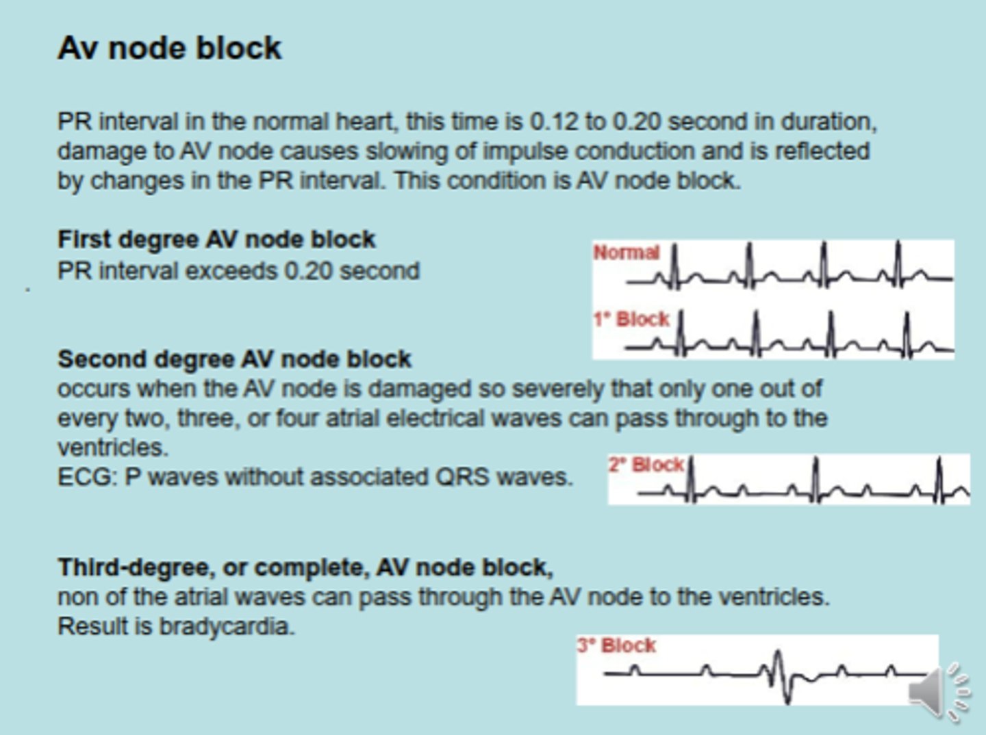
AV blocks are associated with the ________ interval. How long is this interval
PR (normally is 0.12-0.20 s)
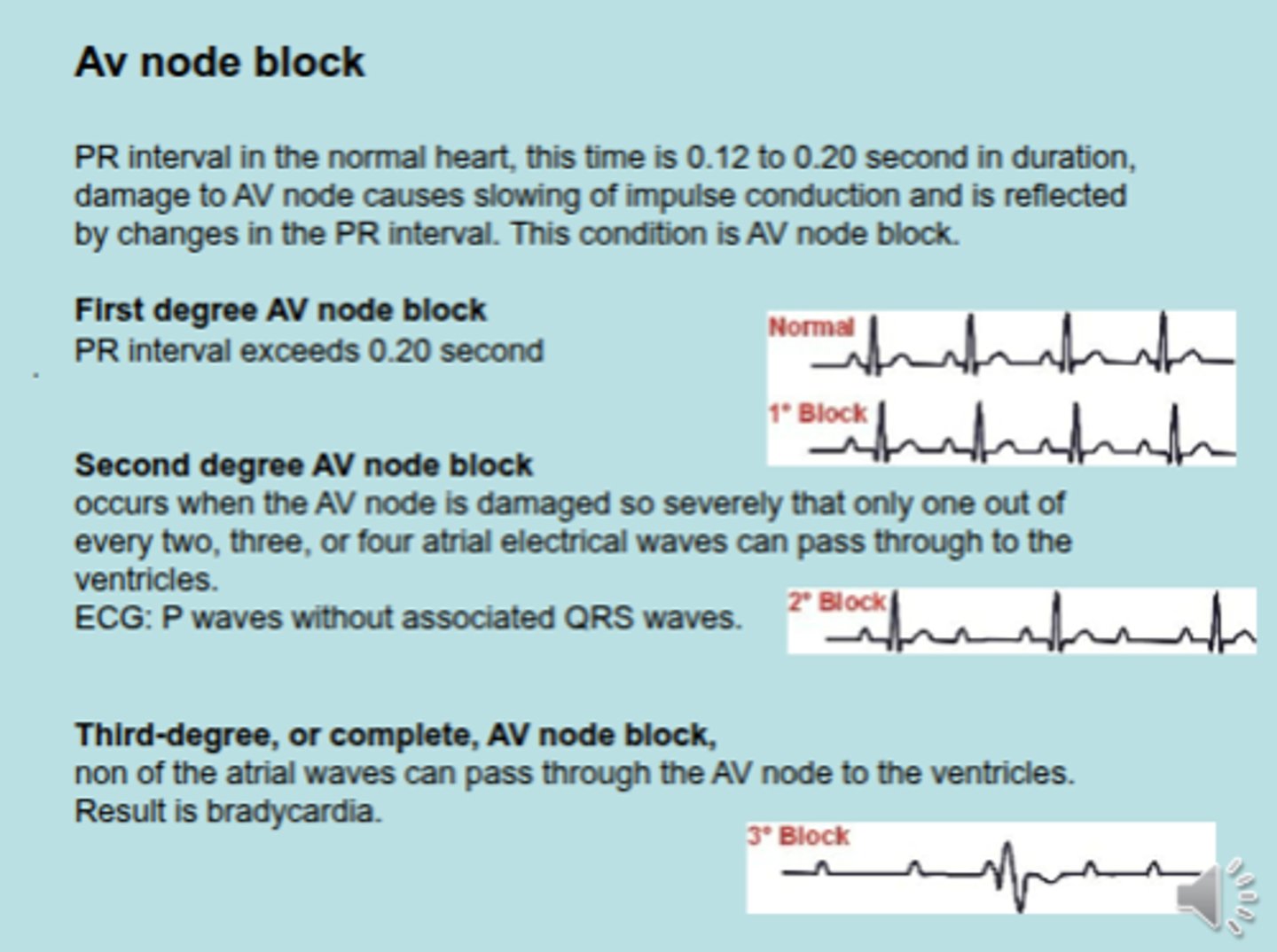
How long is the PR interval in FIRST DEGREE AV NODE BLOCK?
over 0.20 seconds
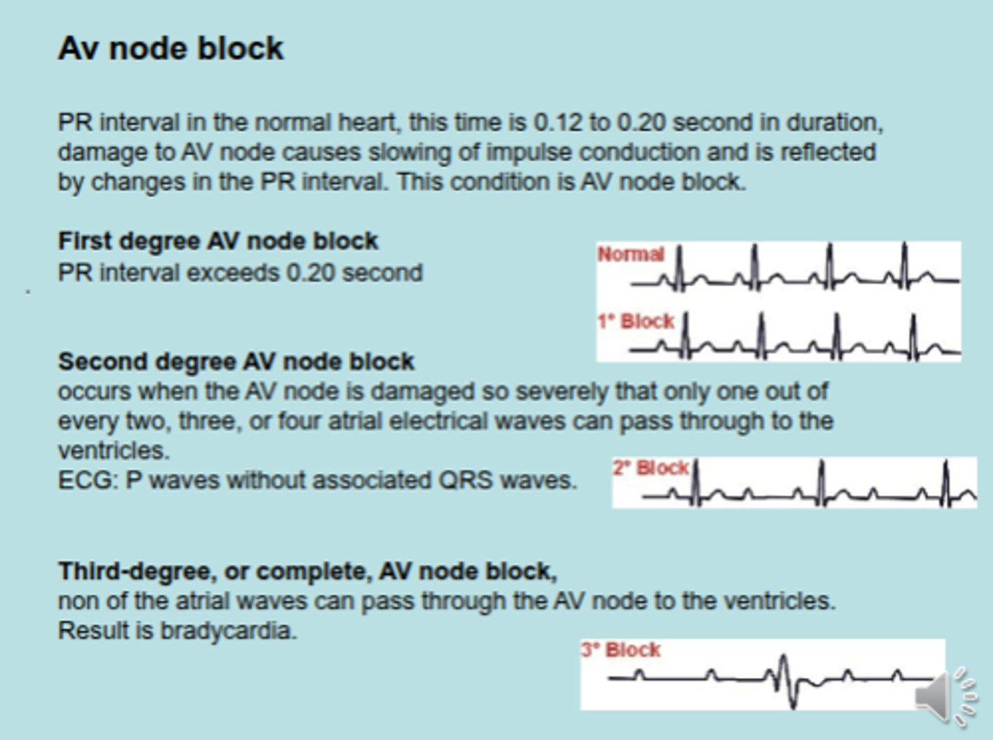
What is characteristic about SECOND DEGREE AV NODE BLOCK?
P waves without associated QRS waves
-
2 P waves --> no QRS
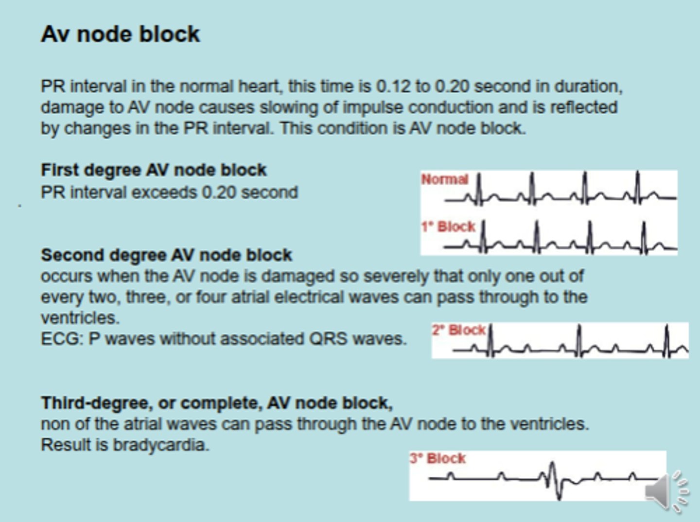
What occurs in THIRD DEGREE/COMPLETE AV NODE BLOCK?
complete interruption between atrium and ventricle because of damaged AV node
-
Result is bradycardia
What can hypercalcemia vs. hypocalcaemia cause?
HYPERCALCEMIA :
arrythmia/ES/VPCs
-
HYPOCALCEMIA :
bradycardia (less contraction)
The heart has 2 types of myocardial cells. Which one has similar features to skeletal muscle cells & which has similar features to nerve cells?
similar to skeletal : contractile cells
(The contractile cells of the heart contain the same contractile proteins actin and myosin arranged in bundles of myofibrils surrounded by a sarcoplasmic reticulum)
-
similar to nerve cells : nodal/conducting cells
How many nuclei do contractile cells have? What do they have a lot of? What joins contractile cells together?
one nuclei
-
lots of mitochondira
-
These cells are extremely efficient at extracting oxygen; they extract roughly 80% of the oxygen from the passingblood—about twice the amount ofother cells. The cells are much shorter, are branched, and are joined together by special structures called intercalated discs
Contractil cells contain ____________________ that bind the cells together, while ________________ allow for the movement of ions and ion currents between the myocardial cell
contain tight junctions that bind the cells together, while gap junctions allow for the movement of ions and ion currents between the myocardial cell
Why are gap junctions good for cardio cells?
Because of the gap junctions, the myocardial cells of the heart can conduct action potentials from cell to cell without the need for nerves
What is the equation of BP?
BP = CO x TPR
What is the amount of end diatolic volume (ESD)?
140 mL
What is the amount of end systolic volume (ESV)?
70 mL
What is the amount of stroke volume?
70 mL
What is the equation for stroke volume?
SV = EDV - ESV
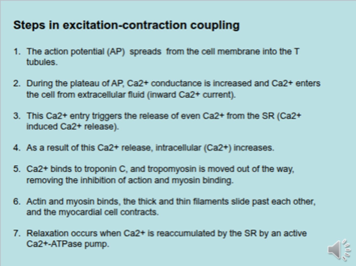
What are the steps in excitation-contraction coupling?
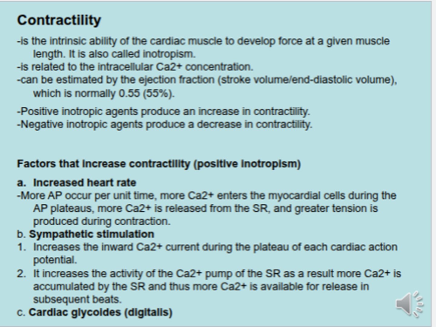
What factors increase contractility of myocardium?
1. hypercalcemia
2. NE/sympathetic (effect B-1 which increases contractility)
3. Digitalis/Digoxin (increases contractility but decreases HR)
What factor decreases contractility (negative inotropism)?
parasympathetic Ach via muscarinic receptors
-
decrease the force of concentration in the atria by decreasing the inward Ca2+ current during the plateau of the cardiac action potential.
-
compressing vagus n. because it is assocaited with parasympathetic NS will decrease HR & contractility
What is preload?
During relaxation phase of ventricle
-
AKA EDV
-
during relaxation of the ventricle each atrium contracts to release content (70ml ) into ventricle. The ventricle already contains 70ml and receives second 70ml from atrium to a total of 140ml
-
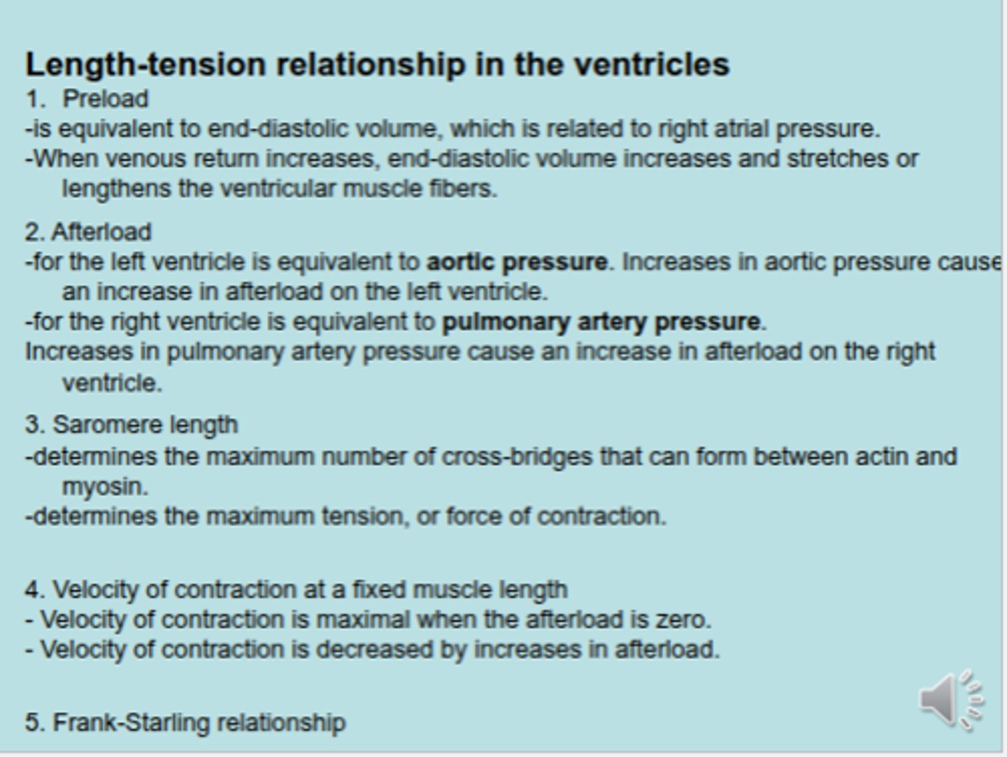
What is afterload?
Pressure from aorta or pulmonary arteries with each ventricle contraction leads to opening of semilunar valves.
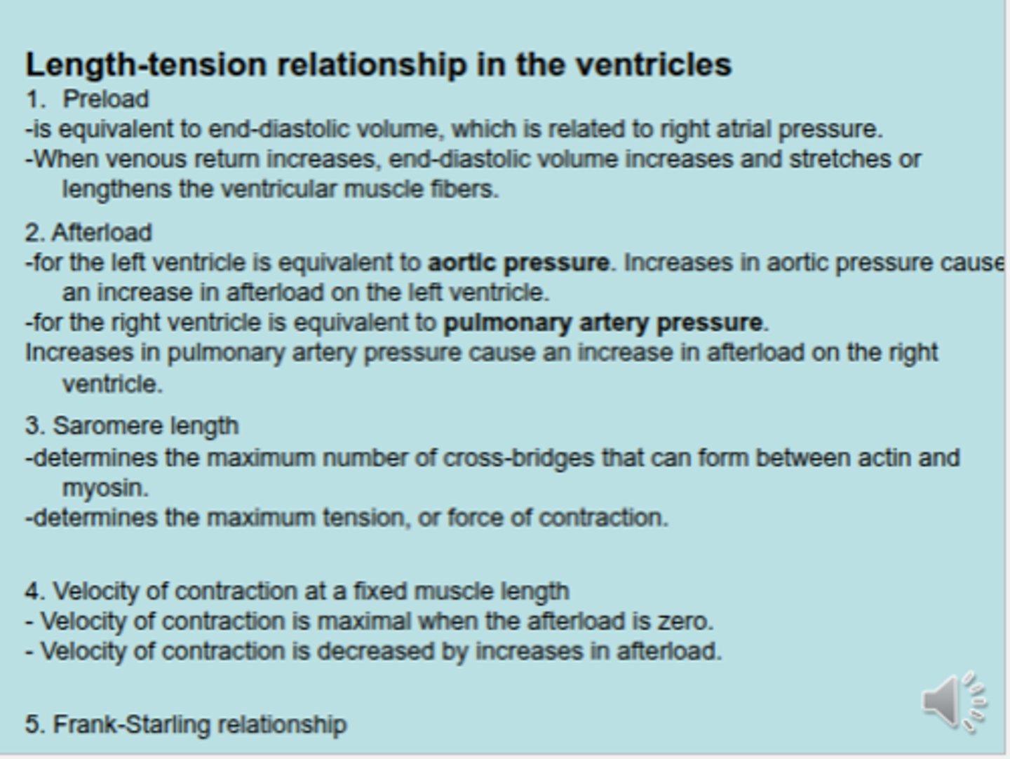
In afterload, the L. ventricle is equivalent to _____________.
aortic pressure
-
Increases in aortic pressure cause an increase in afterload on the L. ventricle
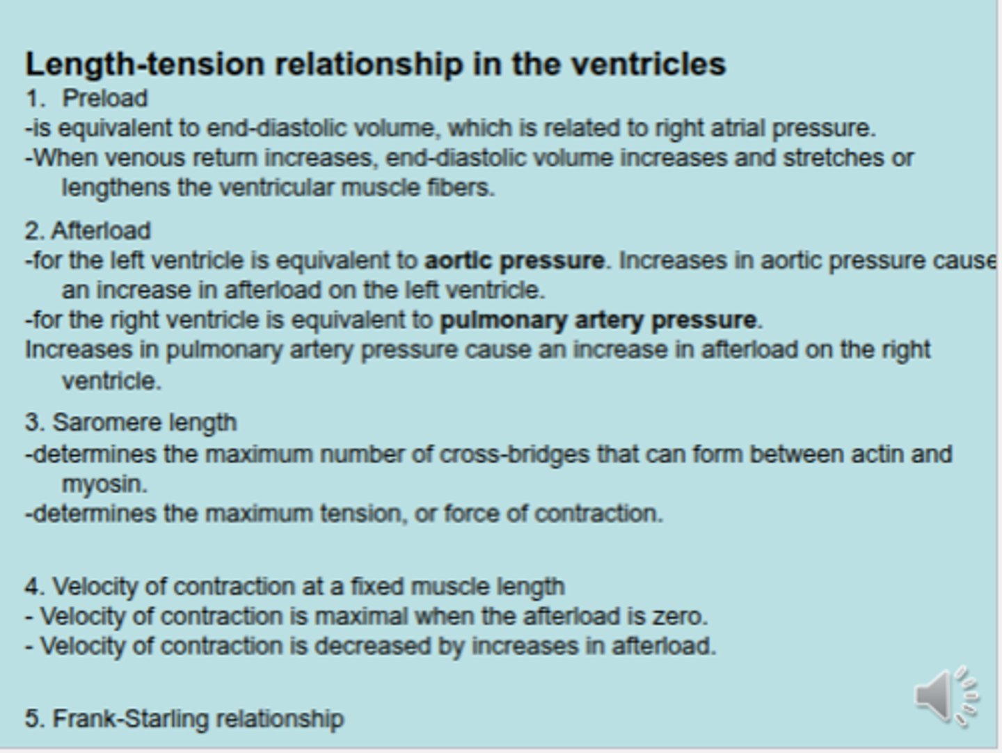
In afterload, the R. ventricle is equivalent to _________________.
pulmonary artery pressure
-
Increases in pulmonary artery pressure cause an increase in afterload on the R. ventricle
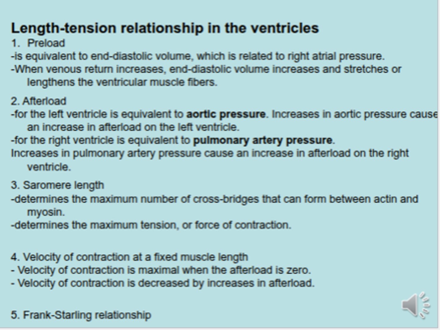
Describes the increases in stroke volume and cardiac output that occur in response
to an increase in venous return or end diastolic volume
Frank-Sterlin relationship
What is the Frank-Sterlin relationship based on?
based on the length-tension relationship in the ventricle. Increases in end-diastolic
volume cause an increase in ventricular fiber length, which produces an increase
in developed tension
What 2 things does the Frank-Sterlin relationship match together?
Cardiac Output (CO) & venous return
The greater the venous return, the greater the _________________.
cardiac output
Changes in contractility shift the Frank-Starling curve ___________ (increased contractility) or ____________ (decreased contractility)
Changes in contractility shift the Frank-Starling curve upward (increased contractility) or downward (decreased contractility
T/F :
Increases in contractility cause an increase in cardiac output for any level of right atrial pressure or end-diastolic volume.
&
Decreases in contractility cause a decrease in cardiac output for any level of right atrial pressure or end-diastolic volume
TRUE!
What is the Frank-Sterling relationship?
Greater venous return increases end diastolic volume and then increases stroke volume to increase cardiac output.
-
Venous return: SVC and IVS returns blood to right atrium and 4 pulmonary veins return to left atrium.
-
EDV: during relaxation phase, the ventricles build up 140 mL of blood. Stroke volume: Ventricles contracts and ejects 70 mL of blood into aorta and 70 mL into pulmonary arteries.
-
Cardiac output: amount of blood released into aorta per minute. 5 L/min
Because of EDV, during the relaxation phase of ventricle, it puts tension on the wall & expands. Because of this it putsincreases length of _______________.
muscle fiber
What two things does sarcomere length determine in length-tension relationship in the ventricles?
1. max # of cross bridges between actin & myosin
2. max tension/force of contraction
-
FROM LEC :
When muscle fibers length Increases, it leads to releasing of calcium from sarcoplasmic reticulum into cytoplasm. Increases sensitivity of troponin C to calcium leading to interwoven of actin and myosin for strong muscle contractions of ventricles to eject 70 mL into aorta with 70 mL of blood into pulmonary arteries (stroke volume).
140-70=70 mL (ESV)
When is velocity of contraction maximal?
when afterload is zero
When is velocity of contraction decreased?
when there is an increase in afterload
The cardiac cycle has 4 steps. Which is isovolumentric contraction?
1-2
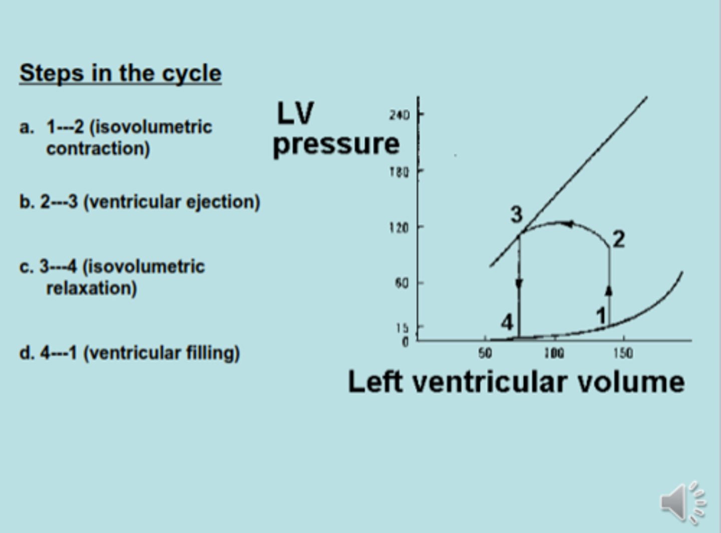
The cardiac cycle has 4 steps. Which is ventricular ejection?
2-3
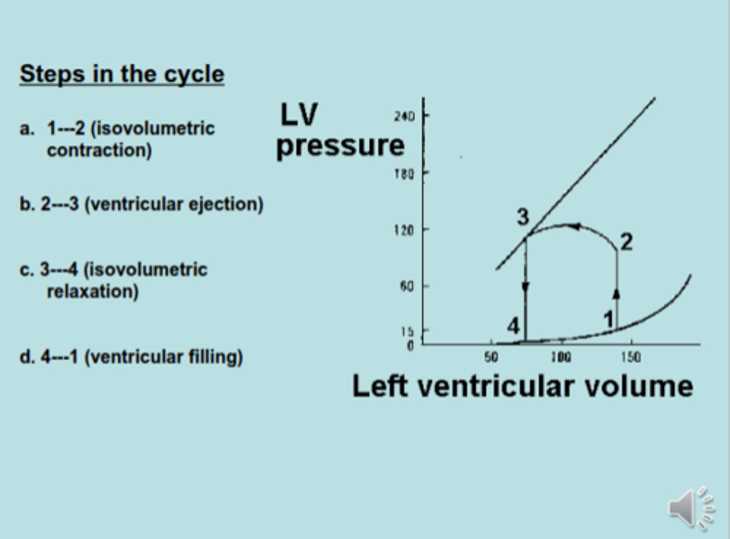
The cardiac cycle has 4 steps. which is isovolumetric relaxation?
3-4
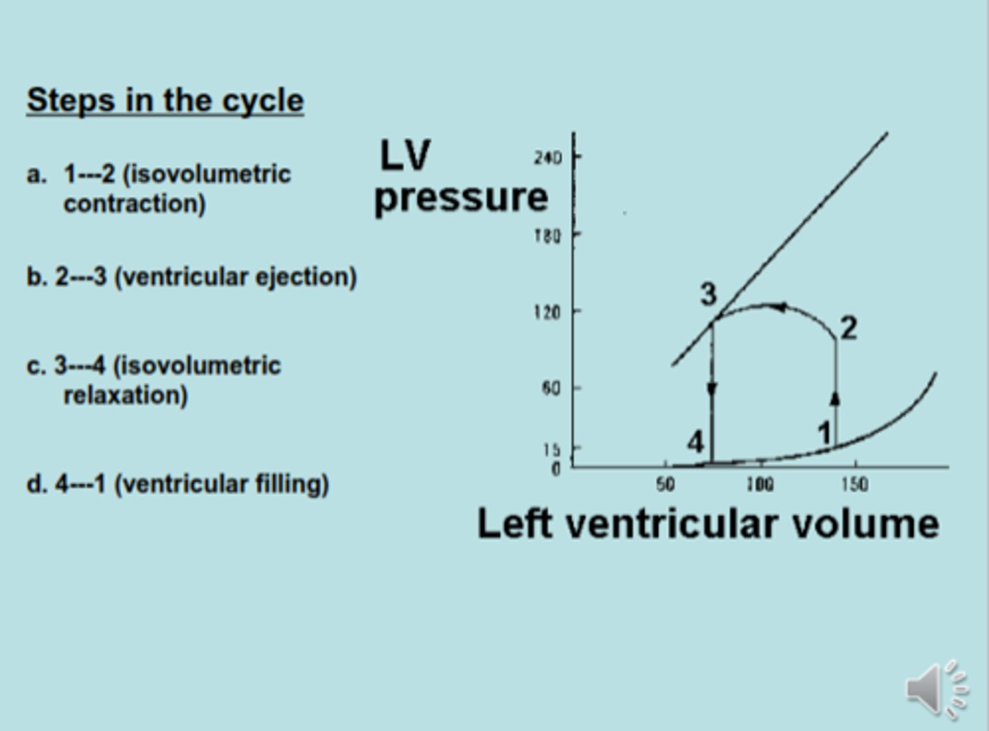
The cardiac cycle has 4 steps. Which is ventricular filling?
4-1
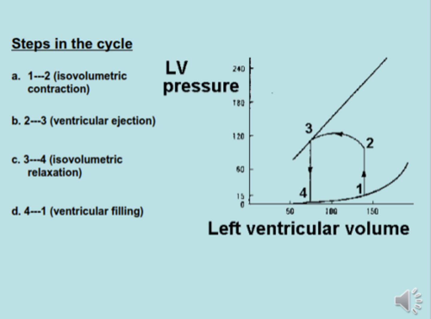
Flip this card for info on the picture on the other side
point 1 is EDV at 140
-
at point 4 is ESV is 70
-
2-3 is stroke volume ejection
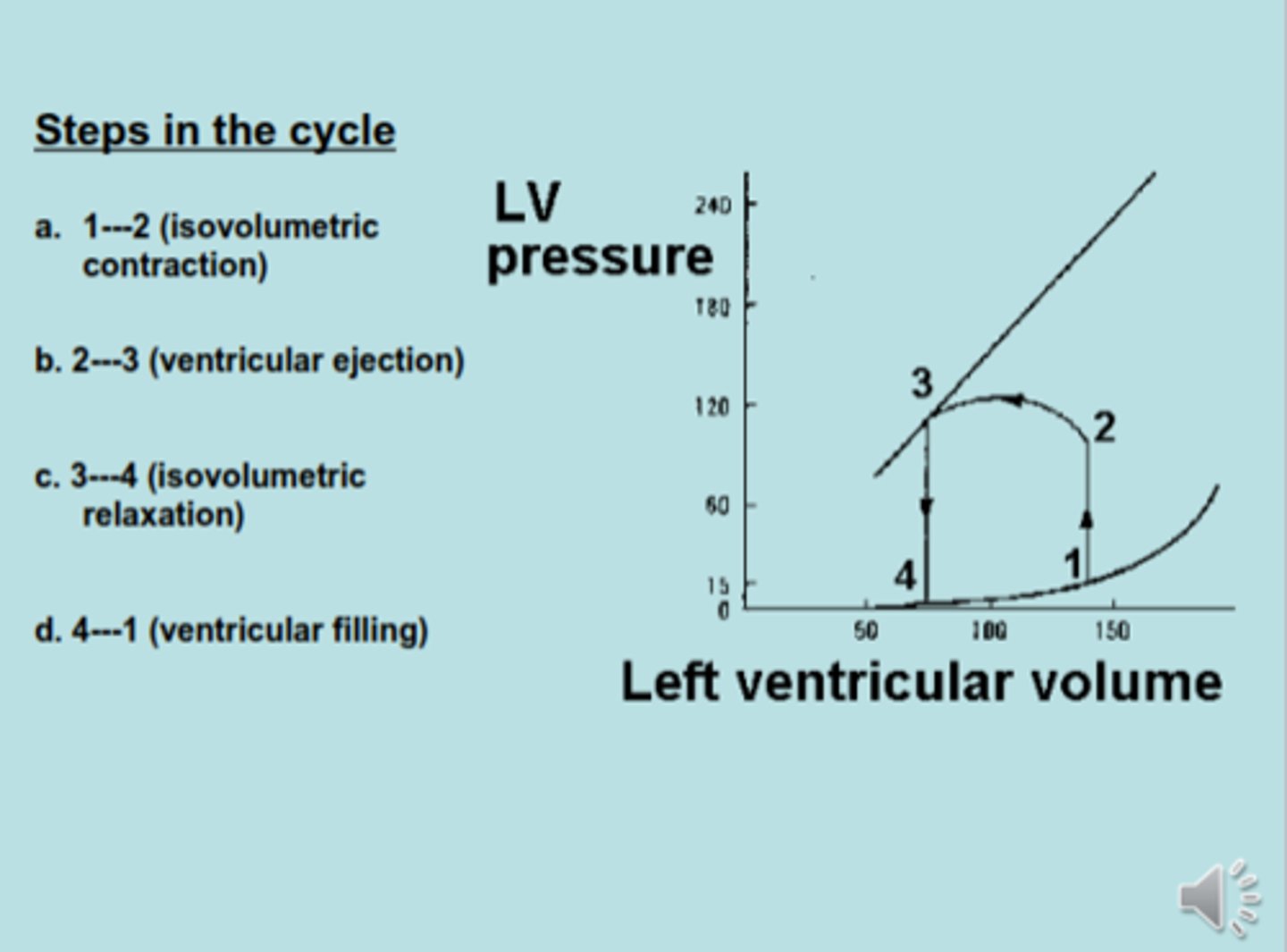
THE CARDIAC CYCLE :
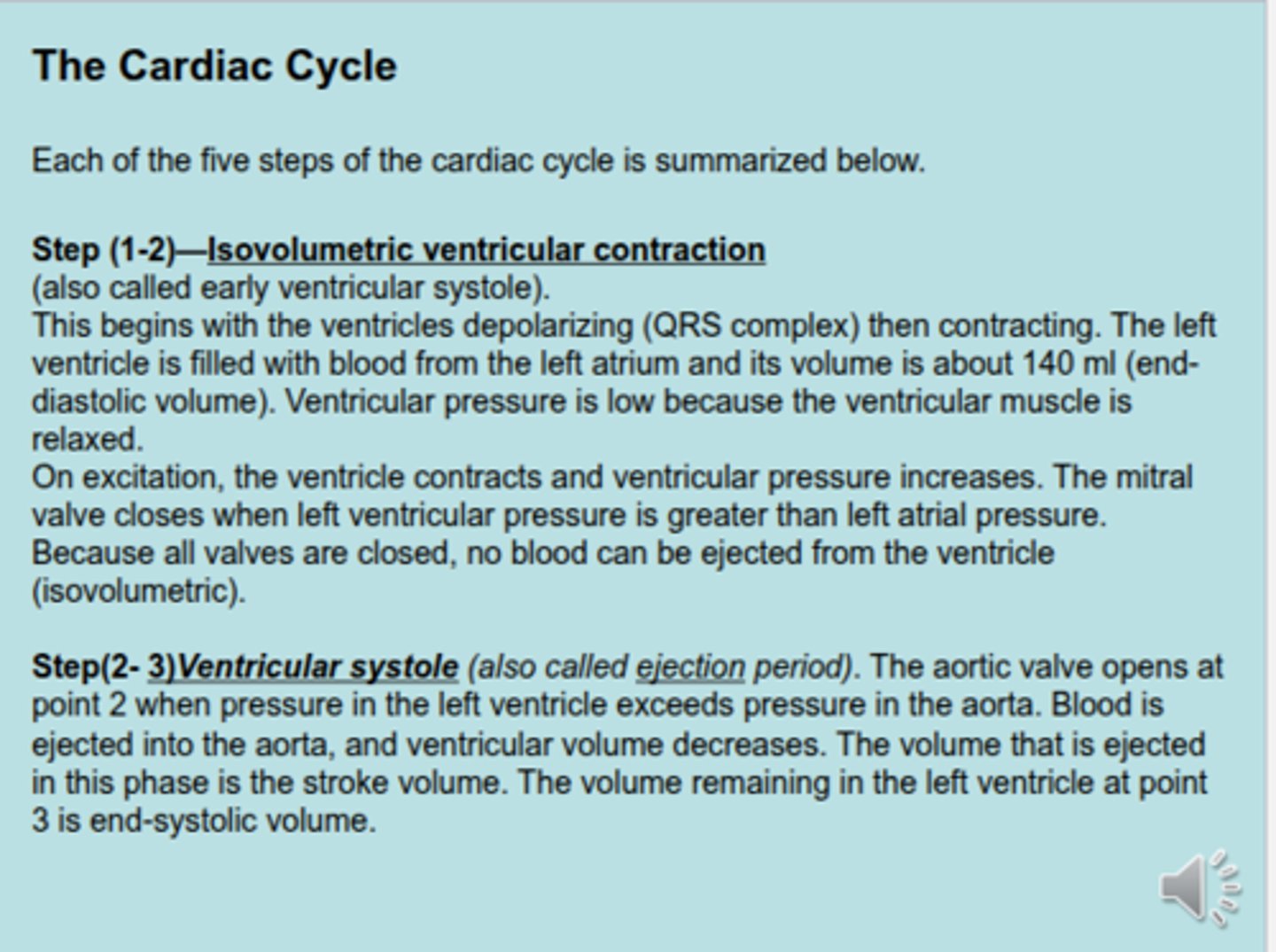
Step 1-2 is isovolumetric ventricular contraction. What occurs in this?
FROM LEC VID :
-at point 1, ventricular pressure is close to 0 & the EDV of blood is 140 mL
- during this phase, all 4 valves are closed. This means there is no change in volume of blood.
THE CARDIAC CYCLE :
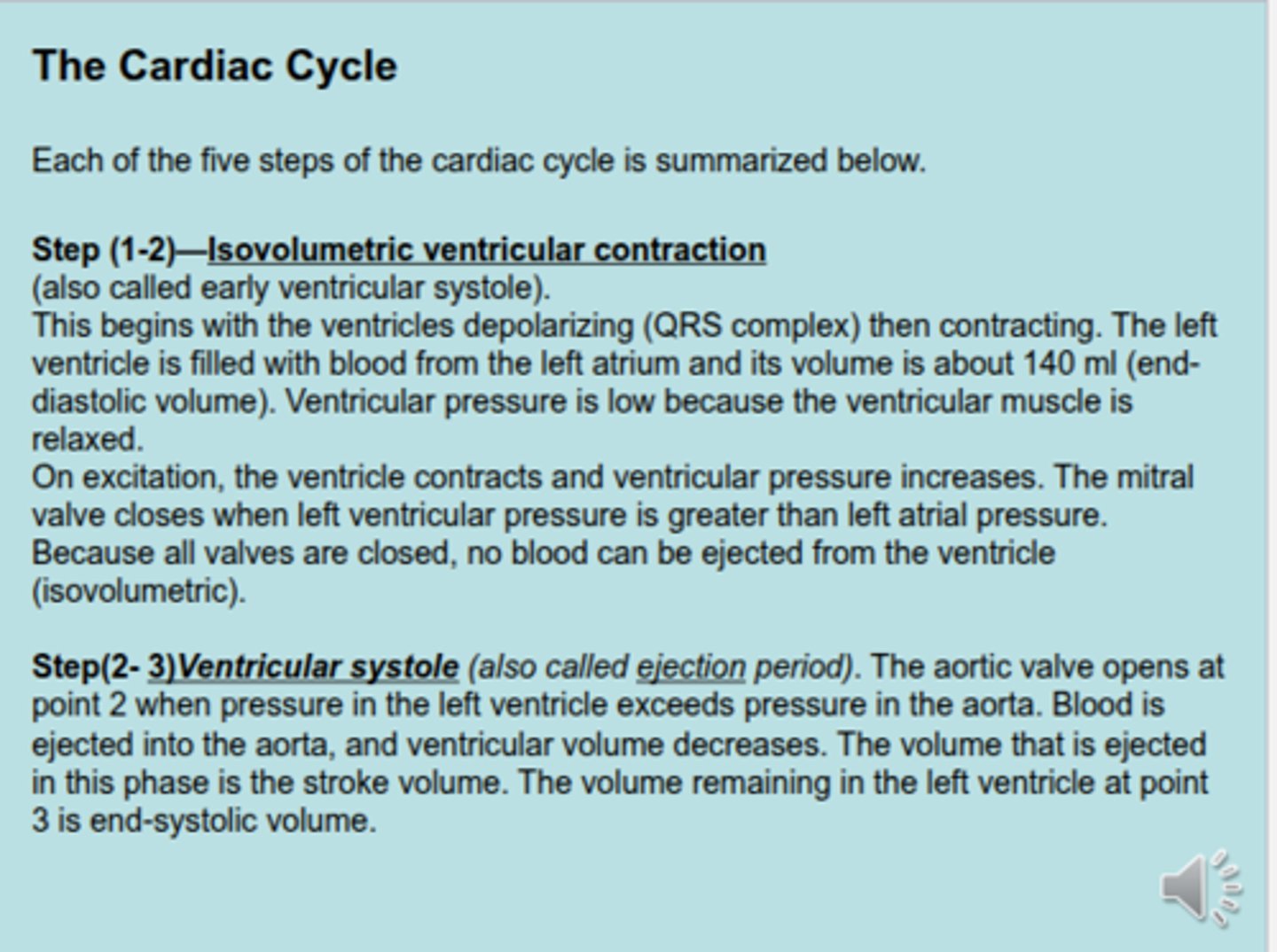
Step 2-3 is ventricular ejection. What occurs in this?
FROM LEC VID :
- ejection of stroke volume, 70 mL goes to aorta; 70 mL goes to pulmonary artery
-pressure & tension increase, which leads to pushing the blood up the semilunar valves to eject
- stroke volume is 70 mL
THE CARDIAC CYCLE :
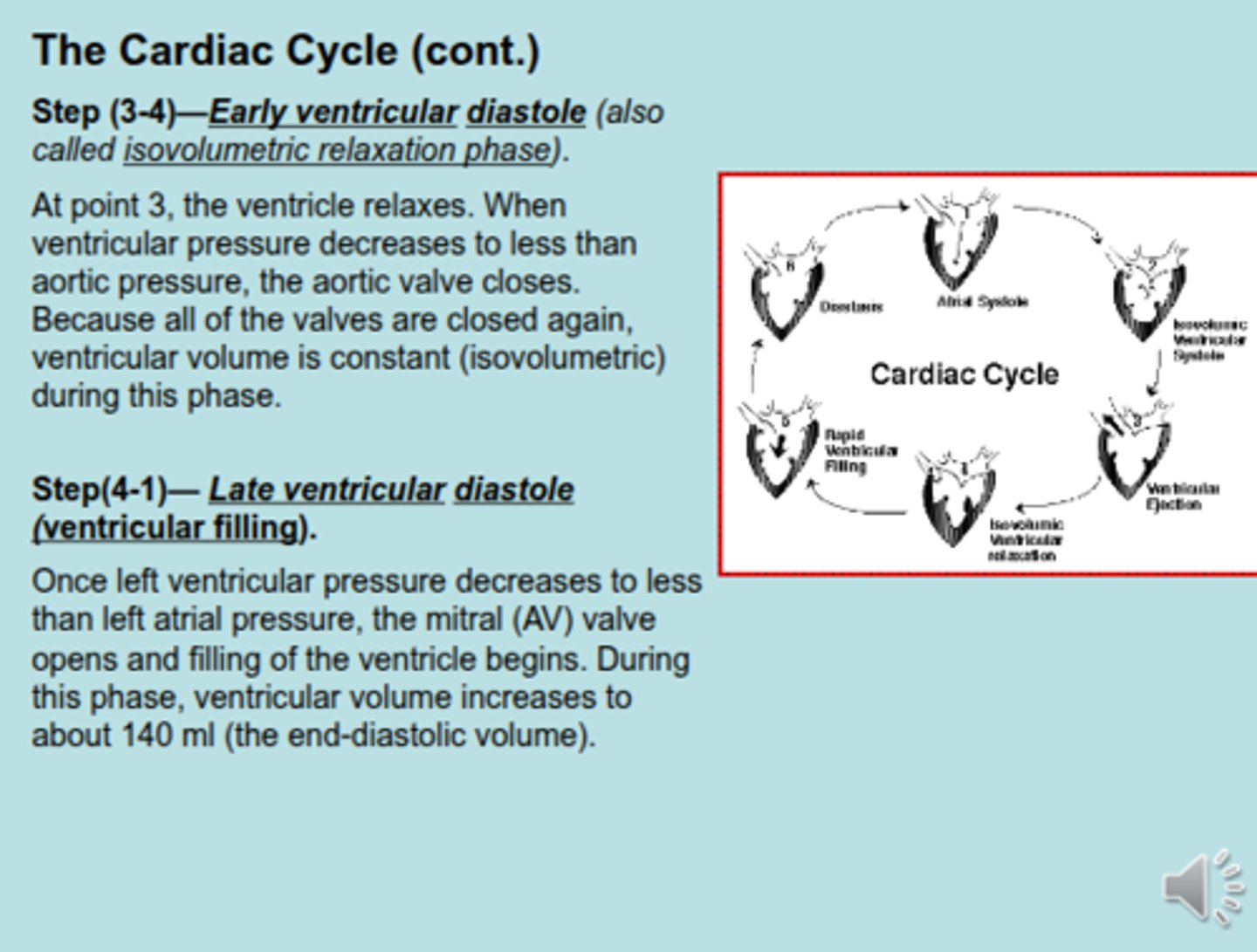
Step 3-4 is isovolumetric relaxation. What occurs in this?
FROM LEC VID :
- pressure in ventricle drops to 0
- atrial pressure here is much higher than ventricular pressure because atria received 70 mL of blood from venous return
-the ventricle is relaxed
- point 4 is end systolic volume (70 mL)
THE CARDIAC CYCLE :
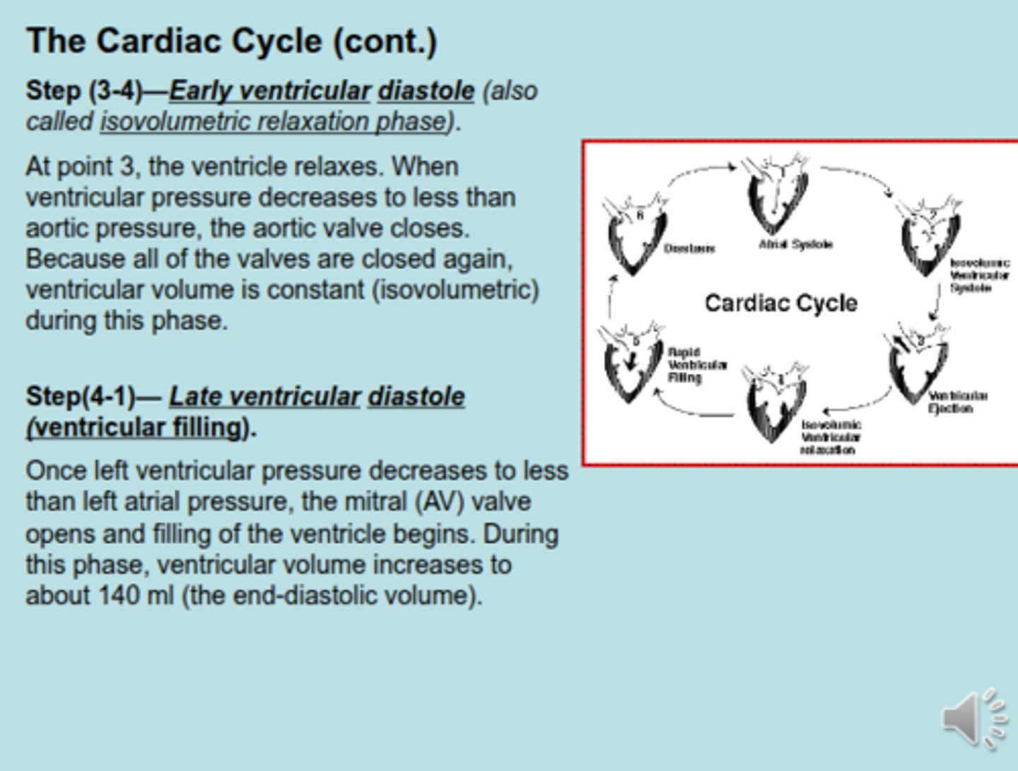
Step 4-1 is ventricular filling. What occurs in this?
FROM LEC VID :
- ventricle is relaxed
- it receives second 70 mL of blood from atrium & it builds up
- 0-4 is 70 mL of blood
- 4-1 second 70 mL
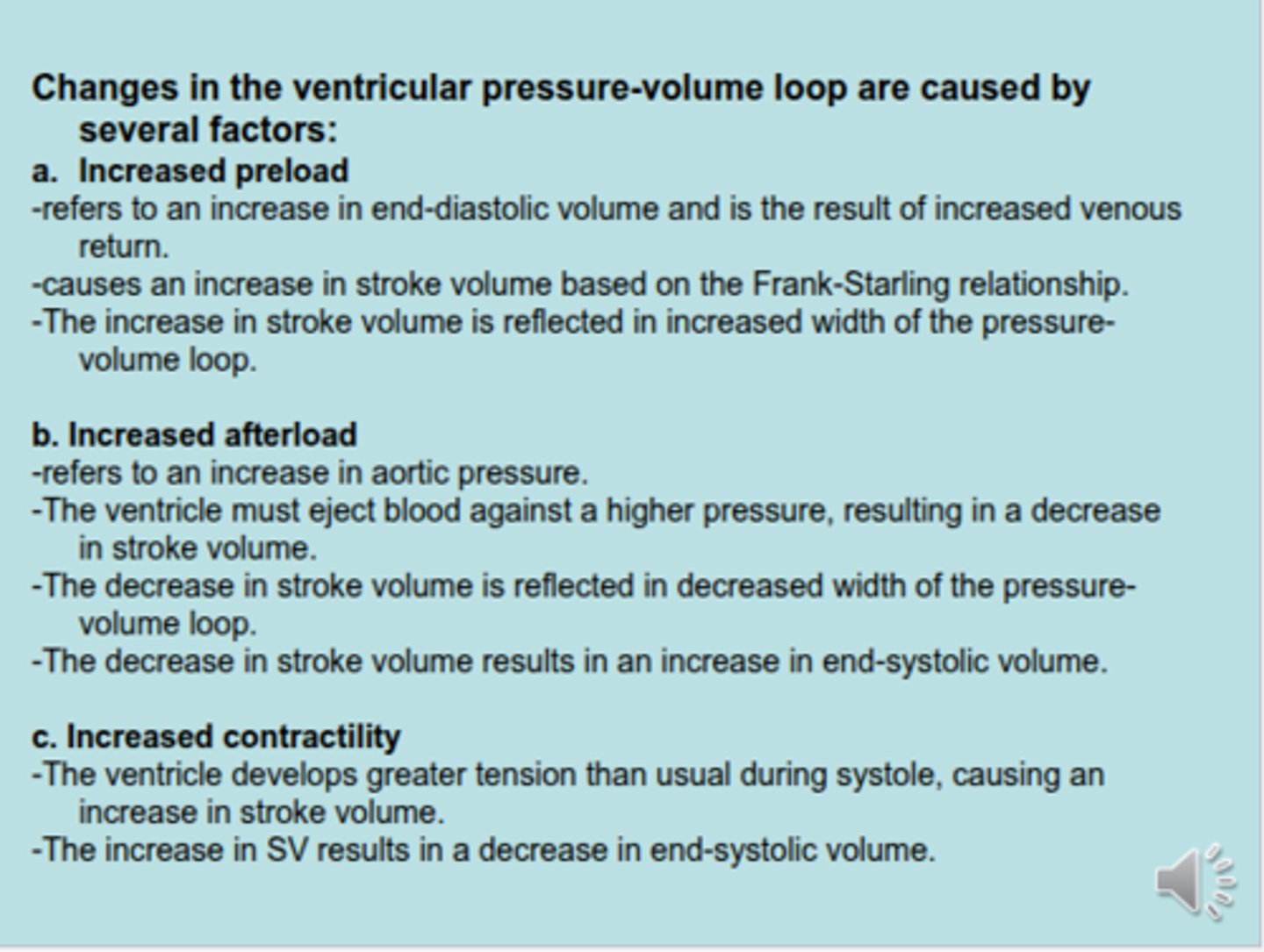
What happens if a person has increased preload?
preload normally is EDV of 140 mL
---
FOR EX : person has 190 mL :
- stroke volume is increased, instead of 70 mL ejection, 90 mL is ejected
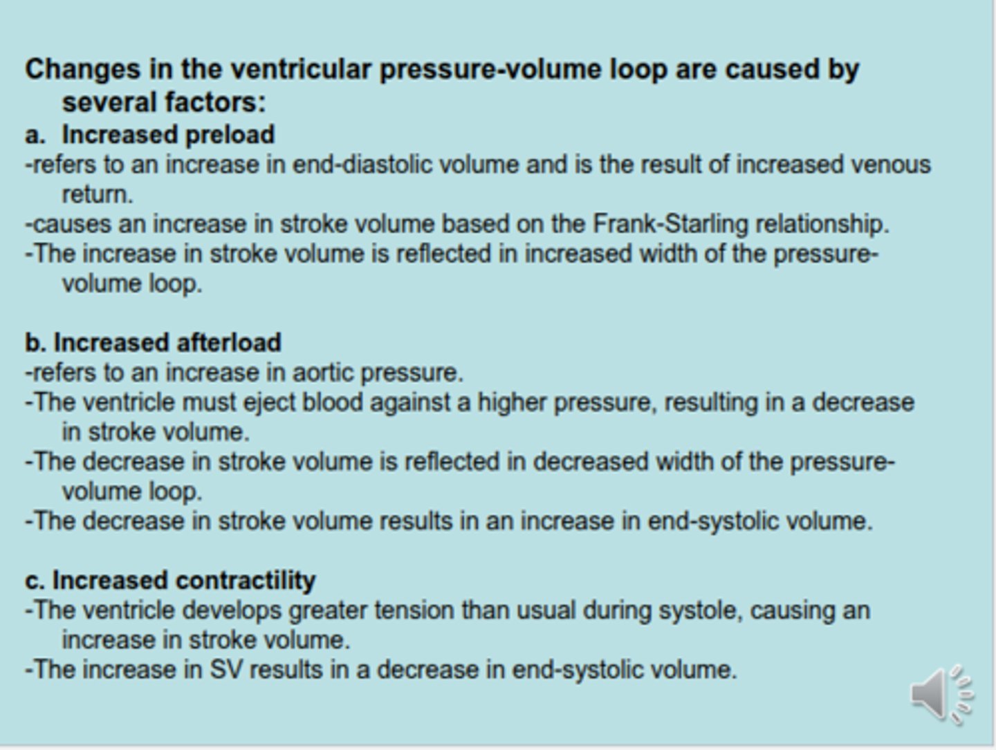
What happens if a person has increased after load?
- aortic pressure is increased
- this decreases stroke volume (instead of 70 mL, maybe its only 45 mL)
- this increases ESV
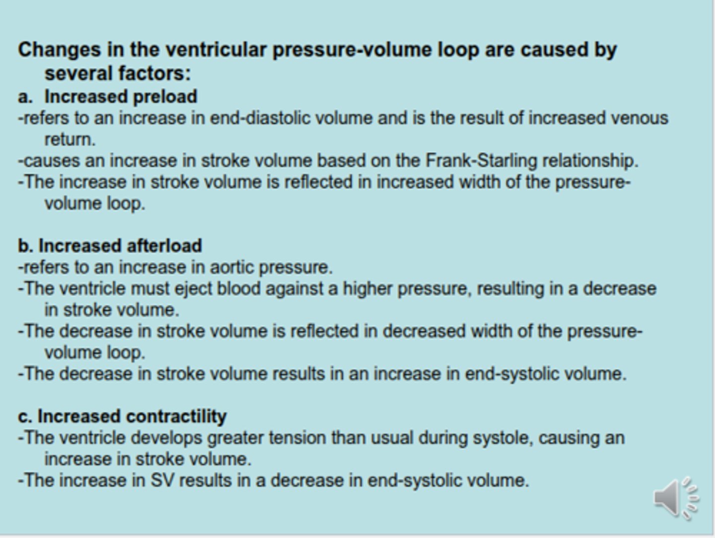
What happens if a person has increased contractility?
- increased stroke volume
- decreased ESV
In what condition would you have increased contractility?
oversecretion of NE (sympathetic)
-
increased NE leads to increased stroke volume, but decreased ESV
the volume ejected from the ventricle on each beat
stroke volume
stroke volume EQ?
SV = EDV - ESV
Cardiac output EQ?
CO = HR x SV
what is ejection fraction?
the fraction of end-diastolic volume that is ejected in each stroke volume
-
is related to contractility
-
normally 0.55 (55%)
What is the formula for Ejection Fraction?
EF = SV/EDV
What is cardiac O2 consumption directly related to?
the amount of tension developed by the ventricles
What is cardiac O2 consumption increased by? (4 things)
1. Increased afterload (increased aortic pressure)
2. Increased size of the heart (cardiomegaly)
3. Increased contractility
4. Increased heart rate
-
Increased contractility & HR depends of blood calcium (hypercalcemia) & over secretion of NE (B-1)