Unit 3 nervous system 1
 Call Kai
 Learn
Learn Practice Test
 Spaced Repetition
 Match
Match Flashcards
 Knowt Play
1/64

Earn XP
Description and Tags
HW #9 Biol 2404 ACC
Name | Mastery | Learn | Test | Matching | Spaced | Call with Kai | Chat |
|---|
No analytics yet
Send a link to your students to track their progress
65 Terms
One general function of the nervous system is to monitor the internal and external environment. This function is also known as
sensory input
The two parts of the central nervous system (CNS) are
brain and spinal cord
The two parts of the peripheral nervous system (PNS) are
nerves and ganglia
One general function of the nervous system is to coordinate the motor response of the body. This function is also known a
motor output
One general function of the nervous system is to process sensory information and decide if the body needs to respond and how. This function is also known as
integration
These are cytoplasmic extensions that receive information from other sources, such as receptors and other neurons.
dendrites
These are cytoplasmic extensions that transmit information to other cells, such as other neurons or effectors.
axons
These are the functional cells of the nervous system that generate electrical signals that are used for communication.
neurons
These are support cells of the nervous system. Although they have many different functions, their main job is to support the functional cells of the nervous system.
neuroglial cells
This is the part of a neuron that contains most organelles and carries out the normal cell functions, such as protein synthesis. It is also called the soma.
cell body
These are the three types of neurons based on function.
association neurons, motor neurons, sensory neurons
These are the three types of neurons based on structure.
bipolar neurons, multipolar neurons, unipolar neurons
This type of neuron conducts motor information from the CNS to the effectors. This is also called a motor neuron.
efferent neuron
This type of neuron is found in the CNS. These neurons are involved in integration. This is also called an interneuron in reflex arcs.
association neuron
This type of neuron conducts sensory information from the sensory receptors to the CNS. This is also called a sensory neuron.
afferent neuron
Schwann cells are neuroglial cells found in the __.
PNS
Satellite cells are neuroglial cells found in the __.
PNS
Ependymal cells are neuroglial cells found in the __.
CNS
Microglial cells are neuroglial cells found in the __.
CNS
Astrocytes are neuroglial cells found in the __.
CNS
Oligodendrocytes are neuroglial cells found in the __.
CNS
These are neuroglial cells that maintain the blood-brain barrier in the CNS.
astrocytes
These are phagocytic neuroglial cells in the CNS.
microglial cells
These are neuroglial cells that move CSF (cerebrospinal fluid) with cilia.
ependymal cells
These are neuroglial cells that support the cell bodies of neurons located in the PNS.
satellite cells
These are neuroglial cells that produce myelin in the central nervous system (CNS).
oligodendrocytes
These are neuroglial cells that produce myelin in the peripheral nervous system (PNS).
Schwann cells
These are collections of cell bodies in the CNS.
nuclei (singular: nucleus)
__ matter is made of myelinated axons.
white
These are groups of axons in the PNS.
nerves (singular: nerve)
These are collections of cell bodies in the PNS.
ganglia (singular: ganglion)
These are groups of axons in the CNS.
tracts (singular: tract)
__ matter is made of cell bodies and unmyelinated axons.
gray

Identify the structural type of neuron labeled “A” in the diagram below.
multipolar neuron

Identify the structural type of neuron labeled “B” in the diagram below.
bipolar neuron
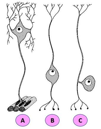
Identify the structural type of neuron labeled “C” in the diagram below.
unipolar neuron

Identify the structure labeled “D” on the diagram of a myelinated neuron.
axon

Identify the structure labeled “E” on the diagram of a myelinated neuron.
axon terminals
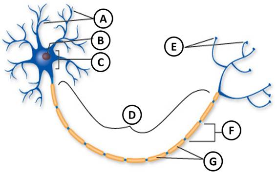
Identify the structure labeled “A” on the diagram of a myelinated neuron.
dendrites

Identify the structure labeled “G” on the diagram of a myelinated neuron.
Schwann cells

Identify the structure labeled “C” on the diagram of a myelinated neuron.
cell body (soma)

Identify the structure labeled “F” on the diagram of a myelinated neuron.
nodes of Ranvier
In a resting neuron, there are more __ ions inside the plasma membrane (cytosol) than outside the plasma membrane (ECF).
potassium
The electrical charge across the plasma membrane of a resting neuron is called the
resting membrane potential
In a resting neuron, there are more __ions outside the plasma membrane (ECF) than inside the membrane (cytosol).
sodium
Neurons typically have a resting membrane potential of __ mV.
-70 mV
These potentials are local changes in membrane potential that occur when chemically-gated receptors on dendrites or the cell body respond to neurotransmitters.
graded potentials
These ions move into the axon when voltage-gated sodium channels open.
sodium
These voltage-gated channels open and close slowly.
potassium channels
These voltage-gated channels open and close quickly.
sodium channels
These potentials are electrical changes in membrane potential that move down the length of an axon. These changes are due to the opening and closing of voltage-gated channels.
action potentials
These ions move out of the axon when voltage-gated potassium channels open.
potassium
This term means the plasma membrane is more positively charged than its resting membrane potential.
depolarization
This term means the plasma membrane is more negatively charged than its resting membrane potential.
hyperpolarization
This term means a positively charged membrane is becoming more negatively charged as it returns to its resting membrane potential.
repolarization
Continuous propagation is __than saltatory conduction.
Option B
slower
Saltatory conduction occurs in axons of
myelinated neurons
Continuous propagation occurs in axons of
unmyelinated neurons

Identify the structures labeled “A” on the synapse drawing below.
axon of presynaptic neuron

Identify the structures labeled “2” on the synapse drawing below.
synaptic vesicle containing neurotransmitters

Identify the structures labeled “4” on the synapse drawing below.
synaptic cleft

Identify the structures labeled “5” on the synapse drawing below.
receptor with attached neurotransmitter molecule

Identify the structures labeled “B” on the synapse drawing below.
dendrite of postsynaptic neuron

Identify the structures labeled “3” on the synapse drawing below.
neurotransmitter molecule

Identify the structures labeled “6” on the synapse drawing below.
synaptic vesicle releasing neurotransmitter by exocytosis