Emergency Medicine EOR (2026): Dermatology (Smarty PANCE)
 Call Kai
 Learn
 Practice Test
 Spaced Repetition
 Match
 Flashcards
 Knowt Play
1/178

There's no tags or description
Looks like no tags are added yet.
179 Terms
What is contact dermatitis?
An inflammatory skin reaction caused by direct exposure to an irritant or allergen, presenting acutely in the ED as a pruritic, erythematous, often vesicular eruption limited to the area of contact.
What distinguishes allergic from irritant contact dermatitis?
Allergic is a delayed type IV hypersensitivity reaction (12-72 hours after exposure) that is intensely pruritic with vesicles and sharp borders; irritant is a direct non-immune chemical injury that occurs within minutes to hours and is more painful/burning than itchy.
What plant antigen causes the classic linear vesicular rash of poison ivy, oak, and sumac?
Urushiol oil; the linear/streaked pattern reflects where the plant brushed the skin, and the fluid in the blisters is NOT contagious.

What is the first-line treatment for localized contact dermatitis in the ED?
Remove/wash off the offending agent, then medium-to-high potency topical corticosteroids (triamcinolone 0.1%) plus oral antihistamines for pruritus and cool compresses.
When should systemic corticosteroids be given for contact dermatitis?
Involvement of >10% BSA, or significant involvement of the face, hands, feet, or genitals; use prednisone 40-60 mg daily tapered over 14-21 days (short 5-day bursts cause rebound).
What is the most common cause of allergic contact dermatitis from jewelry, belt buckles, and snaps?
Nickel
What is a drug eruption?
Any cutaneous adverse reaction to a medication, ranging from a benign morbilliform rash to life-threatening reactions such as DRESS, SJS, and TEN.
What is the most common type of drug eruption and when does it appear?
Morbilliform (exanthematous/maculopapular) eruption, typically appearing 7-14 days after starting a new drug (or within 1-2 days on re-exposure), starting on the trunk and spreading symmetrically.
What ED red flags suggest a severe cutaneous adverse reaction rather than a benign drug rash?
Fever, mucosal involvement, skin pain/tenderness, facial edema, blistering or skin sloughing, positive Nikolsky sign, lymphadenopathy, and end-organ dysfunction (transaminitis, eosinophilia, AKI).
What is DRESS syndrome and what is its classic timing?
Drug Reaction with Eosinophilia and Systemic Symptoms: fever, facial edema, morbilliform rash, lymphadenopathy, eosinophilia, and hepatitis occurring 2-8 WEEKS after drug initiation (later than other drug eruptions).
What drugs most commonly cause DRESS?
Anticonvulsants (phenytoin, carbamazepine, lamotrigine), allopurinol, sulfonamides, vancomycin, and minocycline.
What is a fixed drug eruption?
A well-demarcated round dusky/violaceous plaque that recurs at the exact SAME site with each re-exposure to the culprit drug (classically NSAIDs, sulfonamides, tetracyclines, or barbiturates), often leaving postinflammatory hyperpigmentation.
What is the ED management of a benign morbilliform drug eruption?
Stop the offending drug, give antihistamines and topical steroids for symptom relief, document the allergy, and give clear return precautions for mucosal involvement, blistering, or fever.
What is urticaria?
A mast-cell mediated eruption of transient, pruritic, well-circumscribed edematous wheals with surrounding erythema, in which individual lesions migrate and resolve within 24 hours without residual skin changes.
How is acute urticaria distinguished from chronic urticaria?
Acute lasts less than 6 weeks (usually infection, drug, or food triggered); chronic lasts more than 6 weeks and is most often idiopathic or autoimmune and does not require ED workup.
What is the first-line ED treatment for uncomplicated urticaria?
Second-generation H1 antihistamines (cetirizine, loratadine) at up to 4 times standard dose, with an H2 blocker (famotidine) as adjunct; add a short steroid burst for severe or refractory cases.
When does urticaria mandate epinephrine?
When accompanied by any sign of anaphylaxis: airway involvement (stridor, hoarseness, tongue/lip swelling), respiratory compromise, hypotension, or persistent GI symptoms — give IM epinephrine 0.3-0.5 mg (0.01 mg/kg peds) in the anterolateral thigh.
Why does ACE inhibitor angioedema not respond to epinephrine, steroids, or antihistamines?
It is bradykinin-mediated, not histamine-mediated; management is airway protection first, with icatibant, C1-esterase inhibitor, or FFP as adjuncts and permanent discontinuation of the drug.
What is a key feature that distinguishes urticaria from erythema multiforme or a viral exanthem?
Individual urticarial lesions are migratory and resolve within 24 hours and leave no residual pigmentation; lesions fixed for more than 24 hours or leaving bruising suggest urticarial vasculitis or another diagnosis.
What is bullous pemphigoid?
A chronic autoimmune subepidermal blistering disease of the elderly caused by IgG autoantibodies against hemidesmosomal proteins (BP180/BP230), producing large TENSE bullae on an urticarial base.
What is the classic ED presentation of bullous pemphigoid?
An elderly patient (typically over 60) with intensely pruritic urticarial plaques that evolve into large, tense, fluid-filled bullae on the flexural surfaces, trunk, and thighs; mucosal involvement is uncommon.

How does bullous pemphigoid differ from pemphigus vulgaris?
Pemphigoid = TENSE bullae, subepidermal, NEGATIVE Nikolsky, elderly, mucosa usually spared, better prognosis. Pemphigus vulgaris = FLACCID bullae, intraepidermal, POSITIVE Nikolsky, oral mucosa involved first, higher mortality.
What is the Nikolsky sign and what does it indicate?
Gentle lateral pressure on perilesional skin causing the epidermis to shear off; it is POSITIVE in pemphigus vulgaris, SJS/TEN, and staphylococcal scalded skin syndrome, and NEGATIVE in bullous pemphigoid.

What is the confirmatory test for bullous pemphigoid?
Skin biopsy with direct immunofluorescence showing LINEAR IgG and C3 deposition along the basement membrane zone.
What is the treatment for bullous pemphigoid?
High-potency topical corticosteroids (clobetasol) for localized disease and systemic prednisone for widespread disease, with dermatology referral; admit for extensive disease, secondary infection, or fluid loss.
What is erythema multiforme?
An acute, immune-mediated hypersensitivity reaction characterized by symmetric TARGET (iris) lesions with three distinct zones, most often triggered by infection rather than drugs.
What is the most common trigger of erythema multiforme?
Herpes simplex virus (HSV-1 more than HSV-2) accounts for the majority of cases, especially recurrent EM; Mycoplasma pneumoniae is the most common trigger in children.
Describe the classic erythema multiforme target lesion.
A round lesion with three concentric zones: a dusky/bullous central area, a surrounding pale edematous ring, and an outer erythematous halo — classically appearing acrally on the palms, soles, and extensor extremities and spreading centripetally.

How is EM minor distinguished from EM major?
EM minor has NO or minimal mucosal involvement and no systemic symptoms; EM major involves one or more mucosal surfaces (usually oral) with fever and prodromal symptoms.
How does erythema multiforme differ from SJS/TEN?
EM has raised, well-demarcated TYPICAL targets in an acral distribution with less than 10% BSA and is usually infection-triggered; SJS/TEN has flat atypical targets and dusky macules on the TRUNK with widespread epidermal detachment and is usually drug-triggered.
What is the ED management of erythema multiforme?
Supportive care, treat the underlying trigger (acyclovir for HSV, macrolide for Mycoplasma), antihistamines and topical steroids for symptoms, and oral rinses (magic mouthwash) for mucosal disease; suppressive acyclovir prevents recurrent HSV-associated EM.
What is pilonidal disease?
An acquired condition in which hair and debris become trapped in the natal cleft, producing a sinus tract that may present as an asymptomatic pit, an acutely painful abscess, or a chronically draining sinus.
Where is pilonidal disease located and who is most commonly affected?
The sacrococcygeal region/natal cleft, superior to the anus in the midline; classically young hirsute males aged 15-30, with risk factors of obesity, prolonged sitting, and poor hygiene.
What is the ED management of an acute pilonidal abscess?
Incision and drainage — using an incision made LATERAL to the midline (off the natal cleft) because midline incisions heal poorly; irrigate, break up loculations, and remove hair/debris from the cavity.
Are antibiotics routinely indicated after drainage of a pilonidal abscess?
No — I&D alone is sufficient for most patients; reserve antibiotics for surrounding cellulitis, systemic symptoms, immunosuppression, or diabetes.
What is the definitive treatment for recurrent pilonidal disease?
Surgical excision of the sinus tract with primary or secondary closure (or flap procedures), arranged as outpatient surgical follow-up; recurrence after I&D alone is common.
What discharge instructions reduce pilonidal recurrence?
Local hair removal/depilation, meticulous hygiene of the natal cleft, weight loss, and avoidance of prolonged sitting.
What is toxic epidermal necrolysis?
A life-threatening, almost always drug-induced severe cutaneous adverse reaction causing widespread keratinocyte apoptosis with full-thickness epidermal detachment of greater than 30% of body surface area.
What BSA thresholds separate SJS, SJS/TEN overlap, and TEN?
SJS = less than 10% BSA epidermal detachment; SJS/TEN overlap = 10-30%; TEN = greater than 30%.
What drugs most commonly cause SJS/TEN?
Sulfonamides (TMP-SMX), anticonvulsants (lamotrigine, carbamazepine, phenytoin, phenobarbital), allopurinol, nevirapine, and NSAIDs (oxicam class) — usually 1-3 weeks after initiation.
What are the classic clinical features of TEN?
A prodrome of fever and flu-like symptoms, then painful dusky macules and atypical targets on the trunk/face that coalesce and slough, with POSITIVE Nikolsky sign and involvement of two or more mucosal surfaces (ocular, oral, genital).

What is SCORTEN?
A validated 7-variable mortality prediction score for SJS/TEN (age over 40, malignancy, heart rate over 120, BSA detachment over 10%, BUN over 28, glucose over 252, bicarbonate under 20) calculated within 24 hours of admission.
What is the ED management of TEN?
Immediately STOP the culprit drug, aggressive fluid resuscitation, aggressive analgesia, non-adherent wound care, ophthalmology and dermatology consultation, and transfer to a BURN CENTER or ICU; airway protection if mucosal involvement compromises the airway.
How is TEN distinguished from staphylococcal scalded skin syndrome (SSSS)?
TEN involves MUCOSAL surfaces with full-thickness epidermal necrosis and occurs in adults on medications; SSSS SPARES mucous membranes, involves only the superficial granular layer, occurs in young children, and is treated with anti-staphylococcal antibiotics.
What is a cutaneous abscess?
A localized, walled-off collection of pus within the dermis or subcutaneous tissue presenting as a fluctuant, erythematous, tender nodule that requires drainage rather than antibiotics alone.
What is the definitive treatment for a simple cutaneous abscess?
Incision and drainage — this is the cornerstone of therapy and antibiotics alone will not cure an established abscess.
What is the most common pathogen in cutaneous abscesses?
Staphylococcus aureus, with community-acquired MRSA predominating in many regions.
When should antibiotics be added after incision and drainage?
Surrounding cellulitis, abscess larger than 2 cm, multiple abscesses, systemic signs (SIRS), immunosuppression/diabetes, extremes of age, failed prior drainage, or high-risk locations (face, hand, genitals); TMP-SMX or doxycycline covers MRSA.
What is the role of bedside ultrasound in suspected abscess?
It reliably differentiates abscess (anechoic/hypoechoic fluid collection with posterior acoustic enhancement and "swirl" sign) from cellulitis alone, improving accuracy and preventing unnecessary incisions.
Which abscesses should NOT be drained in the ED without consultation?
Abscesses over the central face/nasolabial triangle (cavernous sinus risk), the hand/palmar space, near major vessels or nerves, perirectal/deep space abscesses, and suspected mycotic aneurysms — these need specialty involvement.
What is a "high-risk" abscess mimic that must be excluded before I&D?
A pulsatile mass, which may be a pseudoaneurysm rather than an abscess — especially in IV drug users with groin swelling; use ultrasound before incising.
What is cellulitis?
An acute bacterial infection of the deep dermis and subcutaneous fat presenting as poorly demarcated erythema, warmth, edema, and tenderness, usually without a drainable collection.
What organisms cause nonpurulent versus purulent cellulitis?
Nonpurulent (diffuse spreading erythema) is usually beta-hemolytic Streptococcus; purulent (associated abscess or drainage) is usually Staphylococcus aureus including MRSA.
What are the first-line antibiotics for nonpurulent cellulitis?
Cephalexin, dicloxacillin, or (if IV) cefazolin — targeting streptococci; routine MRSA coverage is NOT required for nonpurulent cellulitis.
What antibiotics are used for purulent cellulitis or suspected MRSA?
TMP-SMX, doxycycline, or clindamycin for outpatients; vancomycin, linezolid, or daptomycin for inpatients.
How does erysipelas differ from cellulitis?
Erysipelas involves the upper dermis and superficial lymphatics with SHARPLY demarcated, raised, brightly erythematous borders (classic on the face or lower leg), is caused by group A Streptococcus, and often has abrupt fever.
What findings suggest necrotizing fasciitis rather than cellulitis?
Pain out of proportion to exam, rapid progression, systemic toxicity/hypotension, crepitus, bullae, skin anesthesia, dusky or necrotic skin, and gas on imaging; requires emergent surgical consultation, broad-spectrum antibiotics with clindamycin, and resuscitation.
What is the most commonly misdiagnosed mimic of bilateral lower extremity cellulitis?
Stasis dermatitis — cellulitis is almost always UNILATERAL, so bilateral leg erythema without fever should prompt reconsideration (also consider DVT, lymphedema, contact dermatitis, and gout).
What are indications for admission and IV antibiotics in cellulitis?
Systemic toxicity/sepsis, rapid progression, failed outpatient therapy, immunocompromise, significant comorbidities, inability to tolerate oral medication, orbital or perineal involvement, or concern for necrotizing infection.
What is folliculitis?
A superficial infection or inflammation of the hair follicle producing clusters of small erythematous papules and pustules centered on hair follicles.
What organism most commonly causes bacterial folliculitis?
Staphylococcus aureus
What is "hot tub folliculitis" and what causes it?
A pruritic pustular eruption appearing 1-3 days after exposure to inadequately chlorinated hot tubs, pools, or whirlpools, caused by Pseudomonas aeruginosa; it involves areas covered by a swimsuit and is usually SELF-LIMITED without antibiotics.

How do folliculitis, furuncle, and carbuncle differ?
Folliculitis is confined to the follicle; a furuncle (boil) is a deeper abscess of a single follicle extending into subcutaneous tissue; a carbuncle is a coalescence of multiple furuncles with multiple draining sinus tracts and often systemic symptoms.
What is the treatment for uncomplicated bacterial folliculitis?
Warm compresses and good hygiene, plus topical antibiotics (mupirocin or clindamycin); oral antistaphylococcal antibiotics are reserved for extensive, recurrent, or refractory disease.
What is pseudofolliculitis barbae?
"Razor bumps" — a non-infectious foreign-body inflammatory reaction to ingrown curved hairs in the beard area, most common in Black men; treatment is stopping shaving or changing shaving technique, not antibiotics.

What is impetigo?
A highly contagious superficial bacterial skin infection of the epidermis, most common in children, classically producing honey-colored crusted erosions.
What organisms cause impetigo?
Staphylococcus aureus (most common overall and the sole cause of bullous impetigo) and Streptococcus pyogenes (group A strep).
How does nonbullous impetigo present?
Vesicles and pustules that rupture to form the classic golden/HONEY-COLORED crust, most often on the face around the nose and mouth, in children aged 2-5.

How does bullous impetigo present and what causes it?
Flaccid, fluid-filled bullae that rupture leaving a collarette of scale and a shallow erosion, caused by exfoliative toxin-producing Staphylococcus aureus; more common in neonates and infants.

What is the treatment for localized versus widespread impetigo?
Localized: topical mupirocin or retapamulin twice daily for 5 days. Widespread or with systemic symptoms: oral cephalexin or dicloxacillin (or doxycycline/TMP-SMX/clindamycin if MRSA is suspected).
What is the most important complication of streptococcal impetigo?
Post-streptococcal glomerulonephritis; note that antibiotic treatment of impetigo does NOT prevent PSGN, and unlike pharyngitis, impetigo does not cause acute rheumatic fever.
When may a child with impetigo return to school?
After 24 hours of appropriate antibiotic therapy with lesions covered.
What are cutaneous fungal infections?
Superficial infections of the skin, hair, and nails caused by dermatophytes (Trichophyton, Microsporum, Epidermophyton), Candida species, or Malassezia, producing characteristic annular, macerated, or scaly lesions.
What is the classic presentation and bedside test for tinea corporis?
An annular, scaly plaque with a raised advancing border and CENTRAL CLEARING ("ringworm"); KOH preparation of a scraping from the active border shows branching septate hyphae.

Why does tinea capitis require ORAL rather than topical therapy?
Topical agents cannot penetrate the hair follicle; treat with oral griseofulvin or terbinafine for 6-8 weeks, and consider adjunct selenium sulfide or ketoconazole shampoo to reduce transmission.
What is a kerion and how is it managed?
A boggy, tender, inflammatory mass with pustules and alopecia representing a severe hypersensitivity response to tinea capitis; it is NOT an abscess and must not be incised — treat with oral antifungals with or without corticosteroids.

How is candidal intertrigo distinguished from tinea?
Candidal intertrigo occurs in warm moist skin folds with beefy-red macerated plaques and characteristic SATELLITE papules and pustules, and lacks central clearing; treat with topical nystatin or an azole and keep the area dry.
What is the classic KOH finding in tinea versicolor?
A "spaghetti and meatballs" appearance of short hyphae and spores from Malassezia furfur, producing hypopigmented or hyperpigmented scaly macules on the trunk; treat with topical selenium sulfide or ketoconazole.

What is a common ED pitfall when treating a rash with steroids?
Applying topical corticosteroids to a dermatophyte infection produces "tinea incognito" — a modified, less-scaly, atypical, spreading rash; always consider fungal etiology in a rash that worsens with steroids.
What is herpes zoster?
Reactivation of latent varicella-zoster virus within a sensory dorsal root or cranial nerve ganglion, producing a painful, unilateral, dermatomal vesicular eruption that does not cross the midline.
What is the classic presentation of herpes zoster?
A prodrome of pain, burning, or paresthesia for 2-3 days, followed by grouped vesicles on an erythematous base confined to one (or occasionally two adjacent) dermatomes, most commonly thoracic or trigeminal.

What is the antiviral treatment window for herpes zoster and what agents are used?
Antivirals are most effective when started within 72 HOURS of rash onset — valacyclovir 1 g TID, famciclovir 500 mg TID, or acyclovir 800 mg five times daily for 7 days; still treat beyond 72 hours if new lesions are still forming or in high-risk patients.
What is herpes zoster ophthalmicus and what is Hutchinson sign?
Reactivation in the V1 (ophthalmic) division of the trigeminal nerve; Hutchinson sign is vesicles on the TIP or SIDE of the nose (nasociliary branch involvement), which predicts ocular involvement and mandates urgent ophthalmology evaluation and slit-lamp exam.

What is Ramsay Hunt syndrome?
Herpes zoster oticus — VZV reactivation in the geniculate ganglion causing ipsilateral facial nerve palsy, ear pain, and vesicles in the external auditory canal or on the auricle, often with hearing loss, tinnitus, and vertigo; treat with antivirals PLUS corticosteroids.

What isolation precautions are required for herpes zoster?
Localized zoster in an immunocompetent patient requires only CONTACT precautions with lesions covered; DISSEMINATED zoster or zoster in an immunocompromised patient requires AIRBORNE plus contact precautions.
What is postherpetic neuralgia and how is it treated?
Dermatomal pain persisting more than 90 days after rash resolution, most common in patients over 60; treated with gabapentin, pregabalin, tricyclic antidepressants, or topical lidocaine/capsaicin.
What is measles (rubeola)?
A highly contagious airborne paramyxovirus infection producing high fever, a prodrome of cough/coryza/conjunctivitis, pathognomonic Koplik spots, and a descending morbilliform rash.
What is the classic prodrome of measles?
The "3 C's" — Cough, Coryza, and Conjunctivitis — with high fever, occurring 2-4 days before the rash.
What are Koplik spots?
Tiny blue-white papules on an erythematous base on the buccal mucosa opposite the molars — PATHOGNOMONIC for measles, appearing 1-2 days BEFORE the rash and fading as the rash appears.

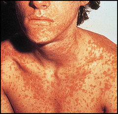
Describe the measles rash.
An erythematous, blanching, maculopapular (morbilliform) rash that begins at the hairline and behind the ears and spreads CEPHALOCAUDALLY to the trunk and extremities, becoming confluent and often desquamating as it fades.
What isolation precautions are required for suspected measles in the ED?
AIRBORNE precautions — immediately mask the patient, place in a negative-pressure room, and notify the health department; the virus remains infectious in air for up to 2 hours after the patient leaves the room.
What treatment reduces measles morbidity and mortality?
Vitamin A supplementation for all children with measles; the remainder of care is supportive as there is no specific antiviral.
What are the major complications of measles?
Otitis media (most common), pneumonia (most common cause of death), diarrhea, acute encephalitis, and the rare late complication subacute sclerosing panencephalitis (SSPE).
What is post-exposure prophylaxis for measles?
MMR vaccine within 72 hours of exposure, or immunoglobulin within 6 days for high-risk contacts (infants under 12 months, pregnant women, immunocompromised patients).
What is rubella?
A generally mild togavirus infection ("German measles" or "3-day measles") producing low-grade fever, tender posterior lymphadenopathy, and a fine pink maculopapular rash — dangerous primarily because of its teratogenic effects.
What lymphadenopathy pattern is characteristic of rubella?
Tender POSTERIOR AURICULAR, posterior cervical, and suboccipital lymphadenopathy, which often precedes the rash.
What are Forchheimer spots?
Pinpoint petechiae on the soft palate seen in rubella; unlike Koplik spots they are NOT pathognomonic and can be seen in other viral illnesses.
How does the rubella rash differ from the measles rash?
Rubella's rash is finer, pinker, less confluent, spreads faster (face to whole body within 24 hours), lasts about 3 days, and is accompanied by only low-grade fever and a milder overall illness.
What is congenital rubella syndrome?
The classic triad of sensorineural DEAFNESS, CATARACTS, and CARDIAC defects (patent ductus arteriosus, pulmonary artery stenosis), plus "blueberry muffin" skin lesions and intellectual disability, resulting from first-trimester maternal infection.
What isolation precautions are needed for rubella and what is a key ED action?
DROPLET precautions; immediately determine pregnancy status of the patient and of exposed contacts and staff, and notify public health.